Section 3.3 Cancer Biology
 Call Kai
 Learn
Learn Practice Test
 Spaced Repetition
 Match
Match Flashcards
 Knowt Play
1/23

There's no tags or description
Looks like no tags are added yet.
Name | Mastery | Learn | Test | Matching | Spaced | Call with Kai | Chat |
|---|
No analytics yet
Send a link to your students to track their progress
24 Terms
Cancer is the Result of
Unregulated Cellular Division
Abnormal gene expression regulation can lead to such issues
Cancer occurs when:
Mutations develop in genes that regulate the cell cycle
Regulatory mechanisms that limit cell division are defective
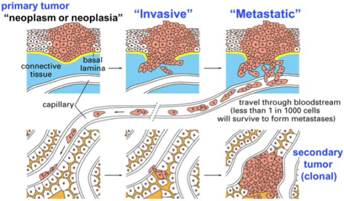
Cells undergo unregulated division; Two types:
Invasive: Tumor breaks through normal barriers between tissues
Metastatic: Can colonize new sites in the body
Primary tumor: “Benign” tumor, cells are replicating slightly faster than it should, still fine as long as it stays localized (Not yet cancer)
Clonal tumors result from metastatic tumors, are clones of the same cells just in a different areas (Ex, Breast cancer cells in lymphatic node)
Cancer kills by hindering normal cell processes
Characteric Changes in Cancer Cells
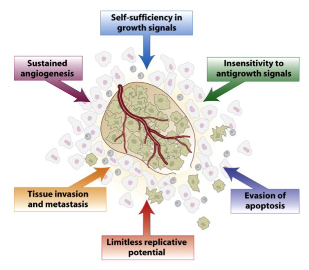
Angiogenesis: Activation of blood vessel growth by tumor; Via secretion of angiogenic growth factors like WECF; Provides more nutrients to growing tumor
Cancer cells are also; self-sufficient in growth signals and do not respond to antigrowth signals, they can also evade apoptosis, have limitless replicative potential (telomerase).
Apoptosis: Programmed cell death; Triggered by irreparable DNA damage and severe stresses
Cancer Incidence Increases With Age
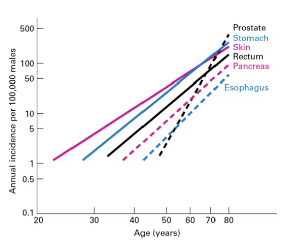
Due to mutation accumulation over a lifetime, such as:
Errors in normal biological processes like chromosome segregation and replication
Spontaneous DNA damage
Exposure to carcinogens or radiations
Inheritance of a mutation (genetic predisposition)
Epigenetic changes that can change gene expression
Cells that undergo constant turnover (i.e. epithelium) have higher development risks (Because pro-growth genes are constantly in euchromatic state)
Cells that do not divide do not develop into cancers (Ex, Heart muscle cells and neurons)
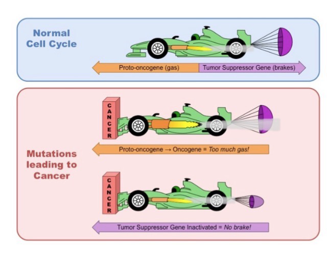
Cancer Pushes Down on the Cell Cycle “Gas Pedal” (One question on activation)
Proto-oncogenes: Normal genes that encodes cell growth proteins
Oncogenes: Mutated proto-oncogenes; Has Activation mutation: Conversion of proto-oncogene to oncogene; this mutation is genetically dominant and will lead to tumor formation
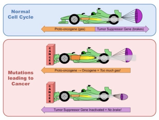
Cancer Doesn’t Have Cell Cycle “Brakes”
Tumor suppressor genes: Encodes regulatory proteins that normally inhibits cell division (Prevents cancer)
Loss of function mutations favors excessive growth (Via signaling pathways like proteins or genetic pathways); Mutations in these genes are genetically recessive
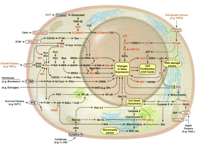
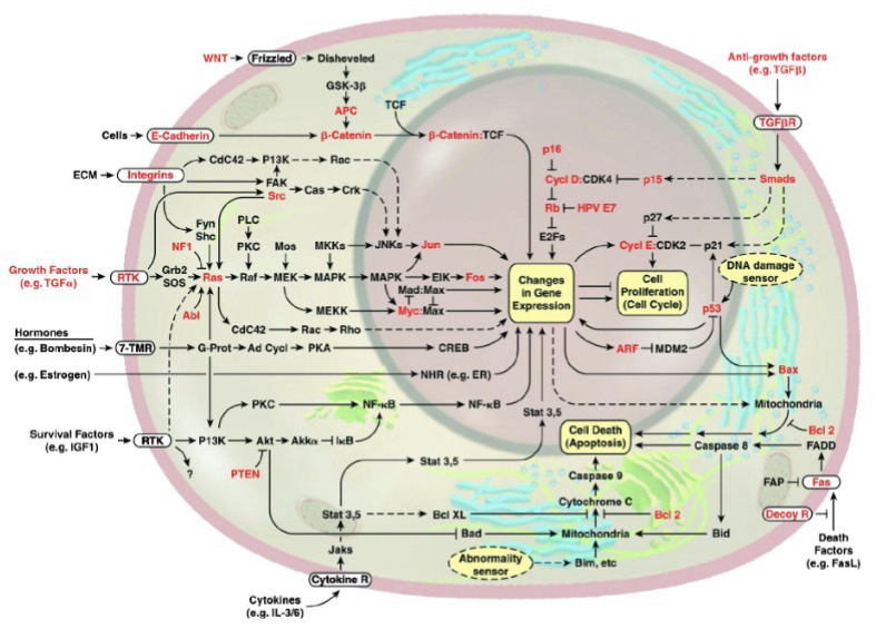
Signaling Pathways are Altered in Cancer
Cancer is driven by genetic and epigenetic alterations which cause constitutive activation of growth pathways and inactivation of tumor suppressors
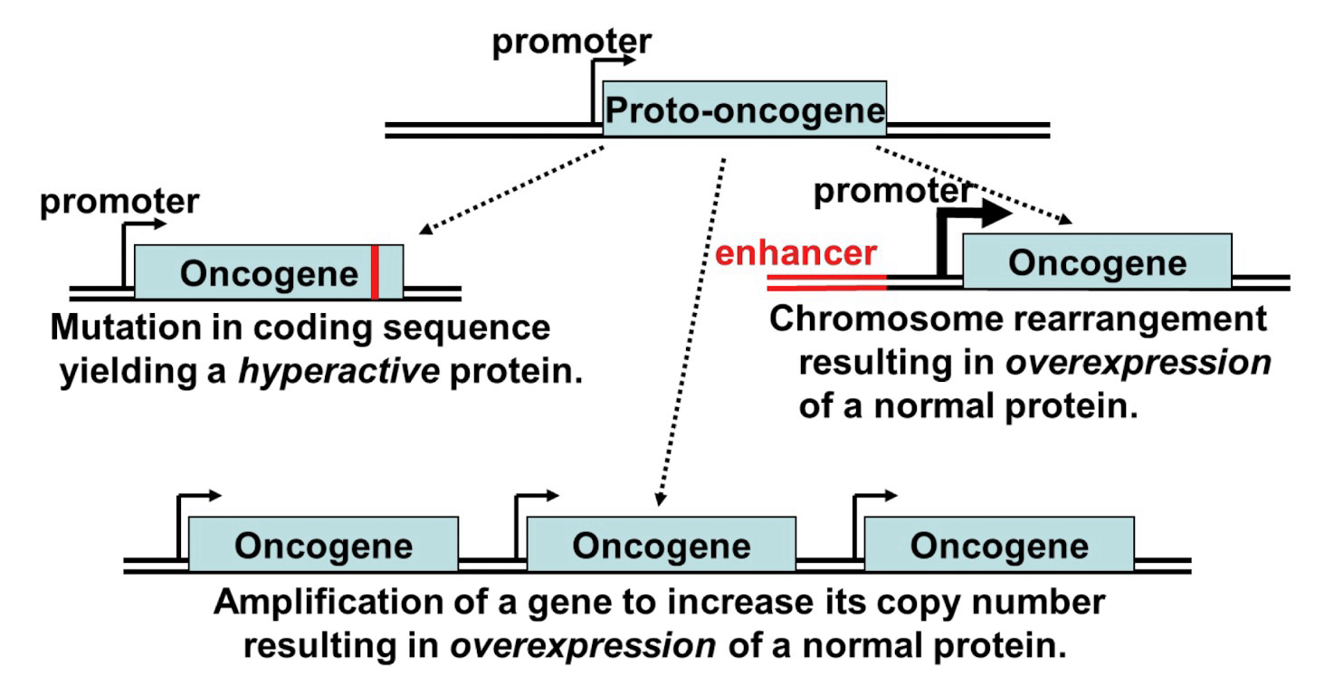
Gain of Function Mutations in Proto-Oncogenes
Activation mutation: Conversion of proto-oncogene to oncogene
Must occur for cancer development
Mutation in the coding of protein may yield hyperactive protein, amplification will increase the copy number resulting in overexpression, chromosome rearrangement results in overexpression
RAS Oncogene
Hyperactive protein example
K-RAS mutations occur in a large percentage of pancreatic, colorectal, and non-small cell lung cancers; Very aggressive tumors; Often at the codon for G12
G12C (Typical nomenclature for mutation; G at position 12 mutates to C) is the most common mutation of RAS in lung cancers; GGT → TGT mutation that stops RAS from hydrolyzing GTP
First K-RAS inhibitor was approved by the FDA in 2021; Sotorasib; Small molecule that only targets G12C RAS, not healthy RAS; Survival rate was 50.8% after one year and 32.5% after two years
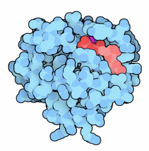
Amplification of the Her2 Gene
Amplification example
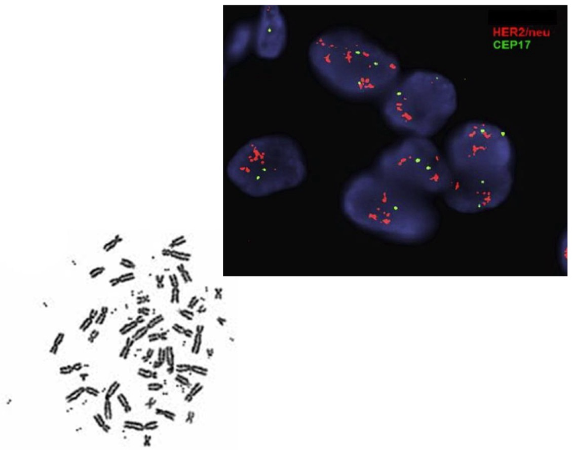
20%-30% of breast cancers overexpress HER2: A receptor tyrosine kinase (RTK) that can activate the RAS-dependent pathway
Amplification: Increase in gene copy number; The result of a localized error in replication (Ex, ORF is replicated several times; cells try to repair but sometimes that results in amplification)
A few too many hundreds of copies of a gene:
Within a chromosome “homogeneous region”
Extrachromosomal DNA “double minute chromosomes”
When repair systems try to remove error but it doesn’t get degraded and the regions that were excised has everything it needs for transcription
Targeted Treatment for HER2+ Cancer
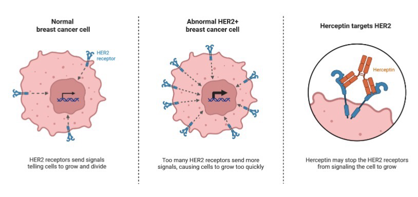
~25% of breast cancers are HER2+; which produces a high amount of HER2 receptor; because there is so many receptors, they have a chance of simply meeting each other and forming a dimer without going through the RTK system pathway
Herceptin is a synthetic antibody that binds to HER2 (Receptor that activates RAS pathway); Traps HER2 in monomer form instead of activated dimer form; This trap also signals and activates immune system to degrade cancerous cells
Herceptin Improves Survival Rates
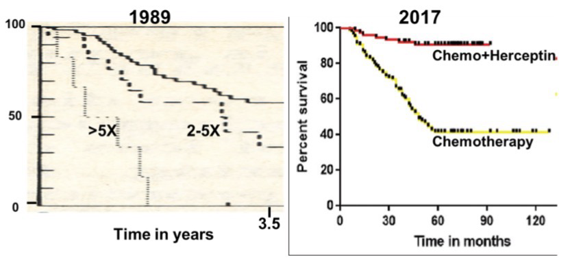
Modern chemotherapy against HER2+ Cancers uses Herceptin in combination with other chemotherapy drugs greatly improved survival; 84% survival rate at 10 years; However, a full one-year course of Herceptin treatment costs around $54,000
Chromosomal Rearrangements in Cancer
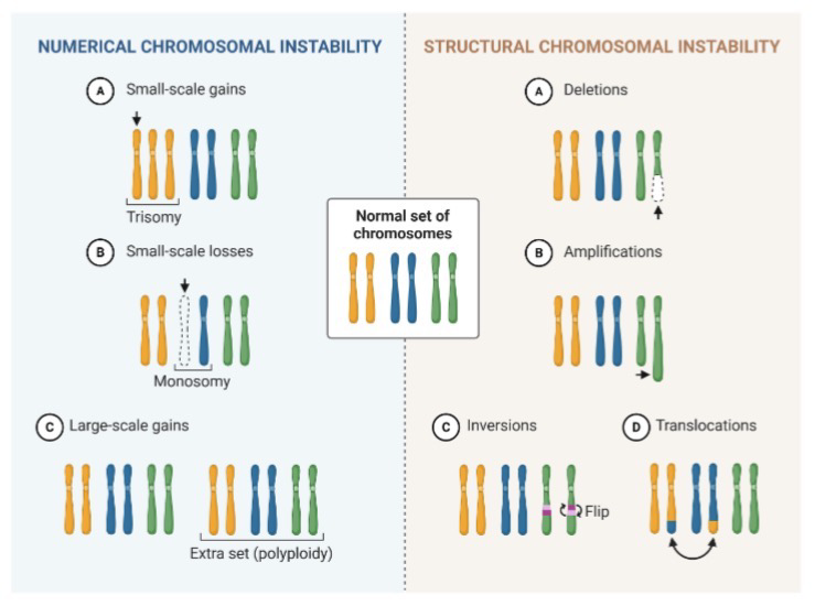
Chromosomal instability is common in cancer cells; Aneuploidy: Abnormal number of chromosomes
Various structural instability can also occur such as deletion, amplification, inversion, and translocation
Enhancer Hijacking in Medulloblastoma
Chromosomal rearrangement example
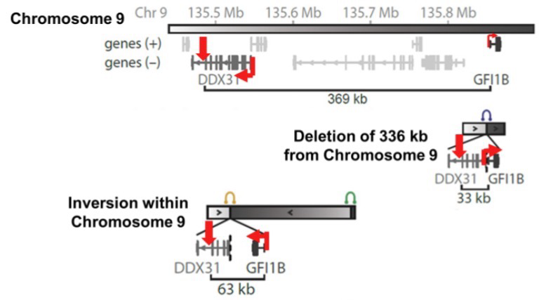
Medulloblastoma is an aggressive tumor; 18% of juvenile brain cancers; 70% of patents are <10 years old
Overexpression of GFI1B transcription factor can be caused by chromosomal rearrangements (Various mutations led to this)
Example mutation:
There’s an enhancer between DDX31 (Highly expressed region) and GFI1B; deletion causes rearrangement and causes overexpression of GFI18B; Inversion can also cause this
Philadelphia Chromosome
Chromosomal rearrangement example
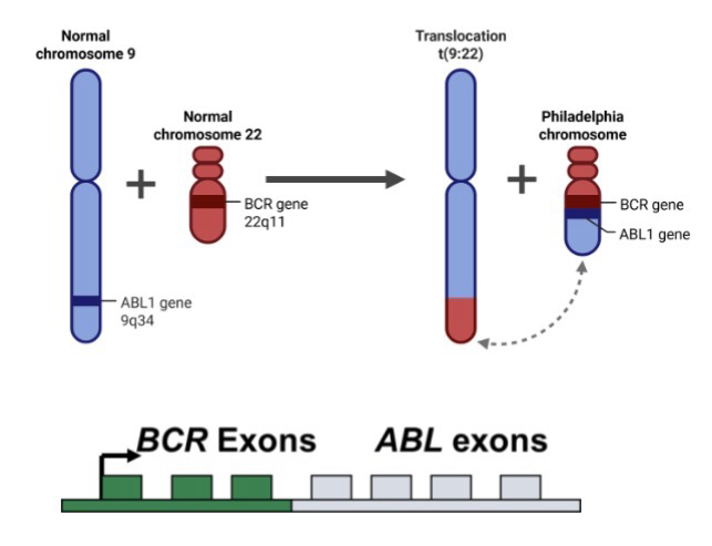
Chromosome 9-22 translocation; Chromosomal rearrangement seen in 95% if chronic myeloid leukemia (CML) patients
Philadelphia chromosome: Fusion of ABL (Pro-growth) gene from chromosome 9 to the BCR (Highly expressed) gene on chromosome 22, resulting in a BCR-ABL fusion protein;
ABL gene on chromosome 9 encodes a protein kinase; the BCR-ABL fusion protein is hyperactive
Gleevec is an inhibitor that binds in the ABL catalytic site; 87% survival rate out to 8 years
Retinoblastoma (Rb)
Rb protein is a checkpoint for G1
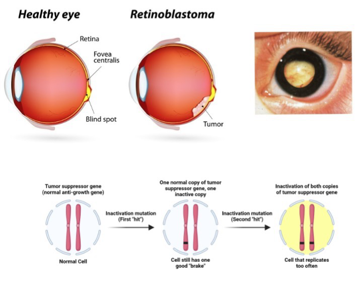
Sporadic retinoblastoma: Patient inherits two good copies of Rb gene; Sporadic tumor in one eye; No additional predisposition to cancer
Familial retinoblastoma: Patient inherits one defective Rb gene; Typically has non-clonal tumors in both eyes; ~85% of gene carriers develop tumors (high penetrance)
During eye development gene errors occur often so Rb mutation is more common
Two-Hit Hypothesis
Two-Hit Hypothesis:
Inactivation of a single copy of a tumor suppressor gene does NOT result in a loss of growth control;
Loss of heterozygosity: Inactivation of the second gene copy results in loss of control
A defective tumor suppressor gene can be inherited (first hit): One functional tumor suppressor is sufficient to prevent cancer (This is recessive)
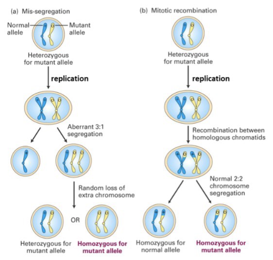
Defects in chromosome segregation, mitotic recombination, or a mutation result in the loss of the functional allele (second hit); Common in retinal development
Check image for
mis-segregation: Incorrect segregation causes development of mutant cells homozygous for mutant allele
mitotic recombination: Recombination can occur between chromosomes in replication, which makes getting two mutant alleles in a cell more common
Epigenetic Silencing of Tumor Suppressor Genes
Alterations in chromatin, such as histone modification and DNA methylation, affect gene expression
Convert transcriptionally active euchromatin into heterochromatin
MLH1 in human: MutL in prokaryotes
CPG island near MHL1 must have low methylation so repair protein can be produced
Mutation causes hypermethylation of CPG region which stops repair protein production and therefore increases development of cancer

The p53 Tumor Suppressor Gene
TP53 is mutated in ~50% of all human cancers; Loss of p53 function results in defective response to DNA damage; Extremely critical in tumor regulation
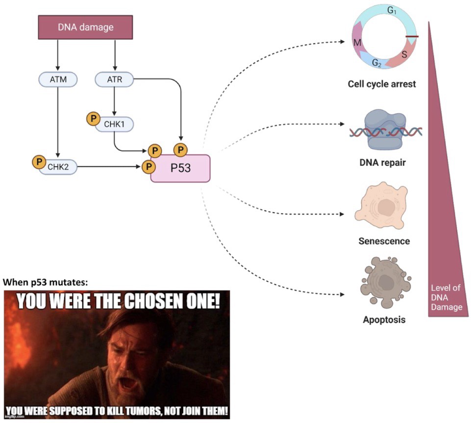
p53: Zinc finger transcription factor; Activated in response to cell stress; including DNA damage; homotetramer (4)
p53 regulates multiple genes that are important for the cellular stress responses such as DNA repair, sustained cell cycle arrest, and apoptosis
Kinases phosphorylate p53 when there is DNA damage, which increases p53 stability; Ataxia Telangiectasia Mutated (ATM) kinase is one
Mutations Seen in p53
First hit caused by deletion, frameshift, nonsense, some missense mutations, or epigenetic silencing
Second hit also results from mutations or epigenetic silencing; Loss of heterozygosity
Some mutations cause issues with only 1 mutated copy (heterozygous)
Some missense mutations have a dominant negative phenotype
Mutations affect the p53 zinc finger motif; but still allows p53 to form a tetramer; This is bad because only one mutated p53 in the four is needed to completely block its function


HPV & Cervical Cancer
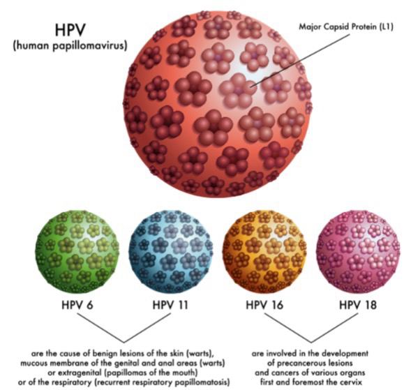
Most human cancers are NOT communicable diseases; Human Papilloma Virus (HVP) is the exception; Most common STD; HPV 16 and 18 cause cancer, HPV 6 and 11 cause milder non-cancerous symptoms
Papanicolaou (“Pap”) test involves scraping cells from the cervical wall, mounting and staining for microscoping examination
Pre-cancerous cells have large nucleus because more replication is occurring
How HPV Inactivates Tumor Suppressors
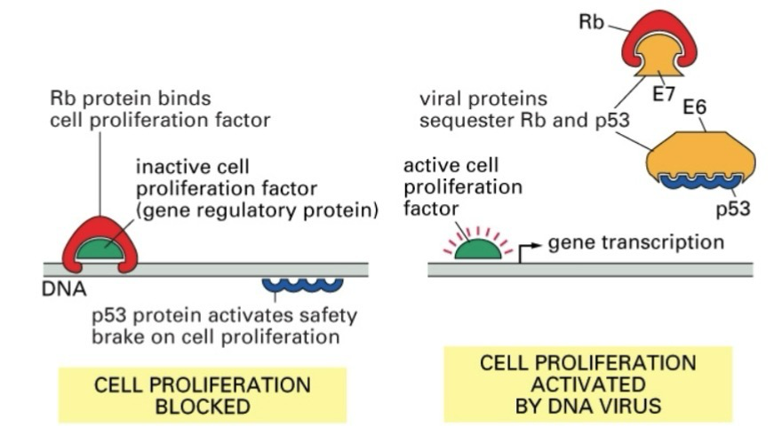
Viruses infect cells and force production of viral proteins
HPV E7 and E6 proteins bind to and sequester pRB and p53, respectively
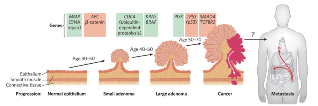
Cancer Development is a Multistep Process
Tumor progression is the conversion of a normal somatic cell into a cancerous tumor cell
The progression from a normal cell to a malignant tumor requires an accumulation of mutations
On average ~6-12 mutations in oncogenes & tumor suppressor genes
No single mutation by itself is sufficient to generate a cancer cell
Once a mutation is established within a genome of a cell, it is permanent, and daughter cells can develop further mutations
Colorectal cancer has several recognizable stages; At least 7 genetic “hits” to become a carcinoma (Malignant tumor)
Signaling Pathways are Altered in Cancer
There are over 200 different types of cancer: Different cell types; Unique genetic changes; Different behaviors; Different pathways involved
Precision medicine aims to match the treatment to the specific pathways affected in each patient’s cancer
Tumor Profiling
Cancer is a disease of the genome; Knowing which genes are altered in a tumor cell allows personalized treatment of a patient’s disease
Important diagnostic and therapeutic tools for cancer are:
Tumor profiling: Sample levels of expression of key cancer-related genes
High throughput DNA sequencing of tumors
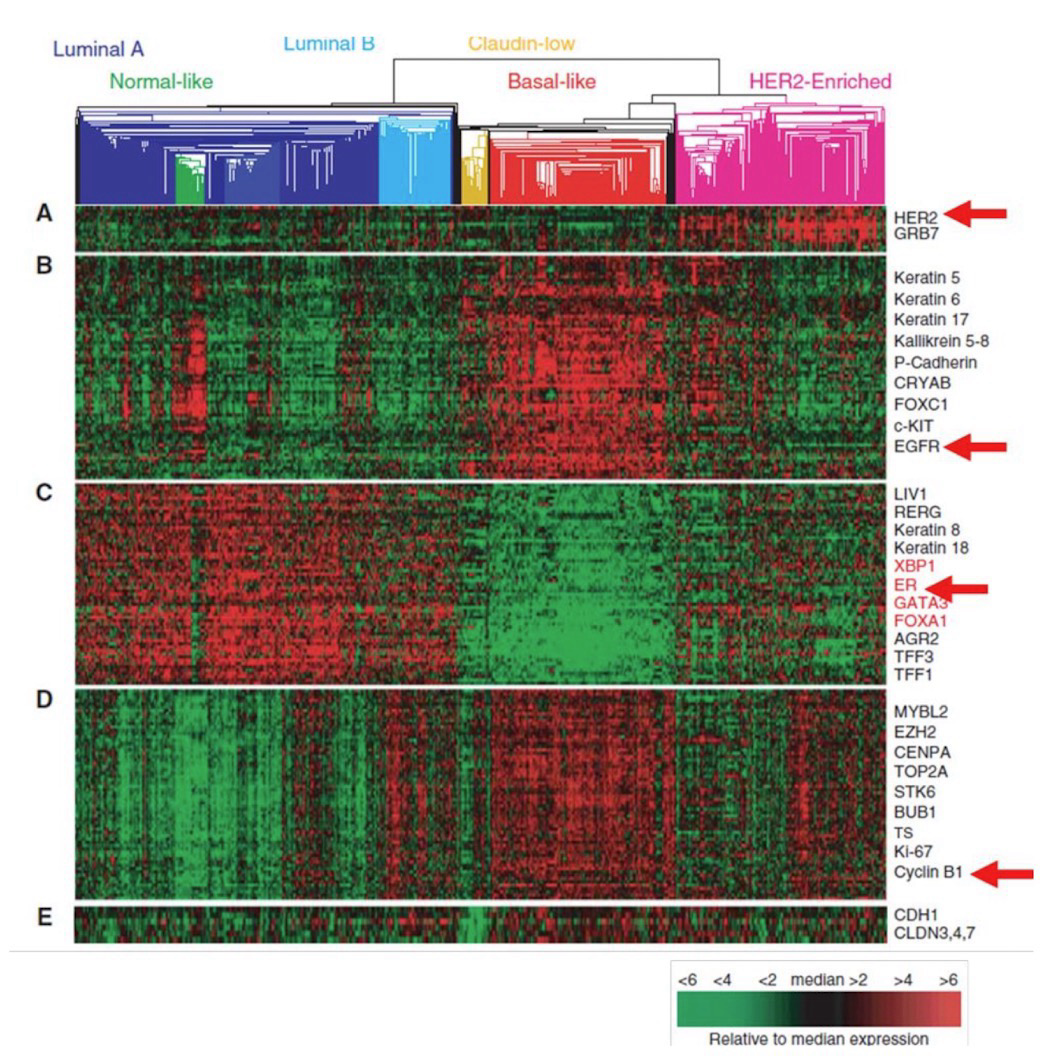
While two patients may have the same diagnosis (i.e. breast cancer), there are often different underlying genetic variations
The study of the expression of proto-oncogenes lets us categorize cancers based on common genetic mutation; Helps inform the treatments for these
Green is good: Normal expression
Red is bad: Abnormal expression