Urinary System - Notes
 Call Kai
 Learn
Learn Practice Test
 Spaced Repetition
 Match
Match Flashcards
 Knowt Play
1/95

There's no tags or description
Looks like no tags are added yet.
Name | Mastery | Learn | Test | Matching | Spaced | Call with Kai | Chat |
|---|
No analytics yet
Send a link to your students to track their progress
96 Terms
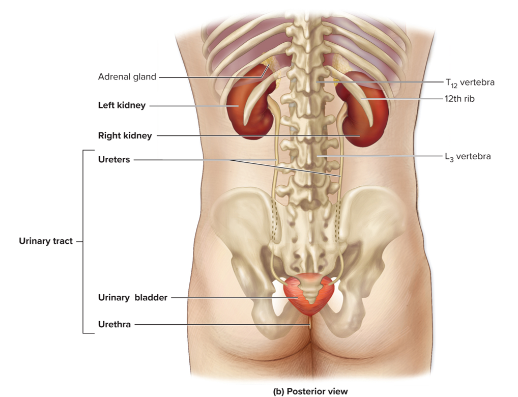
Components of the Urinary System
Kidney
Ureters
Bladder
Urethra
Components of the Urinary System: Kidneys
Filter blood
Remove waste products and convert filtrate into urine
Components of the Urinary System: Urethers
Transport urine
From kidneys to urinary bladder
Components of the Urinary System: Bladder
Expandable muscular sac
Stores as much as 1L urine
Components of the Urinary System: Urethra
Eliminates urine
Anterior View of Urinary System

Posterior of Urinary System
Function of the Urinary System
Filtration
Elimination of Waste
Regulation of Ion Levels
Acid-Base Balance
Blood Pressure Regulation
Hormone Production
Gluconeogensis
Function of the Urinary System: Filtration
Kidneys filter blood and produce urine
Function of the Urinary System: Elimination of Waste
Removes metabolic wastes, toxins, excess substances
Function of the Urinary System: Regulation of Ion Levels
Maintains Na+, K+, and Ca2+ balance
Function of the Urinary System: Acid-Base Balance
Adjusts H+ and HCO3- levels
Function of the Urinary System: Blood Pressure Regulation
Controls blood volume and release renin
Function of the Urinary System: Hormone Production
Calcitriol: Regulates calcium absorption
Erythropoietin (EPO): Stimulates red blood cell production
Function of the Urinary System: Gluconeogenesis
Produces glucose during fasting
Gross Anatomy of the Kidney
Location: Retroperitoneal, lateral to vertebral column
Structure:
Hilum: entry/exit for vessels, nerves, ureter
Fibrous Capsule: Protects kidney
Perinephric Fat: Cushions and support
Renal Fascia & Paranephric Fat: Provide stability

Gross Anatomy of the Kidney: Internal Region
Renal Cortex: Outer layer
Renal Medulla: Contains renal pyramids
Renal Columns: Separate pyramids
Renal Pelvis: Urine collection area

Clinical View: Renal Ptosis
Inferior movement of kidney within abdominal cavity
Due to loss of adipose in elderly or individuals with anorexia nervosa
May kink ureter, which blocks urine flow to urinary bladder
Clinical View: Hydronephrosis
Urine builds up, results in swelling of kidney (hydronephrosis)
Can lead to renal failure
Clinical View: Renal Agenesis
Failure of kidney to develop
Often asymptomatic if unilateral; fatal if bilateral
Clinical View: Pelvic Kidney
Developing kidney fails to migrate from pelvic cavity
Clinical View: Horseshoe Kidney
Inferior parts of left and right kidneys fused
Clinical View: Supernumerary Kidney
Extra kidney develops
All 3 typically asymptomatic
Urine Drainage Pathway
Minor Calyces → Major Calyces → Renal Pelvis → Ureter → Bladder
Nephron Structures: Renal Corpuscle
Glomerulus Capillary: Network for filtration
Glomerular Capsule: Collects filtrate

Nephron Structures: Renal Tubule
Proximal Convoluted Tubule (PCT): Reabsorption of nutrients and water
Nephron Loop: Maintains concentration gradient
Distal Convoluted Tubule (DCT): Regulated secretion and absorption

Nephron Structures: Two Nephron Type
Cortical Nephrons: Short loops, 85% of nephrons
Juxtamedullary Nephrons: Long loops, concentrate urine

Juxtaglomerular Apparatus
Regulates blood pressure and filtration rate
Granular Cells: Secrete renin
Macula Densa: Detects NaCl changes

Histology of Renal Cortex and Medulla

Blood Flow Through the Kidney
Renal Artery → Segmental Arteries → Interlobar Arteries → Arcuate Arteries → Interlobular Arteries → Afferent Arterioles → Glomerulus → Efferent Arterioles → Peritubular Capillaries/Vasa Recta → Veins → Renal Veins

Urine Flow Pathway
Glomerulus → Capsular Space → PCT → Nephron Loop → DCT → Collecting Tubule/Duct → Papillary Duct → Minor & Major Calycles → Renal Pelvis → Ureter → Bladder → Urethra
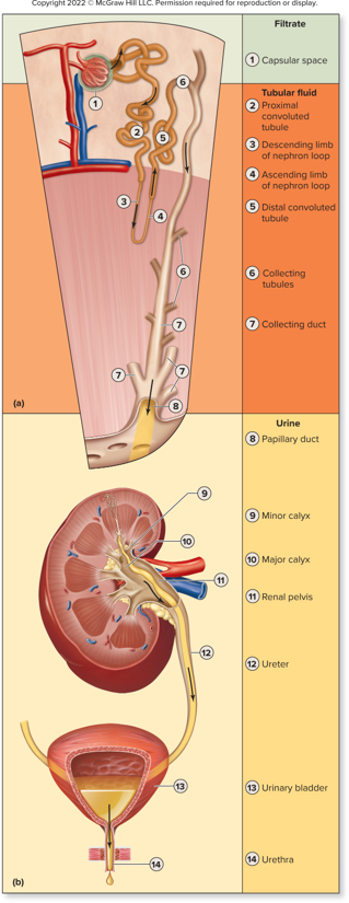
Filtrate
Blood flows through glomerulus
Both water and solutes filtered from blood plasma
Moves across wall of golmerular capillaries and into capsular space
Forms filtrate
Tubular Fluid
New name for filtrate when enters PCT
Flows through
1.PCT
2.Nephron loop
3.DCT
4.Enters collecting tubules
5.Empties into collecting ducts
6.Enters papillary duct within renal papilla; now called urine
Urine Formation
Glomerular filtration
Tubular reabsorption
Tubular secretion

Glomerular Filtration
In glomerular capillaries
Separates some water and dissolved solutes from blood plasma
Water and solutes enter capsular space of renal corpuscle
Due To pressure differences across filtration membrane
Separated fluid is called filtrate
Tubular reabsorption
Movement of components within tubular fluid
Move by diffusion, osmosis, or active transport
Move from lumen or tubules and collecting ducts across walls
Return to blood within peritubular capillaries and vasa recta
All vital solutes and most water reabsorbed
Excess solutes, waste products, some water remaining in tubular fluid
Tubular Secretion
Movement of solutes, usually by active transport
Move out of blood within peritubular and vasa recta capillaries
Move into tubular fluid
Materials moved selectively into tubules to be excreted
Filtration Membrane Layers
Endothelium of glomerulus
Basement membrane of glomerulus
Visceral layer of glomerular capsule

Endothelium of glomerulus
Fenestrated, allows plasma and dissolved substances to pass
Restricts passage of large structures (for example, erythrocytes)

Basement membrane of glomerulus
Glycoprotein and proteoglycan molecules
Restricts passage of large plasma proteins
Visceral layer of glomerular capsule
Outermost layer wrapping around glomerular capillaries
Composed of specialized cells called podocytes
Have long •processes, pedicels
Support capillary wall but don’t completely enclose it
Separated by thin spaces, filtration slits
Restrict passage of most small proteins
Mesangial Cells
Specialized cells positioned between glomerular capillary loops
Phagocytic, contractile, and signaling properties
Three Categories of Substances in Blood
Freely filtered
Not filtered
Limited filtration
Free filtered
Small substances
For example: water, glucose, amino acids, ions
Pass easily through filtration membrane
Not filtered
Formed elements and large proteins
Cannot pass through filtration membrane
Limited filtration
Proteins of intermediate size
Usually blocked from filtration
Due to size or due to negative charge
Regulation of Glomerular Filtration Rate (GFR): Instrinsic Controls
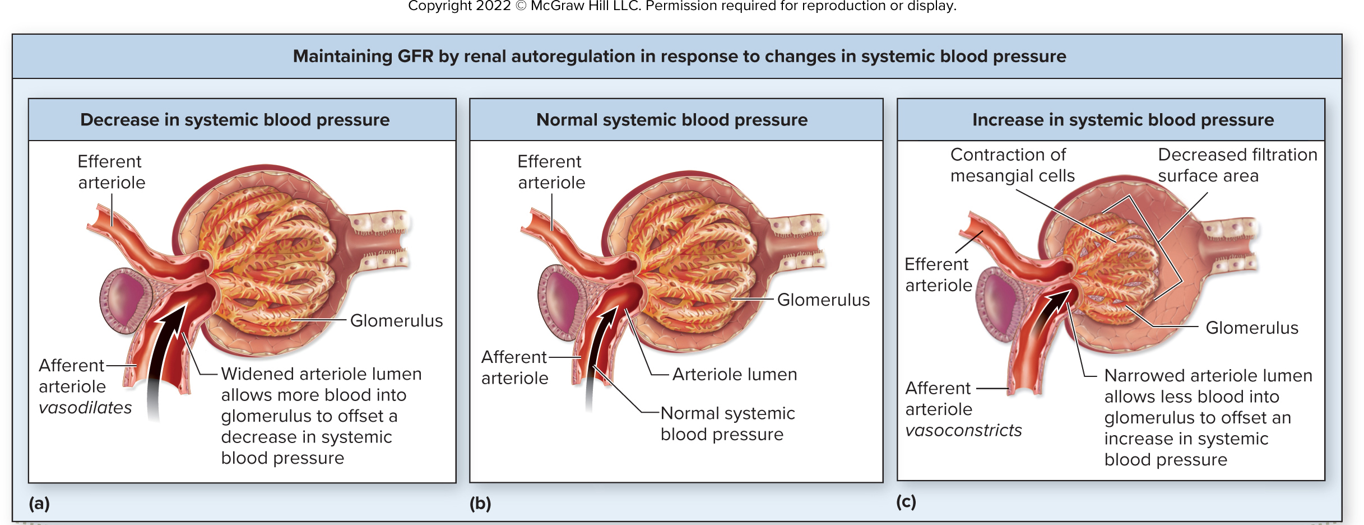
Myogenic response (arteriole stretch response)
Tubulogolmerular feedback (macula densa regulation)
Regulation of Glomerular Filtration Rate (GFR): Extrinsic Controls
Sympathetic Stimulation: Decreases GFR to conserve water
Atrial Natriuretic Peptide (ANP): Increase GFR to eliminate excess fluid
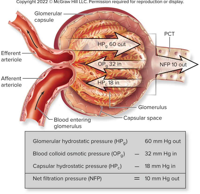
Glomerular Hydrostatic (Blood) Pressure (HPg)
Blood pressure in glomerulus
“Pushes” water and some solutes outo of glomerulus
Pushed into capsular space of renal corpuscle
Higher than blood pressure of other systemic capillaries
Required filtration to occur
Large diameter of afferent arteriole
Smaller diameter of efferent arteriole
Blood colloid osmotic pressure (OPg)
Osmotic pressure exerted by dissolved solutes
For example: plasma proteins
Opposes filtration
Draws fluid back into glomerulus
Capsular hydrostatic pressure (HPg)
Pressure in glomerular capsule due to filtrate
Impede movement of additional fluid
Pressures that Determine Net Filtration Pressure
Renal Autoregulation: Myogenic Response
Reabsorbed Substances
Sodium (Na+): Controlled by aldosterone & ANP
Water: Controlled by ADH (facultative reabsorption)
Potassium (K+): Balance between secretion andreabsorption
Calcium & Phosphate: Regulated by PTH
Bicarbonate (HCO3-) & Hydrogen Ions (H+): pH Balance
Clinical View: Glucosuria
Excretion of glucose in urine
Plasma glucose concentration above 300 mg/dL
Glucose cats as an osmotic diuretic
Pulls water into tubular fluid
Causes loss of fluid in urine
Classic symptom of diabetes, along with frequent urination and thirst
Glucose Reabsorption

Eliminated Waste Products
Urea: Protein metabolism byproduct
Creatinine: From muscle metabolism
Uric Acid: From nucleic acid breakdown
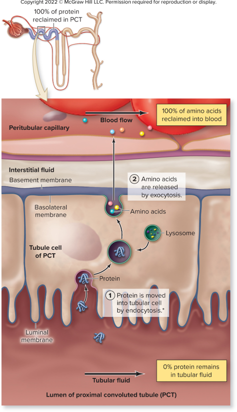
Reclaiming Filtered Protein
Sodium Reabsorption
98% to 100% of Na+ reabsorbed from tubular fluid
Concentration relatively low inside tubule cell
Na+ moves down gradient across luminal membrane into tubular cell
Reabsorption regulated by hormones near end of tubule

Sodium reabsorption: Aldosterone
Steroid hormone produced by adrenal cortex
Stimulates protein synthesis of Na+ channels Na+/K+ pumps
Embedded in plasma membranes of principal cells
Increase in Na+ reabsorption
Water follows by osmosis
Sodium reabsorption: Atrial Natriuretic Peptide
Inhibits reabsorption of Na+ in PCT and collective tubules
Inhibits release of aldosterone
More Na+ and water excreted in urine
Increases GFR
Sodium Transport in PCT

Sodium Transport in DCT, CT, or CD

Water reabsorption
Reabsorbed by paracellular transport between cells
Transcellular transport through water transporter proteins, aquaporins
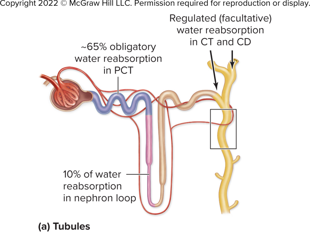
180L filtered daily; all but 1.5L reabsorbed
Tubule permeability varies along its length
65% reabsorbed in PCT
Aquaporins constant number
Water follow Na+ by osmosis, obligatory water reabsorption
Water reabsorption: Nephron Loop
10% of filtered water reabsorbed
Water reabsorption: DCT, collecting tubules, and ducts
Water reabsorption controlled by aldosterone and antidiuretic hormone

Water reabsorption: Aldosterone
Increases Na+/K+ pumps and Na+ channels, increasing both Na+ and water reabsorption
Water reabsorption: Antidiuretic Hormone (ADH)
Binds to principal cells
Adds channels to increase water reabsorption
Concentration gradient within interstitial fluid
Tubular reabsorption, facultative water reabsorption
Results in smaller volume of more concentrated urine
Elevated levels during dehydration (Urine noticeably darker)
Urine range = 1200 mOsm to 50 mOsm
Reabsorption and secretion of potassium
60%-80% reabsorbed in tubular fluid (PCT)
Dependent on movement of Na+
10% to 20% reabsorbed in thick segment of nephron loop
Net secretion or reabsorption in collecting tubule

Movement of Na+
Sodium reabsorbed across luminal membrane, water follows
Increased concentration of remaining solutes in tubular fluid
Creates gradient between tubular fluid and interstitial fluid
K+ moves down gradient from tubular fluid by paracellular route
Allows passive reabsorption of other solutes
Net secretion or reabsorption in collecting tubule
Intercalated cells reabsorb K+ continuously
Principal cells secrete K+ at varying rates
Based on aldosterone level
Stimulates principal cells to secrete K+
Most powerful stimulant for aldosterone, elevated K+ level
Calcium and Phosphate Balance
60% in blood goes into filtrate
Remainder bound to protein and prevented from filtration
90% to 95% filtered as blood passes through glomerular capillaries

Parathyroid Hormone (PTH)
Regulates excretion of Ca2+ and PO43−
Inhibits PO43- reabsorption in PCT
Stimulates Ca2+ reabsorption in DCT
Less phosphate available to form calcium phosphate
Calcium deposition in bone decreased
Ca 2+ blood levels increased

Bicarbonate ions, hydrogen ions, and pH
Moves freely across filtration membrane
If filtered HCO3− not reabsorbed, blood too acidic
80% to 90% reclaimed from tubular fluid
Remaining 10% to 20% taken up from thick segment of ascending limb
Filtered HCO3 − “replaced” not reabsorbed
pH of urine and blood regulated in collecting tubules

Substances with Regulated Reabsorption: pH (acidic)
If acidic blood, then
Synthesized HCO3− reabsorbed into blood
H+ excreted within filtrate by type A intercalated cells
Increase blood pH and decrease urine pH
Substances with Regulated Reabsorption: pH (alkaline)
If alkaline blood, then
Type B intercalated cells active
Secret HCO3- and reabsorb H+
Lower blood pH and increase urine pH
Nitrogenous waste
Metabolic waste containing nitrogen
Main nitrogenous waste products:
Urea
Uric acid
Creatinine
Substances Eliminated as Waste: Urea
Molecule produced from protein breakdown
Both reabsorbed and secreted
50% excreted in the urine
Helps establish concentration gradient in the interstitial fluid
Substances Eliminated as Waste: Uric acid
Produced from nucleic acid breakdown in liver
Both reabsorbed and secreted
Substances Eliminated as Waste: Creatinine
Produced from creatinine metabolism in muscle
Only secreted
Elimination of Drugs and Bioactive substances
Most secretion occurring in PCT
Certain Drugs: penicillin, sulfonamides, aspirin
Other metabolic wastes: urobilin, hormone, metabolites
Some hormones: human chorionic gonadotropin, epinephrine
Urinary Tract & Micturition: Structures
Ureters: Transport urine from kidneys to bladder
Urinary Bladder:
Stores urine (up to 1L)
Detrusor Muscle: Contracts during urination
Urethra:
Female: only urine transport
Male: Longer, transports urine & semen
Micturition (Urination Process)
Storage Reflex: Sympathetic control inhibits urination
Micturition Reflex: Parasympathetic stimulation contracts bladder and reflaxes sphincters
Voluntary Control: Somatic motor control of external urethral sphincter
Common Disorders
UTI (Urinary Tract Infection): Bacterial infection
Kidney Stones: Mineral crystallization
Renal Failure:: Loss of kidney function; may require dialysis
Each nephron loop has two limbs, a(n) ____ limb that extends from the cortex to the medulla and a(n) ____ limb that returns back into the renal cortex
descending, ascending
At least 20%-25% of the resting ____ normally flows through the kidney via the renal artery
cardiac output
Filtrate is produced due to the difference between ___ pressure of the blood in the glomerulus and the opposing pressures of the osmotic blood pressure and fluid pressure in the capsular space.
hydrostatic
A mechanism that is effective in maintaining a normal glomerular blood pressure only if the systemic mean arterial pressure remains between 80 and 180 mm Hg is called ____ autoregulation
renal
Sympathetic stimulation causes the release of renin which will result in the subsequent production of ______ which will result in a(n) ______ in the GFR.
angiotensin II; decrease
Some substances are not normal components of urine because they are completely reabsorbed. Choose the substances that satisfy this requirement under healthy conditions.
filtered plasma proteins
lactate
amino acids
glucose
All substances that have regulated reabsorption
K+
Ca2+
Na+
Water
Water is reabsorbed through specific water transporter proteins called ___
Aquaporins
As tubular fluid moves through the collecting tubules and ducts, water reabsorption is controlled primarily by ____ and ____ hormones
aldosterone; antidiuretic
Urine volume can be several liters per day if there is a decrease in the release of ____
ADH
Ureters enter the bladder through the ___ wall of the bladder
posterolateral
The layer of the utretr called the ____ consists of inner and outer layers of smooth msucle
mmuscularis