chp. 16: cell signaling
 Call Kai
 Learn
Learn Practice Test
 Spaced Repetition
 Match
Match Flashcards
 Knowt Play
1/133

There's no tags or description
Looks like no tags are added yet.
Name | Mastery | Learn | Test | Matching | Spaced | Call with Kai | Chat |
|---|
No analytics yet
Send a link to your students to track their progress
134 Terms
what r the usual concentrations of Na+ and K+ in cell
Na+ is the most plentiful positively charged ion outside the cell, while K+ is the most plentiful inside
what r usual Ca2+ conc’n in cell?
maintain low Ca2+ conc’n in cytosol, and high Ca2+ conc’n outside of cell (like Na+) and inside ER lumen!
signal transduction
Conversion of an impulse or stimulus from one physical or chemical form to another.
A signaling cell releases an extracellular signal molecule that binds to a receptor on a target cell, triggering intracellular signal molecule changes (signal transduction) that alter cell behavior.
extracellular signal molecules
Any molecule present outside the cell that can elicit a response inside the cell when the molecule binds to a receptor.
can be endocrine, paracrine, neuronal, autocrine, and contact-dependent signals
signals can also be
Large/hydrophilic → bind cell-surface receptors
Small/hydrophobic → cross membrane → bind intracellular receptors
endocrine signaling
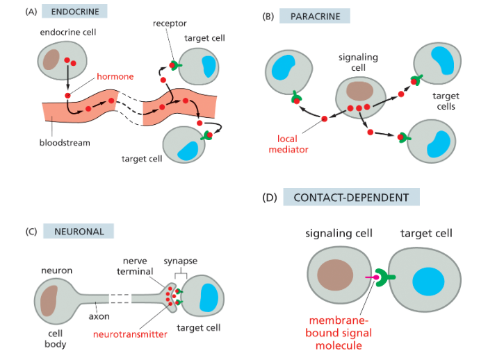
an extracellular signal that is a hormone is released into the bloodstream and act on distant target cells throughout the body.
broad and public
ex of hormones: cortisol, epinephrine, insulin, testosterone, T4
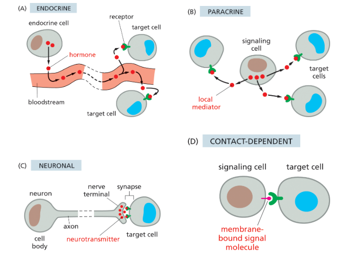
paracrine signaling
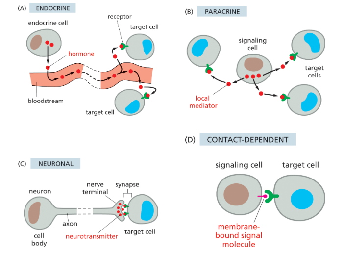
Extracellular signals diffuse locally to affect nearby cells.
act as local mediators
ex: nitric oxide, epidermal growth factor, platelet derived growth factor, histamine
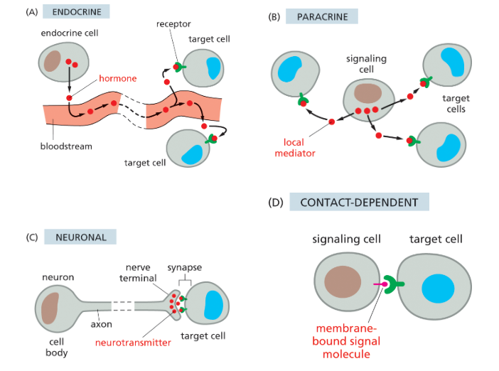
local mediators
A secreted extracellular signal molecule that acts at a short range on adjacent cells.
paracrine signaling
ex: nitric oxide, epidermal growth factor, platelet derived growth factor, histamine
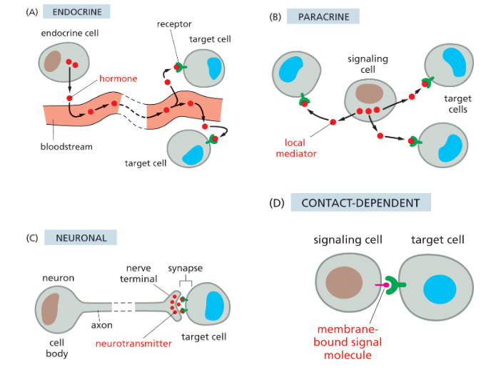
autocrine signaling
A cell releases signals that act on itself.
a form of paracrine signaling cuz it responds to its own local mediators
most cancer cells do this!!
neuronal signaling
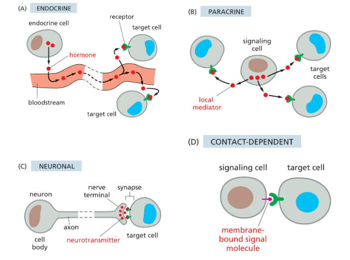
Electrical signals travel along neurons, triggering neurotransmitter release to specific target cells.
delivered quickly long distances over private lines
uses neurotransmitters!
EX: acetylcholine, y-aminobutric acid (GABA)
neurotransmitter
Small signaling molecule secreted by a nerve cell at a synapse to transmit information to a postsynaptic cell.
used in neuronal signaling
EX: acetylcholine, y-aminobutric acid (GABA)
contact dependent signaling
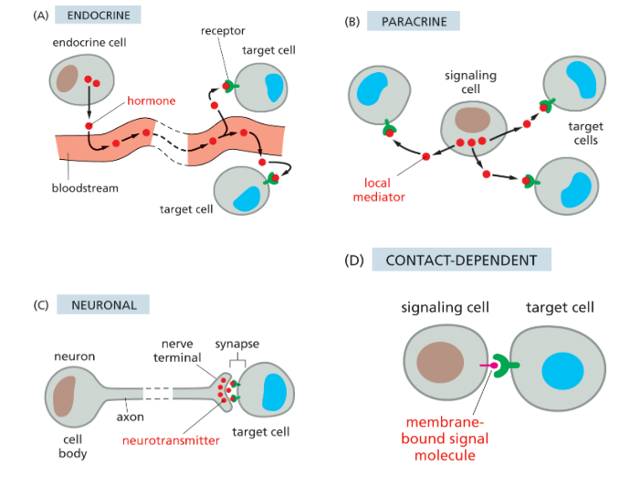
Cells must physically touch; a membrane-bound signal binds a receptor on another cell.
most intimate and short range, doesn’t req release of of a secreted molec.
allows adjacent cells that are initially similar to become specialized to form different cell types (ex: Delta)
receptor
Protein that recognizes and responds to a specific signal molecule.
if cell doesn’t have specific receptor for a signal, it won’t respond to it
2 types:
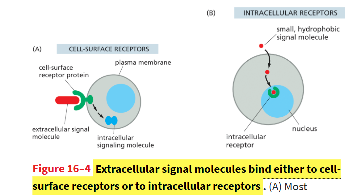
cell-surface receptors
bind to large hydrophilic signals that cant enter cell and generate intracellular signaling molec in target cell
intracellular receptors
small hydrophobic signals can bind to these receptors in nucleus or cytosol
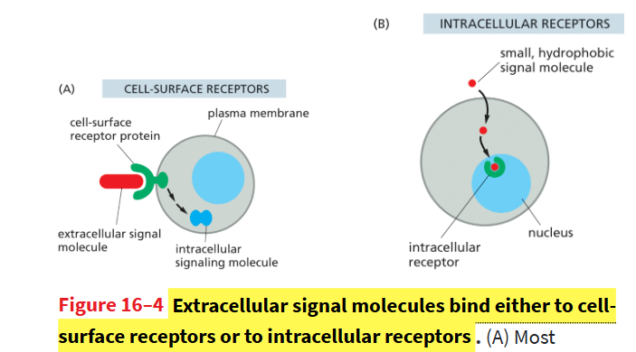
cell-surface receptors
transmembrane proteins, extracellular domain binds signal, cytoplasmic domain interacts with cytoplasmic proteins to induce response
bind signals that r too large/hydrophilic to cross cell membrane
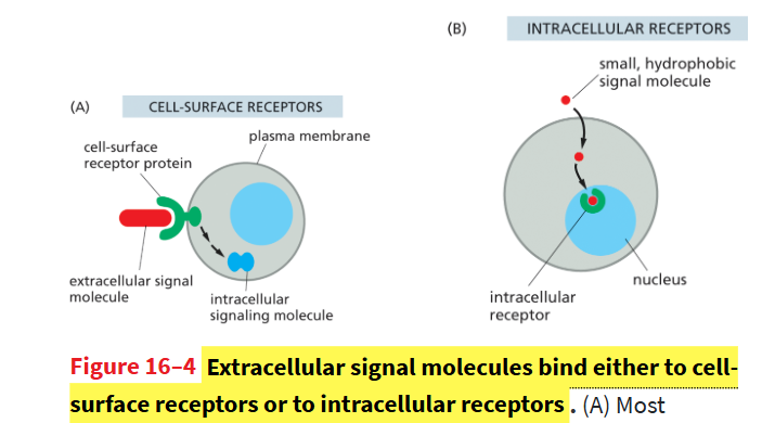
intracellular receptors
cytoplasmic or nuclear receptors that r often transcription factors
bind signals that r small/hydrophobic enough to cross cell membrane and bind to these receptors in cytoplasm or nucleus. they regulate gene transcription!
effector proteins
Carry out the final response by having direct effect on target cell (change gene expression, metabolism, movement, etc.).
this allows diff cells to respond to the same signal in diff ways since they’ll have diff effector proteins + signaling pathways
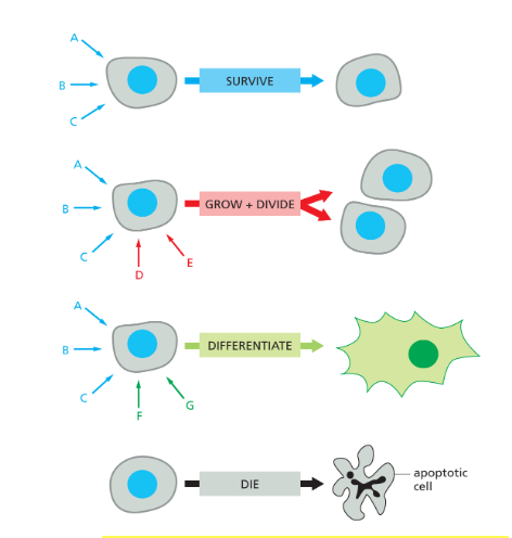
How do cells respond to multiple signals?
Signals combine and interact to produce a specific response
A combination of signals can evoke a response that is different from the sum of the effects that each signal would trigger on its own.
if a cell lacks a survival signal, itll undergo apoptosis
what do excitory neurotrasnmitters do?
bring the membrane closer to the threshold potential, increasing the probability of action potentials
makes membrane less negative by bringing the membrane closer to the threshold, so less additional depolarization is needed to trigger an action potential.
ex: acetycholine
what do inhibitory neurotransmitters do?
they hyperpolarize the membrane, decreasing the probability of action potentials
hyperpolarization makes the membrane more negative, so a larger stimulus is needed to reach threshold that would fire an action potential
ex: y-Aminobutyric acid (GABA)
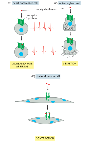
How can acetylcholine (a neurotransmitter) cause different effects in different cells?
in skeletal cells: Causes muscle contraction by opening ligand-gated Na⁺ channels → depolarization → action potential.
in cardiac pacemaker cells: released by parasympathetic neurons and Slows heart rate by reducing frequency of cardiac muscle contraction and an increase in salivary secretions
Why are some cell signaling responses fast and others slow?
Fast responses use existing proteins, while slow responses require gene expression and new protein synthesis.
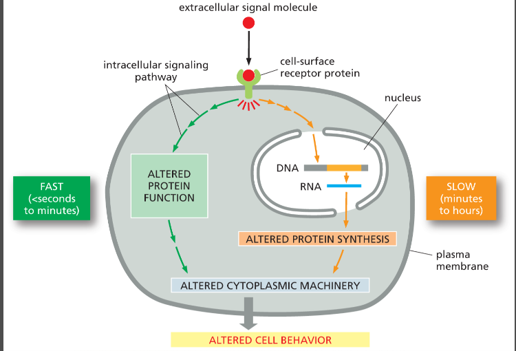
What characterizes a fast cellular response to a signal?
Occurs in seconds–minutes by changing the activity of proteins already in the cell.
ex: acetylcholine stimulating skeletal cel
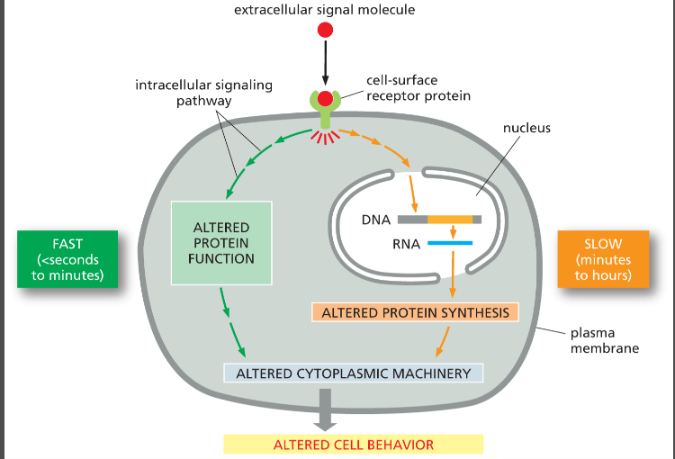
What characterizes a slow cellular response to a signal?
Takes hours because it involves changes in gene expression and making new proteins.
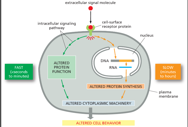
intracellular signaling pathways
A set of proteins and small-molecule second messengers that interact with each other to relay a signal from the cell membrane to its final destination in the cytoplasm or nucleus.
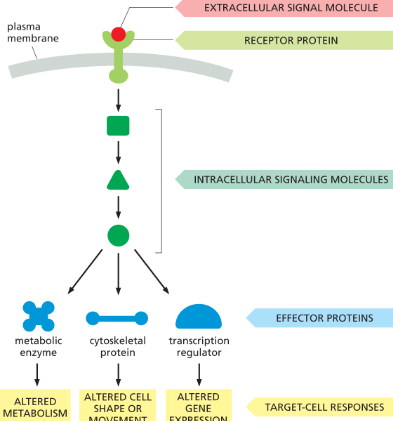
what performs the first step in signal transduction?
the receptor protein!
it recognizes an extracellular signal molecule and generates a different type of intracellular signal molecule in response
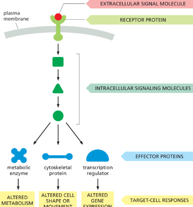
What are five things intracellular signaling proteins can do?
Relay, amplify, integrate, distribute, and provide feedback.
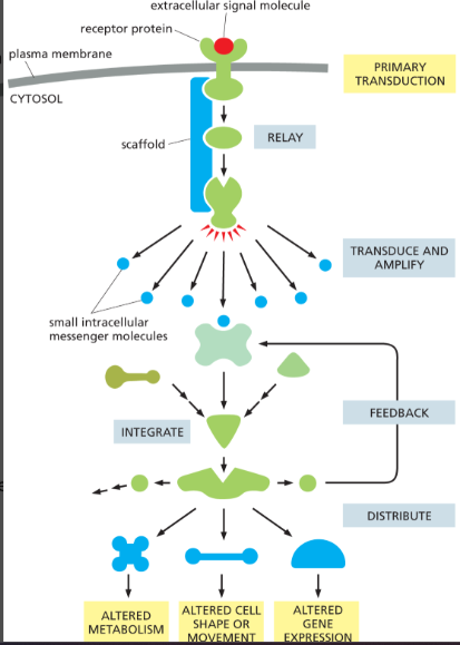
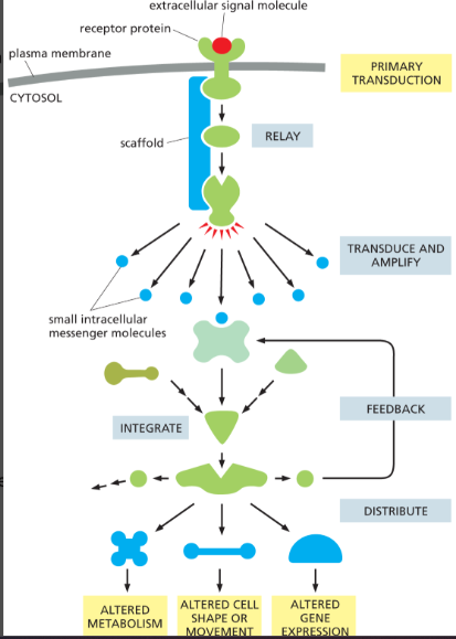
intracellular signals can relay
relay the signal onwards to help it spread in cell
scaffold proteins can help by bringing together components needed to propagate signal
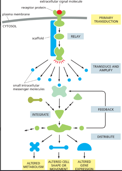
scaffold protein
helps intracellular signals relay info by holding multiple signaling proteins together in one place, allowing them to interact faster, more efficiently, and more specifically.
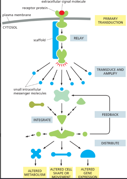
intracellular signals can amplify
can amplify signal received and make it stronger to evoke large intracellular response
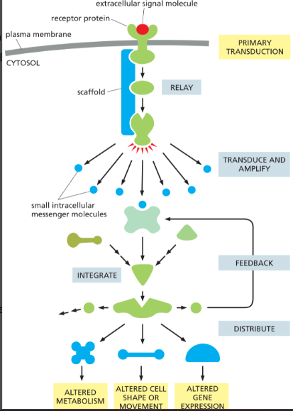
intracellular signals can integrate
a signaling protein combines inputs from multiple pathways and processes them together before producing one coordinated response onwards
intracellular signals can distribute signals
can distribute one signal to multiple effector proteins which creates a complex response
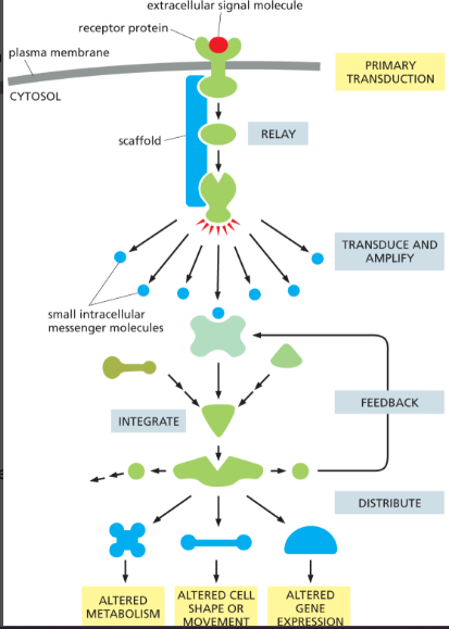
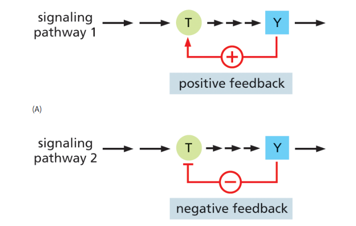
intracellular signals can engage in feedback
regulates activity of components upstream in signaling pathway
positive feedback: A downstream signal increases earlier steps, strengthening the response.
generates all-or-none switch like responses
negative feedback: A downstream signal inhibits earlier steps, reducing the response.
can oscillate on/off as conc’ns rise and fall
what r the 2 main ways signaling proteins r switched on/off?
regulation by phosphorylation
done by kinases and phosphatases
regulation by GTP binding/hydrolysis
monomeric G-proteins and trimeric G-proteins
both regulated by GAP and GEF proteins (but GAP/GEF mainly focus on monomeric G-proteins)
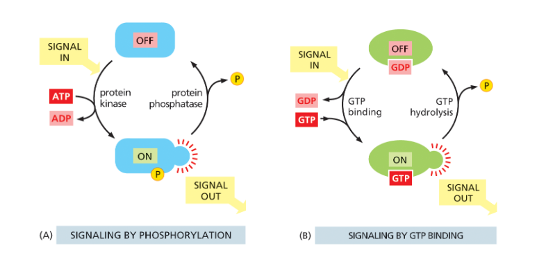
How are phosphorylation switch proteins turned on/off?
protein kinases adding a phosphate group turns it ON
protein phosphatases removing the phosphate group turn it OFF
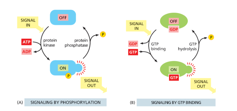
What are the two main types of protein kinases?
Serine/threonine kinases (phosphorylate serines or threonines)
tyrosine kinases. (phosphorylate a.a tyrosine)
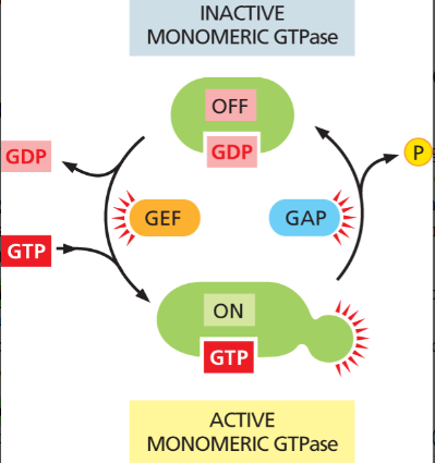
How do GTP-binding proteins act as switches?
They are ON when bound to GTP and OFF when bound to GDP. (they will hydrolyze GTP)
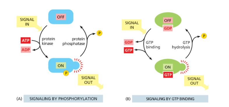
What are the two main types of GTP-binding proteins in cell signaling?
Trimeric (heterotrimeric) G proteins – made of 3 subunits, activated by GPCRs, and relay signals from cell-surface receptors.
Monomeric (small) GTPases – single proteins regulated by GEFs (turn ON) and GAPs (turn OFF), often involved in intracellular signaling and trafficking.
what do GEFS/GAPS do?
GEFS- Activate GTP-binding proteins by promoting GDP → GTP exchange.
GAPS- Inactivate GTP-binding proteins by stimulating GTP hydrolysis.
used to regulate monomeric (small) GTPases, not the main regulators of trimeric (heterotrimeric) G proteins, which are instead controlled by GPCRs (G-protein-coupled receptors) but can still technically regulate them
What are the three main classes of cell-surface receptors?
Ion-channel-coupled receptors
converts molecular signal → electrical signal
G-protein-coupled receptors (GPCRs)
activates trimeric G-protein
Enzyme-coupled receptors
activate cytoplasmic domain or associated enzyme
they all act as targets for drugs as well
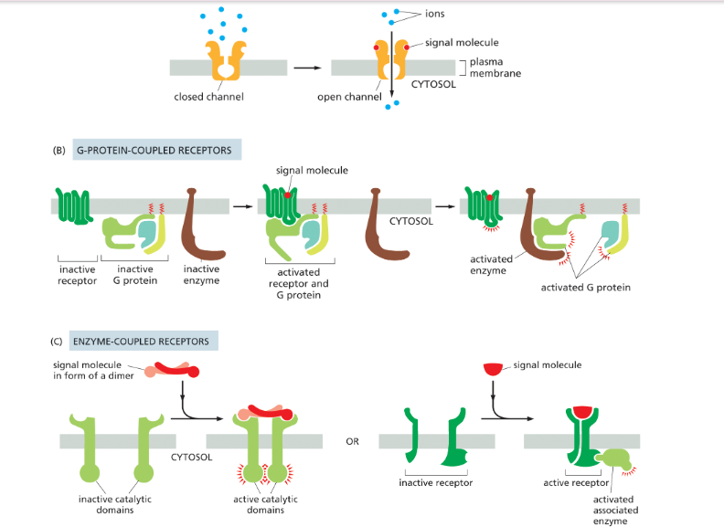
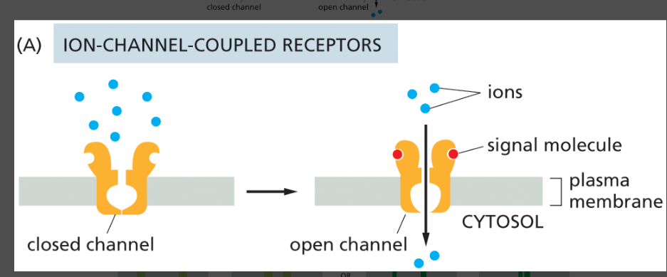
ion-channel-coupled receptors
opens in response to binding an extracellular signal molecule. These channels are also called transmitter-gated ion channels. (change memebrane potential)
produce electrical current by changing membrane potential
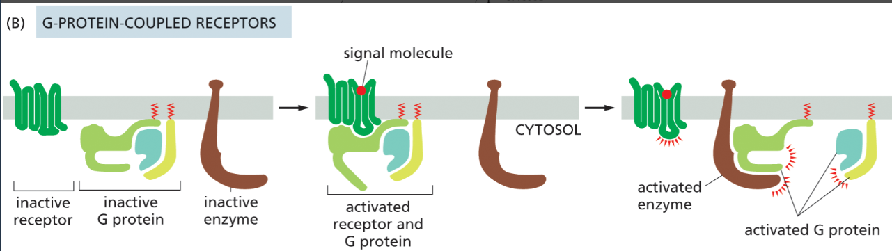
G-protein-coupled receptors (GPCRs)
binds its extracellular signal molecule, the activated receptor signals to a trimeric G protein on the cytosolic side of the plasma membrane, which then turns on (or off) an enzyme (or an ion channel; not shown) in the same membrane, activating an intracellular signaling cascade
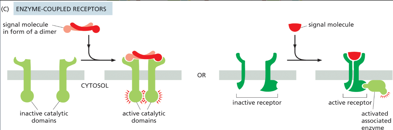
enzyme-coupled receptors
They either act as enzymes themselves or activate associated enzymes, triggering signaling pathways inside the cell when an extracellular signal binds.
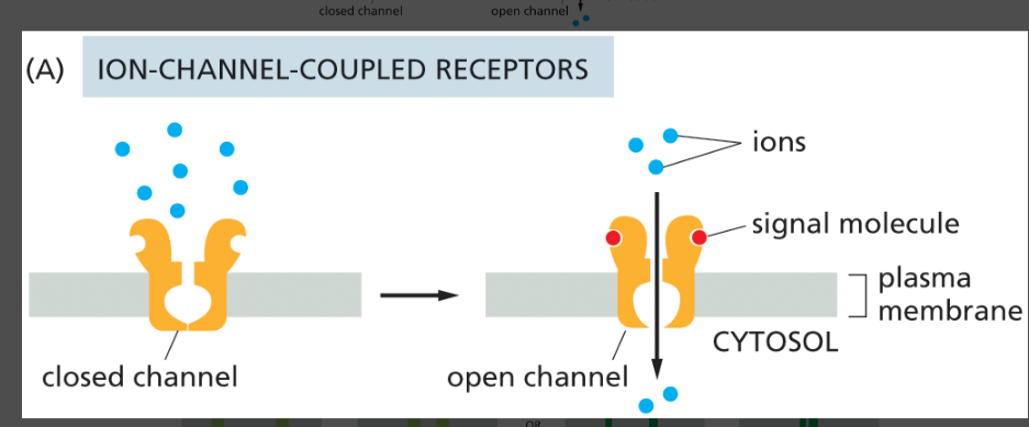
How do ion-channel-coupled receptors work?
A neurotransmitter binds → receptor changes shape → ion channel opens → ions flow across membrane.
They convert chemical signals (neurotransmitters) → electrical signals (changes in membrane potential).
Ions like Na⁺, K⁺, or Ca²⁺ move across the membrane
they r used in neurons and easily excitable cells like muscle cells cuz they work fast since they dont rlly use secondary messengers.
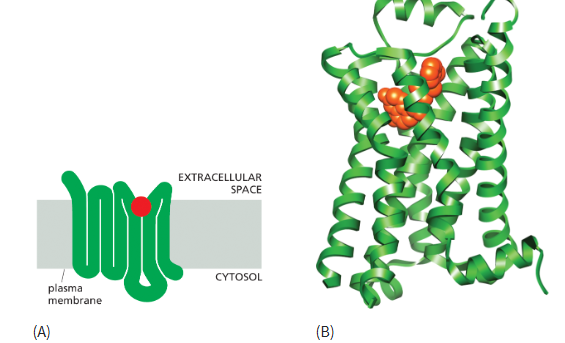
structure of a G-protein coupled receptor (GCPR)
made of polypeptide chain that passes through membrane 7x
extracellular region produce ligand binding site
cytoplasmic regions interact w/ and activate trimeric G-proteins
G-protein (trimeric GTP-binding protein)
activated by GTP and has 3 subunits: alpha, beta, gamma
in inactive form: all 3 subunits associate together
in inactive form: alpha subunit binds GTP and dissassociates from beta and gamma subunit
beta and gamma units remain associated
the 2 diff complexes r both active and interact w different targets
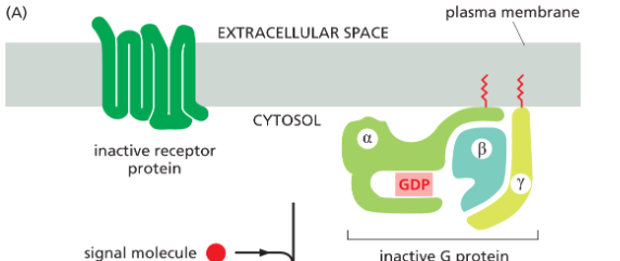
how does the G-protein attach to the plasma membrane?
both the α and γ subunits of the G protein have covalently attached lipid molecules that help anchor the subunits to the plasma membrane.
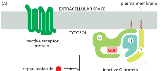
inactive state of G-protein
alpha subunit has GDP bound to it and all 3 subunits (alpha, beta, gamma) r stuck together
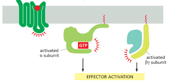
active state of G-protein
When an extracellular signal molecule binds to its receptor, the altered receptor activates a G protein by causing the α subunit to decrease its affinity for GDP, which is then exchanged for a molecule of GTP.
so receptor is the GEF for the G-protein!!
alpha subunit binds GTP and dissociates from beta and gamma subunit
beta and gamma units remain associated
the 2 diff complexes r both active and interact w different targets
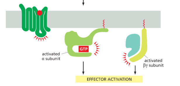
what controls how long a G-protein is active?
after both subunits (alpha and combined beta and gamma) interact w/ downstream signaling targets, the α subunit has an intrinsic GTPase activity, and it hydrolyzes its bound GTP to GDP, returning the whole G protein to its original, inactive conformation
all 3 regions will reassociate again
usually only a few seconds before alpha deactivates after being activated
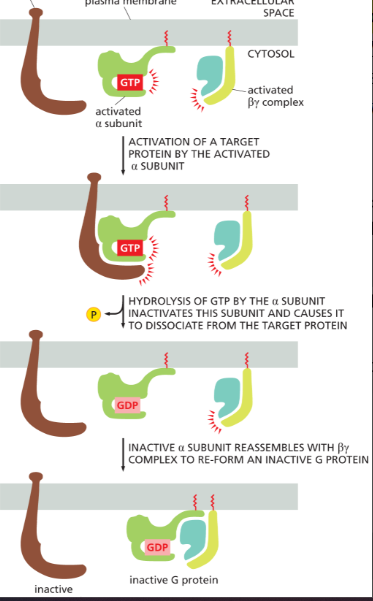
What would happen if the α subunit of a G protein had reduced affinity for GDP but normal affinity for GTP?
The G protein would activate more easily and stay active longer, because GDP would dissociate more readily and be replaced by abundant GTP. This would cause excessive, prolonged signaling (gain-of-function effect).
What are Gs and Gi G-proteins?
They are trimeric G proteins that regulate adenylyl cyclase in opposite ways:
Gs (“stimulating”) → activates adenylyl cyclase → increases cAMP
Gi (“inhibitory”) → inhibits adenylyl cyclase → decreases cAMP
Gs G-protein
stimulates/activates adenyl cyclase → increases cAMP
Gi G-protein
inhibits adenylyl cyclase → decreases cAMP
What are the targets of G-proteins subunits?
target either enzymes or ion channels in the plasma membrane, changing their activity to produce intracellular signaling responses
Ion channels → rapid, immediate changes in cell behavior.
Enzymes → slower, more complex signaling through production of second messengers.
How does acetylcholine slow the heart via a G protein?
Acetylcholine activates a GPCR → G protein is activated → βγ subunit opens K⁺ channels → heart pacemaker cell becomes harder to excite → heart rate slows.
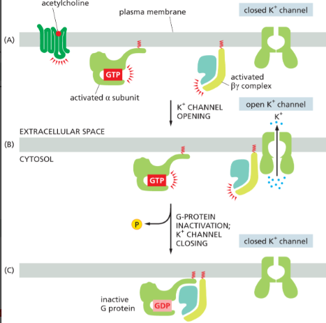
Why does opening K⁺ channels by acetylcholine receptor slow the heartbeat?
Increased K⁺ permeability makes the membrane more negative (hyperpolarized), making it harder to reach the threshold for an action potential to activate so therefore slows down heart
By-complex opens K+ channels
secondary messengers
Small intracellular signaling molecules produced after GPCR activation that relay and amplify signals inside the cell.
ex: cAMP, IP₃, DAG, and Ca²⁺.
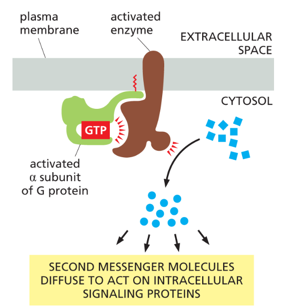
What are the two main enzymes activated by G proteins?
Adenylyl cyclase and phospholipase C.
adenyl cyclase produes cAMP
phospholipase C produces Inositol trisphosphate (IP₃) and diacylglycerol (DAG).
enzymes activated by Gproteins will increase conc’n of small intracellular signaling molecules
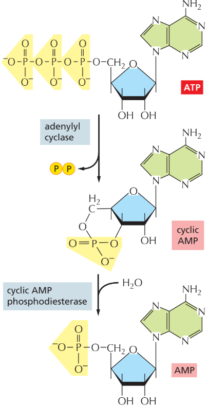
adenyl cyclase
Enzyme that catalyzes the formation of cyclic AMP from ATP; an important component in some intracellular signaling pathways.
activated by the alpha subunit of a G-protein! adenyl cyclase will then produce cAMP
cylic AMP (cAMP)
Small intracellular signaling molecule generated from ATP in response to hormonal stimulation of cell-surface receptors.
a secondary messanger produced by adenyl cyclase
it is degraded by cAMP phosphodiesterase
is water-soluble and can carry signal throughout cell (nucleus, cytosol)
cAMP will active the enzyme PKA!
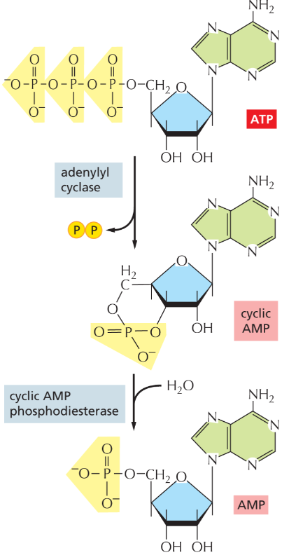
cylic AMP phosphodiesterase
converts cAMP to ordinary AMP which degrades it
continuously active so cAMP lvls r constanctly changing
cyclic-AMP-dependent protein kinase (PKA)
Enzyme that phosphorylates target proteins (serines/threonines on proteins) in response to a rise in intracellular cyclic AMP concentration.
activated by cAMP which was activated by adenyl cyclase
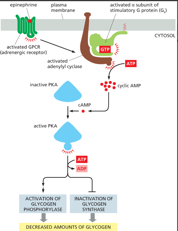
can be used by epinephrine to decrease glycogen lvls in cell (fast), or can enter nucleus and regulate transcriptional regulators (slow)
How does cAMP activate PKA?
PKA is normally bound to a regulatory protein that inhibits it. but then cAMP binds the regulatory subunit, causing a conformational change that releases active PKA.
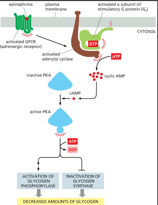
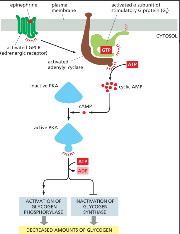
How does epinephrine decrease glycogen lvls in skeletal muscle cells?
GPCR → Gs → adenylyl cyclase → ↑cAMP → PKA → activates glycogen breakdown enzymes and inhibits glycogen synthesis.
PKA phosphorylates and inactivates glycogen synthase
occurs rapidly!
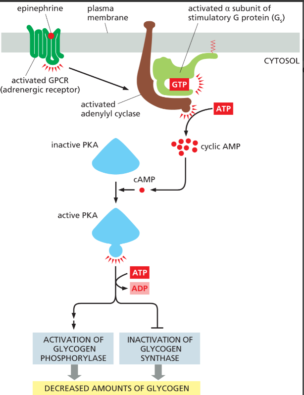
How can cAMP signaling lead to changes in gene expression?
PKA enters the nucleus and phosphorylates transcription regulators that activate gene transcription.
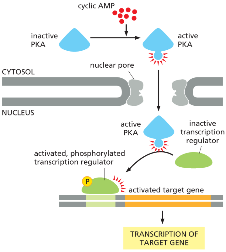
What are the fast vs. slow roles of PKA in cAMP signaling?
Fast (seconds): In skeletal muscle, PKA phosphorylates and activates a kinase that activates glycogen phosphorylase (↑ glycogen breakdown) and phosphorylates and inactivates glycogen synthase (↓ glycogen synthesis), rapidly increasing glucose availability for ATP production during fight-or-flight responses (e.g., epinephrine signaling).
Slow (minutes–hours): PKA moves into the nucleus and phosphorylates transcription regulators, which activate transcription of specific target genes, leading to new protein synthesis involved in long-term responses such as hormone production (e.g., in endocrine cells) and processes like learning and memory in neurons.
how can cAMP activate gene expression?
PKA will be activated by a rise in cAMP and can enter the nucleus and phosphorylate transcriptional regulators to stimulate transcription of target genes
phospholipase C
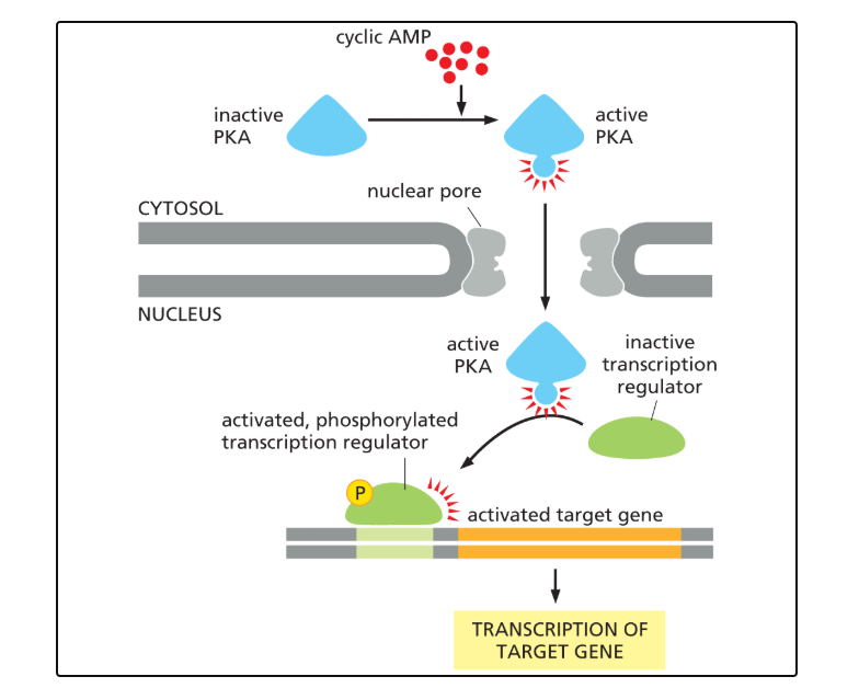
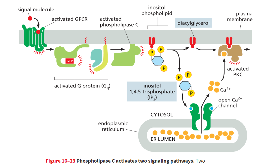
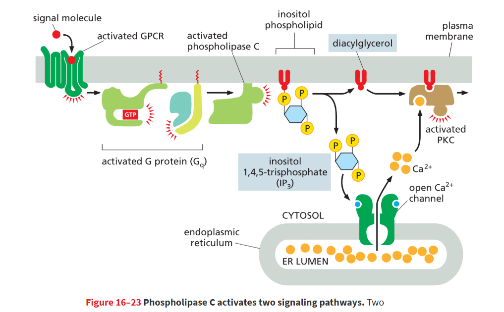
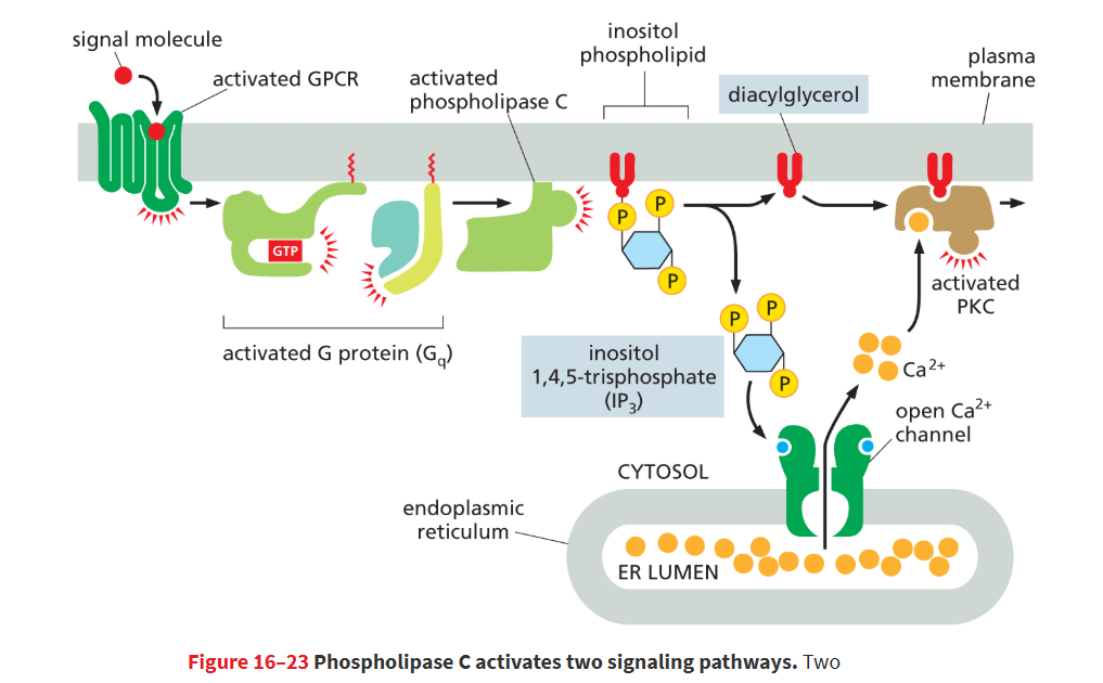
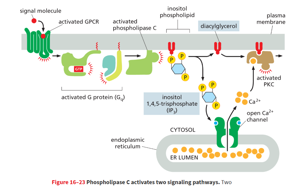
one of the enzymes activated by a G-protein that produces 2 secondary messengers: inositol 1,4,5-triphosphate (IP3) and diacylglycerol (DAG)
it produces these 2 messengers by cleaving an inositol phospholipid
Gq
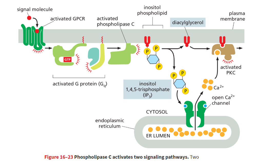
a G protein that activates phospholipase C instead of adenyl cyclase
inositol phospholipid (PIP2)
a lipid molecule with inositol sugar on its head in the plasma membrane that is cleaved by phospholipase C and cleavage yields two small messenger molecules, IP3 and diacylglycerol.
inositol 1,4,5-trisphosphate (IP3)
Small intracellular secondary signaling molecule that triggers the release of Ca2+ from the endoplasmic reticulum into the cytosol
produced when phospholipase C cleaves an inositol phospholipid
it is a water-soluble sugar phosphate that’s released into cytosol where it opens Ca2+ channel in ER membrane
diacylglycerol (DAG)
Small secondary messenger molecule produced by the cleavage of membrane inositol phospholipids in response to extracellular signals. Helps activate protein kinase C.
produced when phospholipase C cleaves an inositol phospholipid
it is a lipid that remains embedded in plasma membrane that recruits PKC from the cytosol to membrane
protein kinase C (PKC)
it is an enzyme that was recruited by diacylglycerol (DAG), it enters plasma membrane
it must bind Ca2+ and (DAG), it phosphorylates target proteins (serines/threonines) in response to rise in the conc’n of DAG and Ca2+
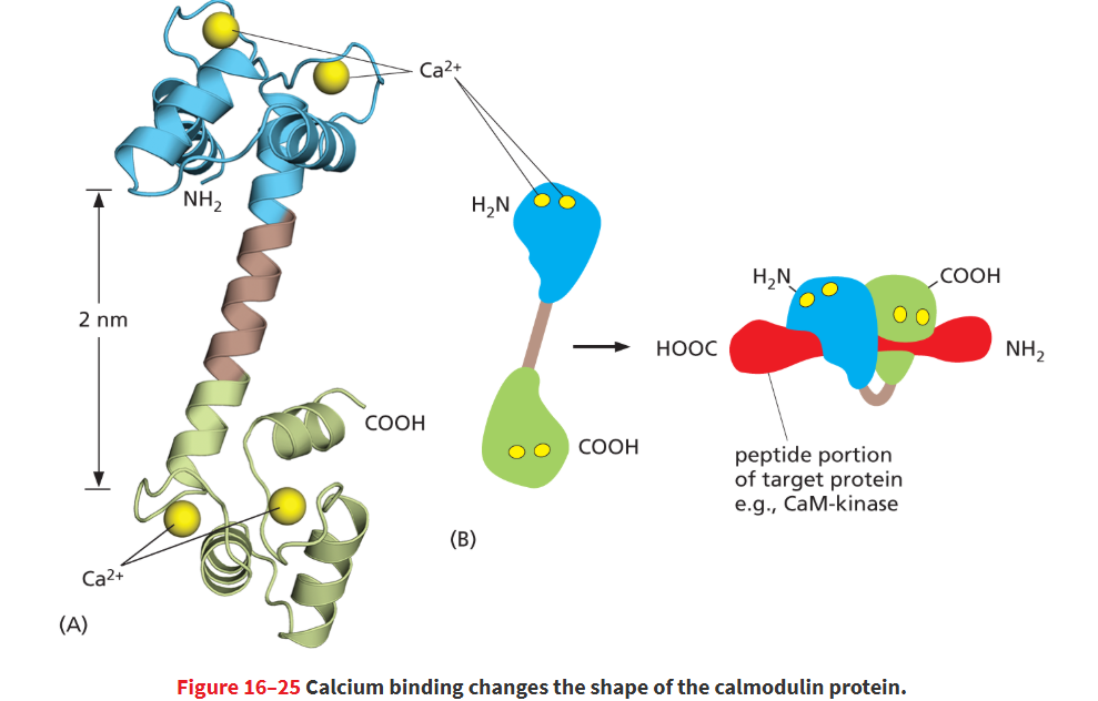
calmodulin
Small Ca2+-binding protein that modifies the activity of many target proteins in response to changes in Ca2+ concentration
activated by binding 4 Ca2+ ions
calcium binding causes a shape change in calmodulin that allow it to wrap around its target protein
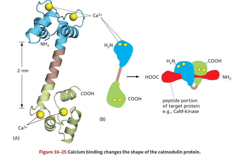
Ca2+/calmodulin-dependent protein kinase (CaM-kinase)
Enzyme that phosphorylates target proteins in response to an increase in Ca2+ ion concentration through its interaction with the Ca2+-binding protein calmodulin.
this is a class of target proteins for calmodulin to bind to!
involved in processes like learning, memory
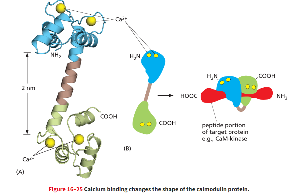
calmodulin structure
Calmodulin has a dumbbell shape, with two globular ends connected by a long α helix. Each of the globular ends has two Ca2+-binding sites.
in total 4 calciums bind and then a shape change will let the alpha helix portion wrap around the target protein which is usually a CaM-kinase that will go onto phosphorylate target proteins
nitric oxide
a secondary messenger in response to GCPR activation that is small/hydrophobic enough to pass across the membrane and carry a signal directly to nearby cells.
paracrine
Acetylcholine binds to a GPCR on the endothelial cell surface, resulting in activation of Gq and the release of Ca2+ inside the cell. Ca2+ then stimulates nitric oxide synthase, which produces NO from the amino acid arginine.
NO will diffuse into nearby smooth muscle cell and allow cell to relax
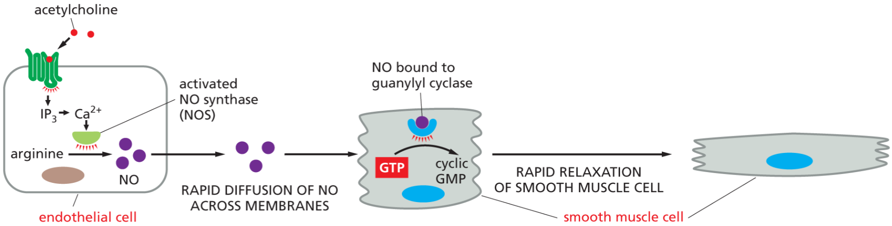
guanyl cyclase
a target protein in smooth muscle cells which NO binds to.
it then stimulates the formation of cyclic GMP → GTP
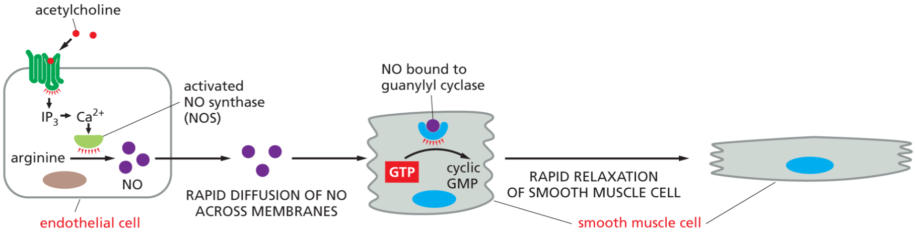
What does NO do in smooth muscle cells?
Activates guanylyl cyclase → increases cGMP → causes smooth muscle relaxation and vasodilation.
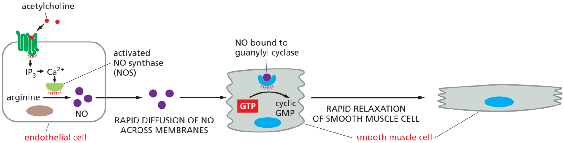
What is the key second messenger in NO signaling?
cyclic GMP which is made from guanyl cyclase binding to NO
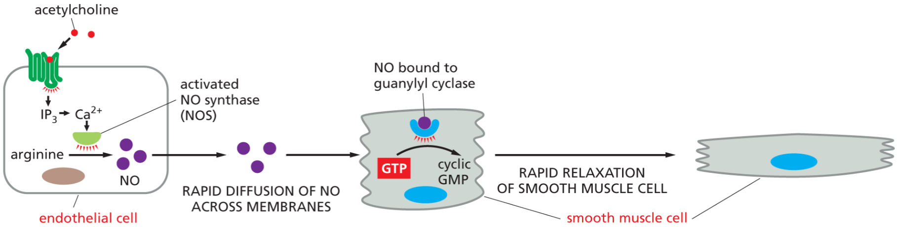
How does IP₃ increase intracellular Ca²⁺ concentration?
IP₃ diffuses through the cytosol and binds IP₃-gated Ca²⁺ channels on the ER membrane, opening them. Because Ca²⁺ concentration is high in the ER and very low in the cytosol, Ca²⁺ rapidly flows into the cytosol, causing a sharp increase in intracellular Ca²⁺.
how does Ca2+ play a role in fertilization?
When the sperm binds and fuses with the egg, it activates signaling pathways that open Ca²⁺ channels in the egg’s ER which spreads through the egg to block additional sperm and triggers embryonic development.
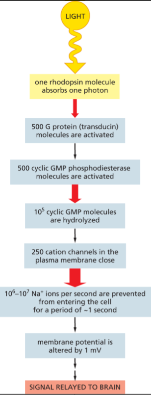
Rhodopsin
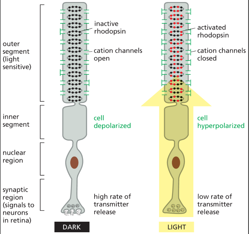
a G-protein coupled light receptor that activates a G-protein called transducin when stimulated by light.
transducin
the alpha subunit of a G-protein thats activated when light strikes rhodopsin. transducin will turn on an enzyme called cGMP phosphodiesterase which breaks down cGMP → GMP.
this will cause cation channels to close and change the voltage gradient
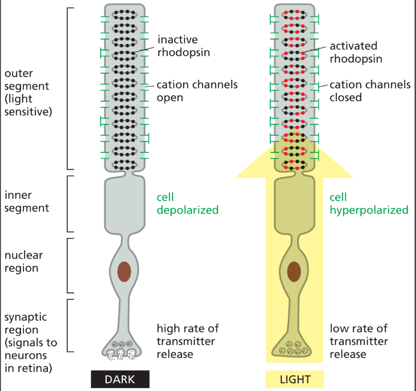
cGMP phosphodiesterase
cGMP phosphodiesterase is the enzyme that terminates cGMP signaling by breaking cGMP into GMP, especially in photoreceptor cells during light detection.
its activated by transducin and will cause cGMP levels drop → channels close → electrical signal is generated
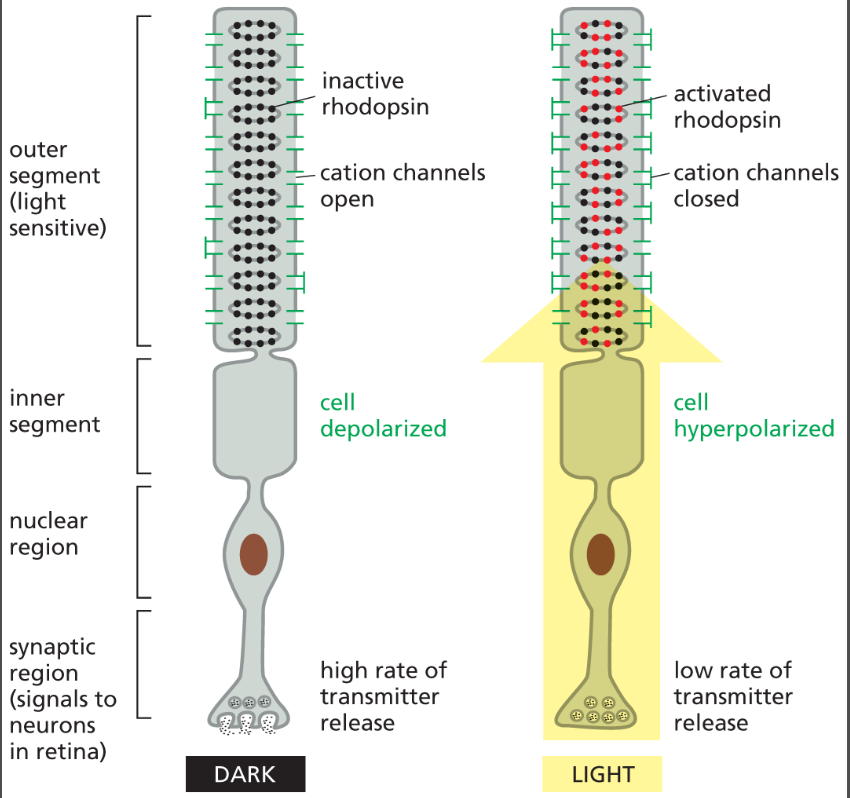
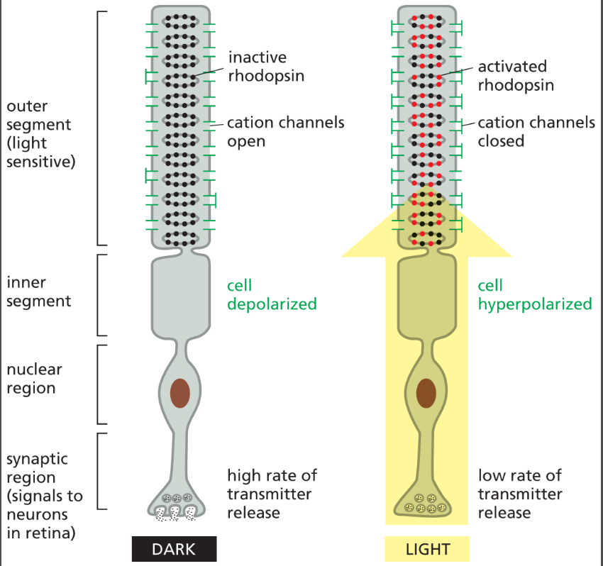
What is the role of cGMP in photoreceptor cells in the dark?
cGMP is continuously produced and binds to cation channels (Na⁺/Ca²⁺ channels), keeping cation channels open and maintaining the “dark current” (cell is depolarized in darkness).
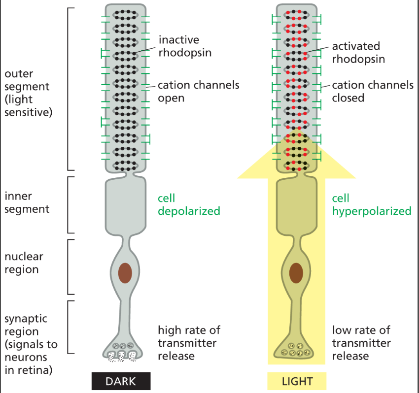
What happens to cGMP levels when light activates rhodopsin?
Activated transducin stimulates cGMP phosphodiesterase, which breaks down cGMP → GMP, causing cGMP levels to fall sharply.
cGMP levels drop → channels close → electrical signal is generated from light
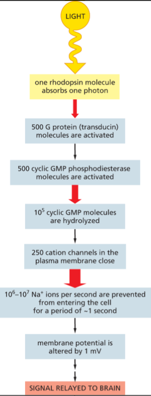
How does light ultimately change the electrical state of a photoreceptor cell?
↓ cGMP → closure of cGMP-gated cation channels → ↓ Na⁺ influx → membrane hyperpolarization → altered neurotransmitter release → signal sent to brain.
What is receptor-level adaptation in GPCR signaling?
GPCRs can be inactivated, desensitized, internalized, or degraded (lysosomal breakdown) so they stop responding to ligand stimulation.
adaptation allows them to prevent overstimulation and allows cells to respond appropriately across a wide range of signal intensities.
What is the GPCR signaling pathway in rod photoreceptor cells in the dark vs. in light?
Dark (no light):
Guanylyl cyclase active → high cGMP → cGMP binds cation channels → Na⁺/Ca²⁺ channels OPEN → cell is depolarized → continuous neurotransmitter release to brain
Light:
Light activates rhodopsin → activates transducin (G protein) → activates cGMP phosphodiesterase → cGMP ↓ (broken down to GMP) → cation channels CLOSE → Na⁺ influx stops → cell hyperpolarizes → neurotransmitter release decreases → signal sent to brain
cGMP
a small intracellular signaling molecule (second messenger) that transmits signals inside cells.
Made from GTP (guanosine triphosphate) by the enzyme guanylyl cyclase
regulates cellular responses like vision and smooth muscle relaxation by controlling protein activity and ion channels.
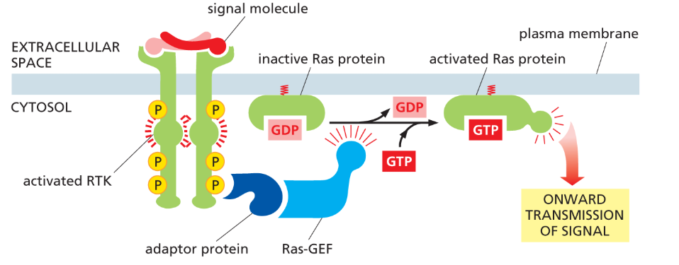
receptor tyrosine kinases (RTKs)
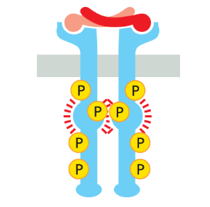
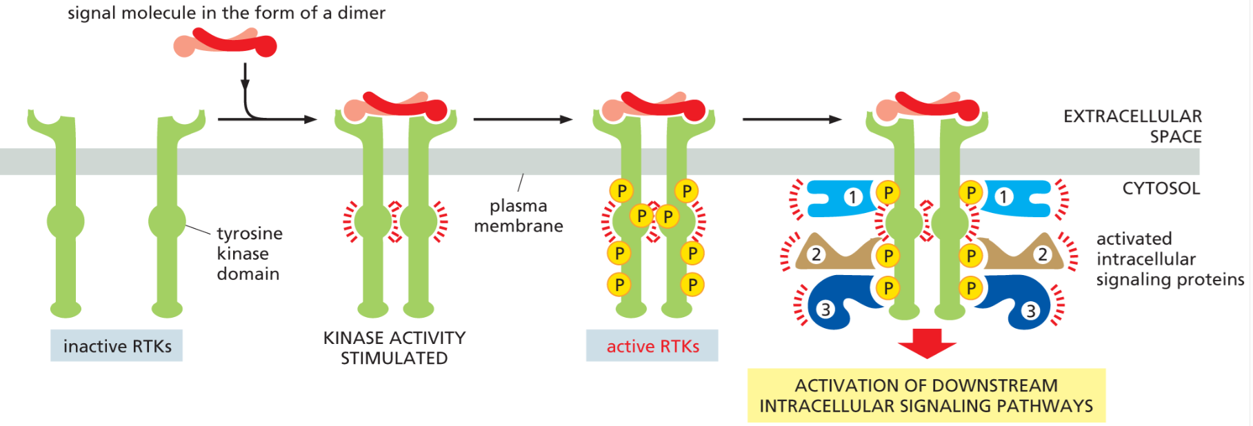
single alpha helix pass transmembrane protein where cytoplasmic domain is a tyrosine kinase, which gets activated by a ligand binding to the receptor’s extracellular domain
a type of enzyme-associated receptor where it is itself the enzyme
phosphorylate tyrosine on target proteins and themselves!
What are enzyme-coupled receptors and how are they different from GPCRs?
Enzyme-coupled receptors are single-pass transmembrane proteins whose cytosolic domain is either an enzyme or binds to an enzyme. Unlike GPCRs, they do not use G proteins; instead, they directly activate intracellular enzymes and signaling proteins, often leading to changes in gene expression, cell growth, differentiation, and survival.
What is the basic activation mechanism of RTKs?
Ligand binding causes receptor dimerization, which brings two kinase domains together. Each receptor phosphorylates the other (cross-phosphorylation) on tyrosine residues, activating them.
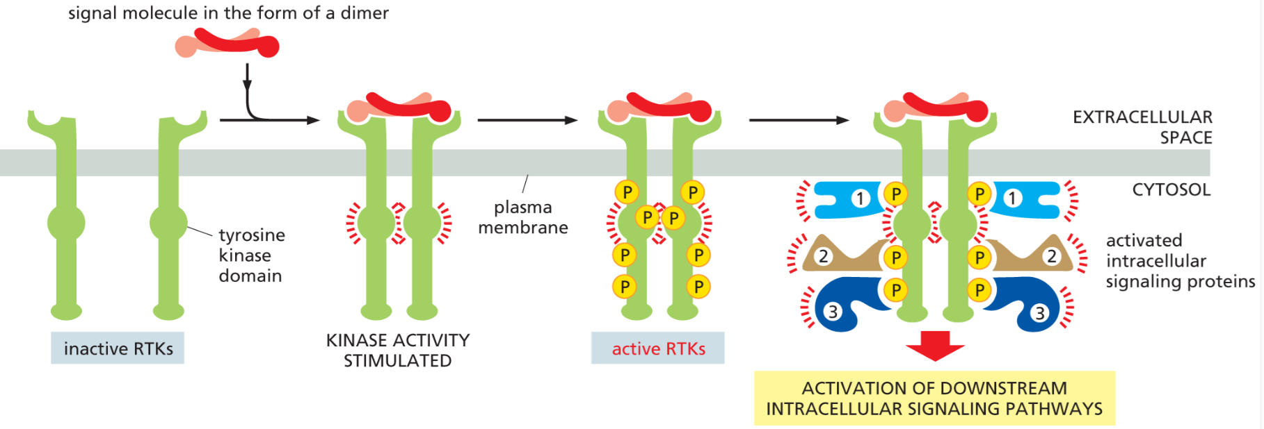
What happens after RTKs are phosphorylated?
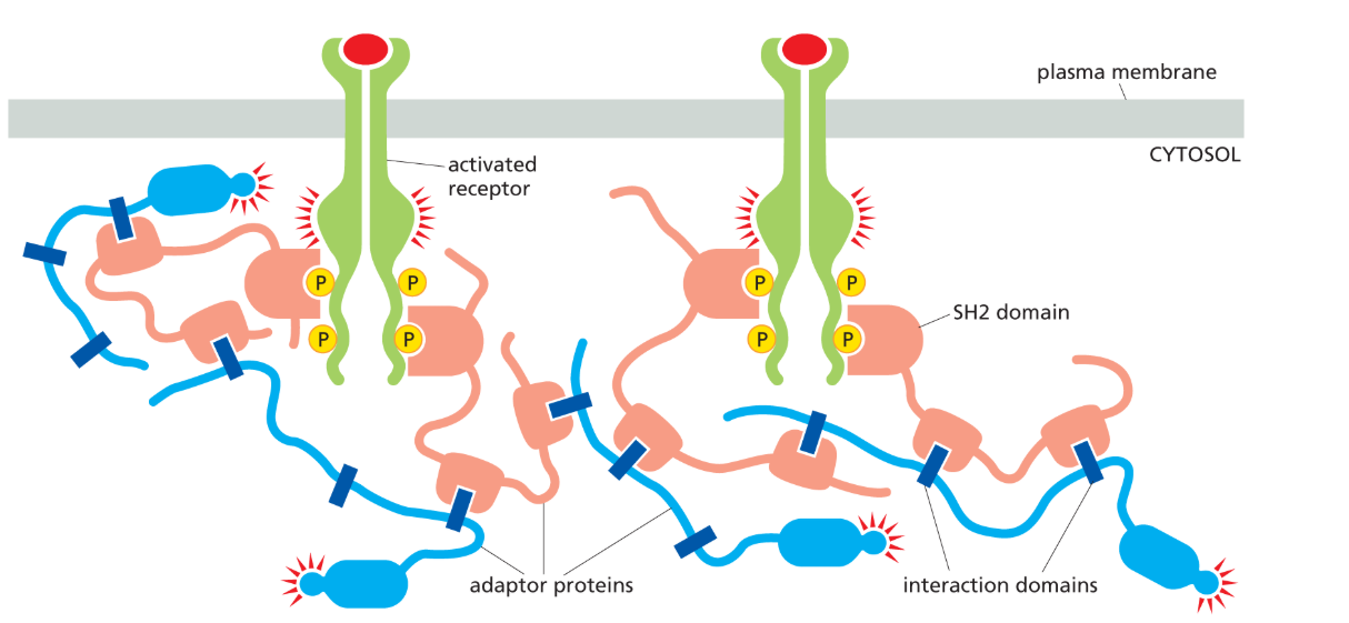
The phosphorylated tyrosines become docking sites for intracellular signaling proteins, which bind and form a large signaling complex at the cytosolic tail of the receptor.
SH2 domains
protein interaction modules that specifically recognize and bind phosphorylated tyrosines on activated RTKs, allowing signaling proteins to assemble at the receptor.
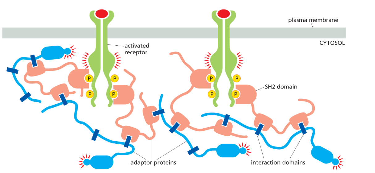
SH3 domains
binds proline rich repeats
helps assemble signaling complexes at plasma membrane and works together w/ SH2 domains
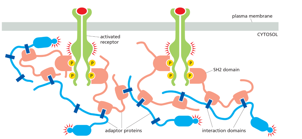
biomolecular condensates
Large, gel-like, membraneless clusters of signaling proteins formed by many weak interactions between adaptor proteins and receptors, which organize and amplify signaling at the membrane.
How is Receptor tyrosine kinase signaling turned off?
Tyrosine phosphatases (remove phosphate groups from tyrosines)
Endocytosis and lysosomal degradation of the receptor
Disassembly of signaling complexes
Ras
a small GTP-binding protein that helps relay signals from cell-surface receptors to the nucleus. Many human cancers contain an overactive mutant form of the protein.
most receptor tyrosine kinases activate Ras!
it is attached covalently by a lipid tail to cystolic face of plasma membrane
it resembles the alpha subunit of a trimeric G protein
gets activated by GTP and help of GEF/GAP!
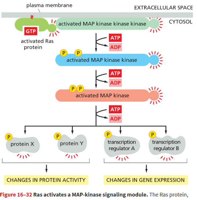
MAP-kinase signaling module
Set of 3 functionally interlinked protein kinases that allows cells to respond to extracellular signal molecules that stimulate proliferation; includes a mitogen-activated protein kinase (MAP kinase), a MAP kinase kinase, and a MAP kinase kinase kinase.
activated by Ras
Ras → MAP kinase kinase kinase (MAPKKK) → MAP kinase kinase (MAPKK) → MAP kinase (MAPK) → effector proteins (including transcription regulators)