Lids & Injection Grading
1/83
There's no tags or description
Looks like no tags are added yet.
Name | Mastery | Learn | Test | Matching | Spaced |
|---|
No study sessions yet.
84 Terms
allergic conjunctivitis, all CL wearers, suspected foreign body
give examples of indications for lid eversion
recent history of trauma/surgery to the lid
when is lid eversion contraindicated?
lids, tear film, corneal epithelium
what are the non-immune responses of the cornea?
heat, redness, swelling, pain, loss of function
what are the classic signs of inflammation?
hypertrophy
increase in cell size
hyperplasia
increase in cell number; cancer risk
metaplasia
reversible changes in which 1 adult cell is replaced by another; cancer risk
dysplasia
loss of uniformity of cells & their architectural orientation; cancer risk
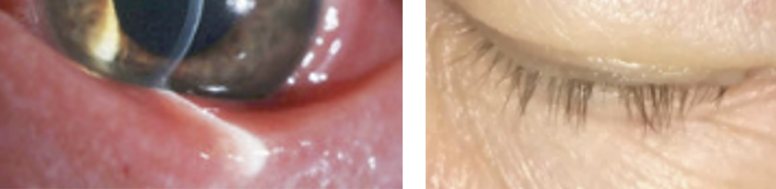
madarosis
loss of eyelashes due to chronic inflammation or trauma
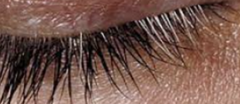
poliosis
whitening of lashes due to chronic inflammation
trichiasis
misdirection of lashes; oftentimes on the lower lid; due to age/trauma
entropion
lashes are pulled into the eye
ectropion
outward turning of the eyelid margin, inferior lid margin or lacrimal puncta does not contact the globe
distichiasis
partial or complete second row of lashes
anterior blepharitis
characterized by scales around the base of the lashes, redness, & thickened hypertrophic lid margins
generally bacterial in origin
sx: itchiness, burning, foreign body sensation, tearing, crusting, associated with lash findings
staphylococcal
anterior blepharitis characterized by thin, honey-colored flakes (collarettes) among eyelashes
seborrheic
anterior blepharitis characterized by dandruff-like flakes or accumulations of oily sebaceous material among the eyelashes; most common blepharitis form
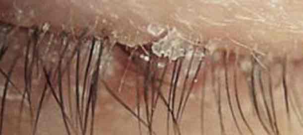
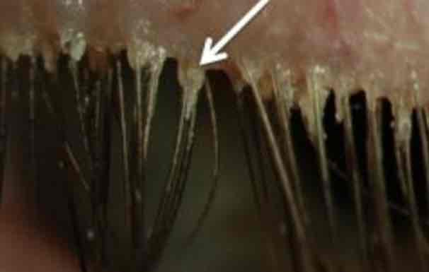
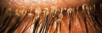
demodectic
anterior blepharitis caused by a mite; presence of a waxy-appearing, cylindrical cuffs around bases of lashes
posterior blepharitis
blepharitis caused by meibomian gland irritation/inflammation
frequently associated with seborrhea
staph and hypersensitivity response
mild-moderate inflammatory appearance to the posterior lid margin & palpebral conj
papillary response
tear film disturbances
meibomian gland
linear & 3-4mm in length traversing the posterior lid perpendicularly from the lid margin to the opposite edge of the tarsus
tubulo-acinar architecture with saccular arrangements of acini & a ductal system that communicates with orifices near the mucocutaneous junction of the lid
meibocytes
functional unit of the meibomian gland; synthesizes & secretes lipids into precorneal tear film
meibum
prevents the tear evaporation & thus desiccation of the ocular surface, acts as a hydrophobic barrier to the inward movement of environmental & organic agents, lubricates the ocular surface to prevent irritation while promoting a clear ocular image
more
the _____ color fringes present in tear film, interferometry, the thicker the tear film is
greater than 100nm
what is the normal thickness of the lipid layer of the tear film?
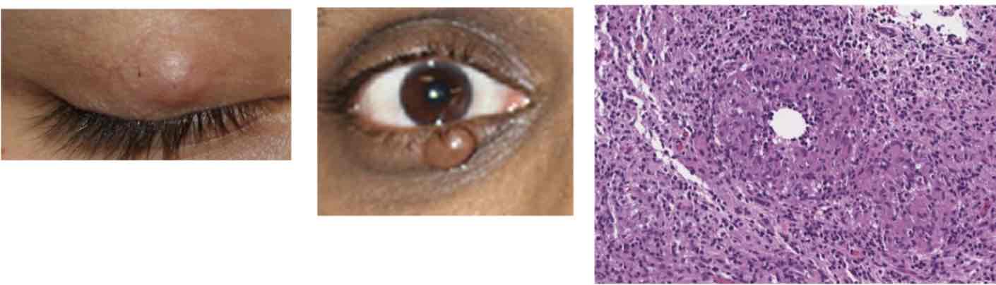
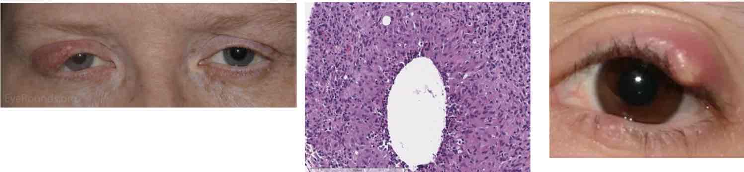
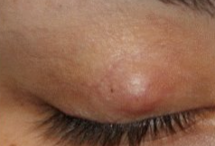
chalazion
presents with a hard, painless nodule in the eyelid that slowly enlarges over the course of weeks-months
can result from a hordeolum or can occur independently
associated with rosacea & blepharitis
specimens show zonula lipogranulomatous inflammation centered on clear spaces previously filled with lipid
mixed inflammatory infiltrate that consists of neutrophils, plasma cells, lymphocytes, epithelioid histiocytes, & multinucleate giant cells
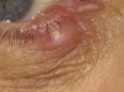
hordeolum
acute, purulent inflammatory process of any gland in the eyelid that presents as a discrete, warm, erythematous, painful pustule over the course of a few days
pathology shows a small, purulent abscess consisting of neutrophils & necrotic cellular debris on a hair follicle & its adjacent gland
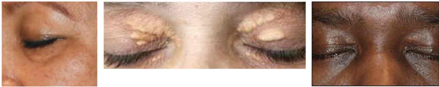
xanthelasma
tumor consisting of intracellular accumulation of lipid with lipid-laden macrophages within the dermis
presents as multiple soft, yellowish plaques commonly found near the medial canthi of the upper & lower lids
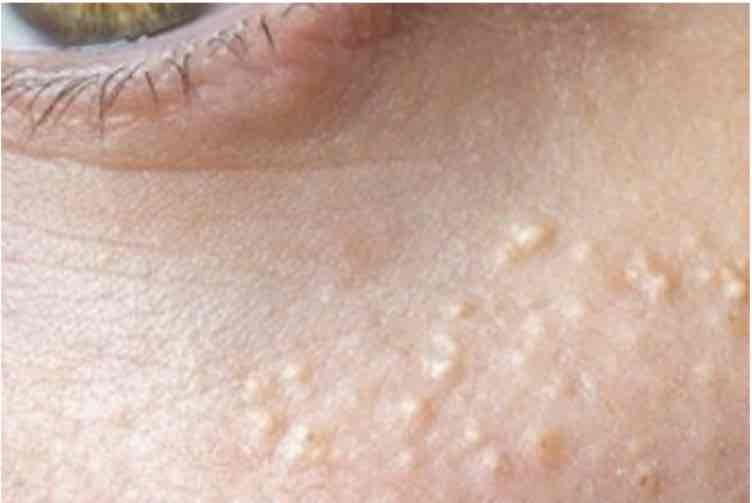
milia
very common in young people & newborns
dead skin cells/keratins trapped w/in the base of a hair follicle or sweat gland
small raised bump that looks like a pimple
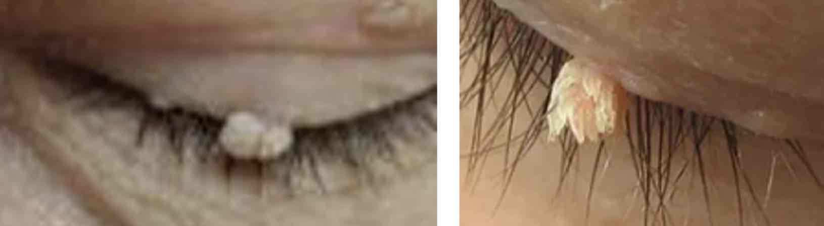
papilloma
one of the most common eyelid tumors
occurs in middle-aged or elderly patients
benign, painless, & carries little to no risk for growth into cancer
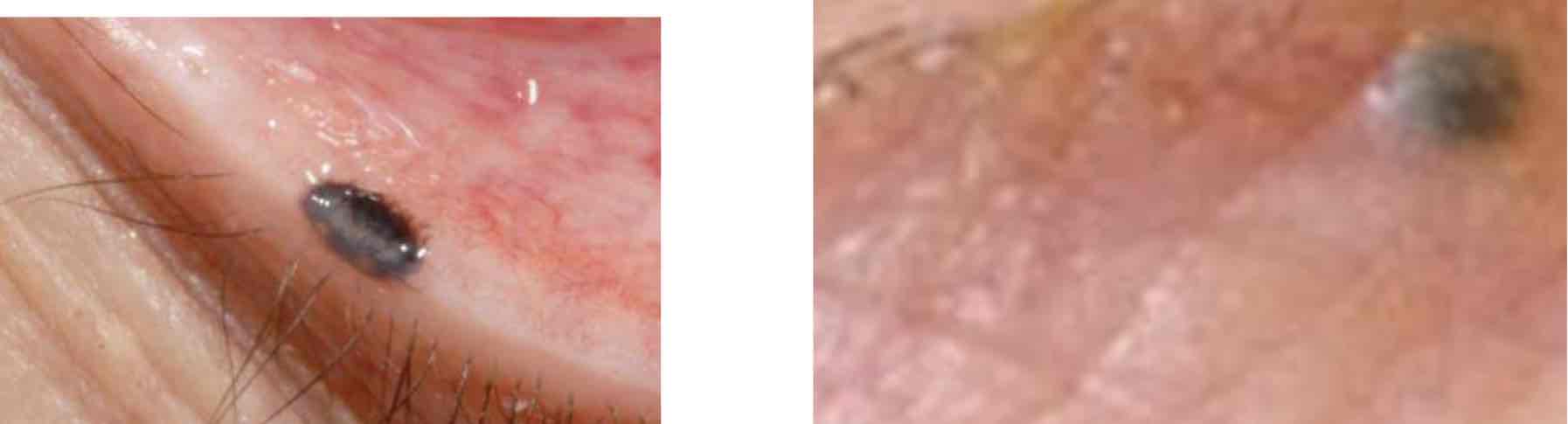
nevus
freckle
congenital or acquired
stable
no
can tumors cross the lid?
no
can tumors cross from the left eye to the right eye?
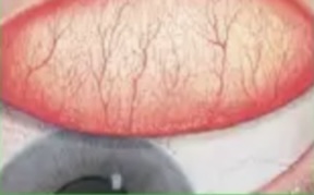
0
grade the papilla
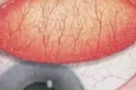
1
grade the papilla
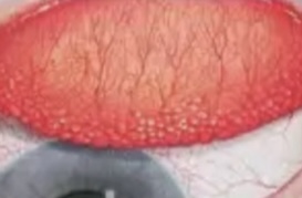
2
grade the papilla
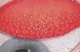
3
grade the papilla
4
grade the papilla
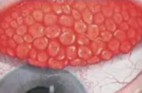
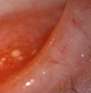
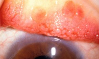
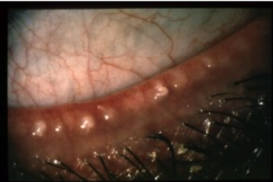
granulomata
what is shown in the pic?
calcium deposit
what is shown in the pic?
hordeolum
what is shown in the pic?
endophthalmitis w/ hypopyon
what is shown in the pic?
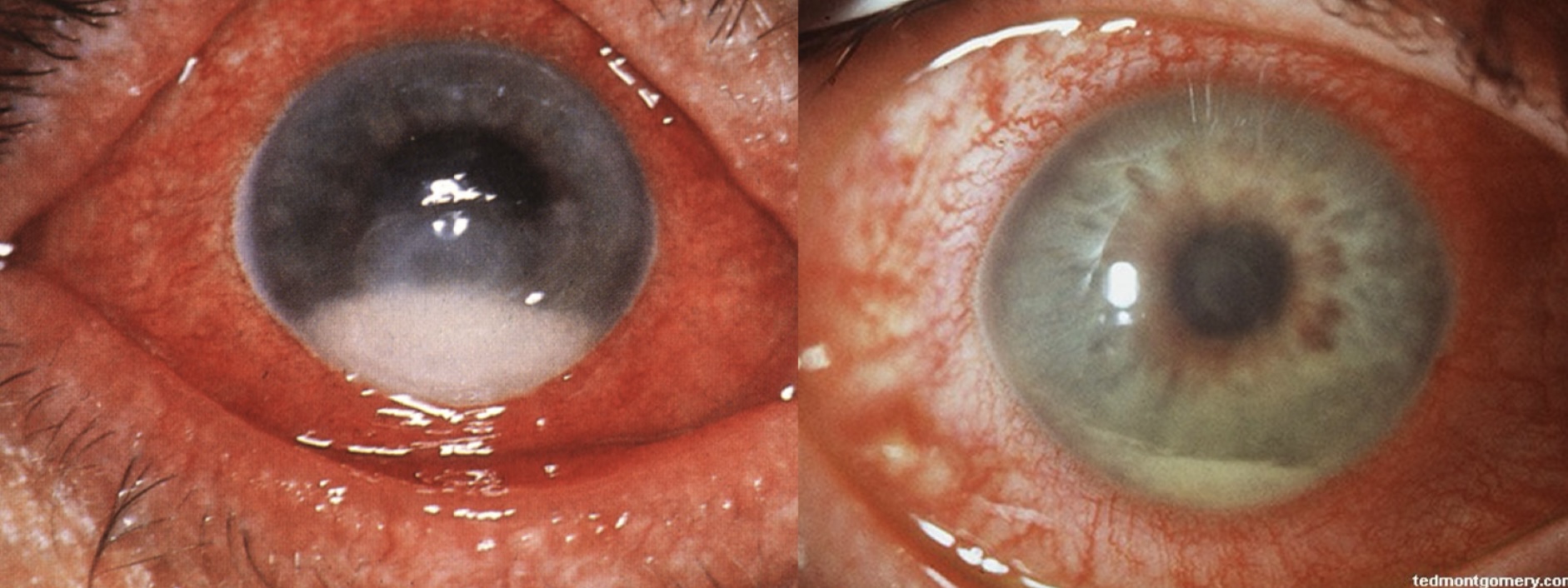
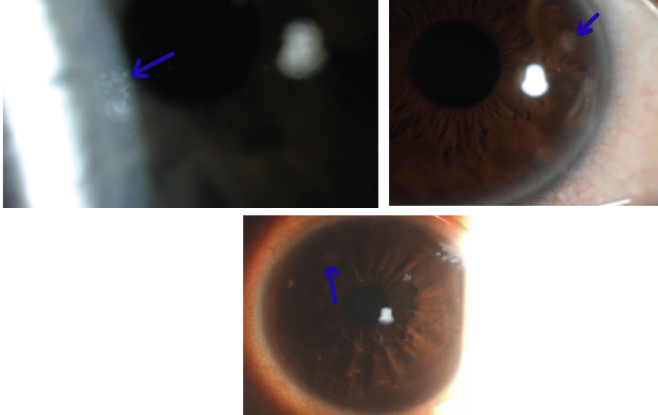
corneal ulcer
what is shown in the pic?
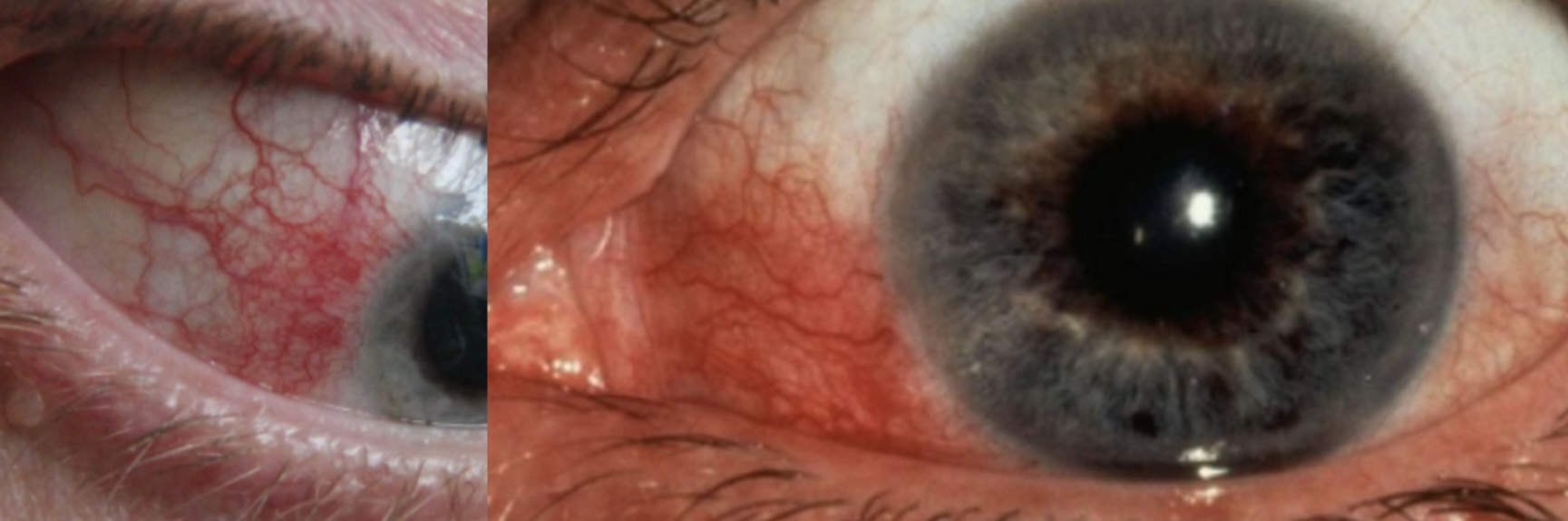
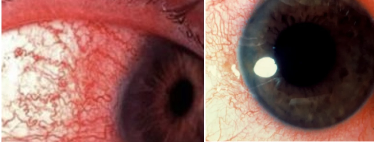
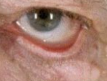
diffuse redness
what is shown in the pic?
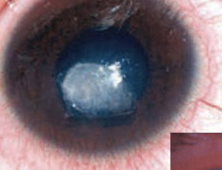
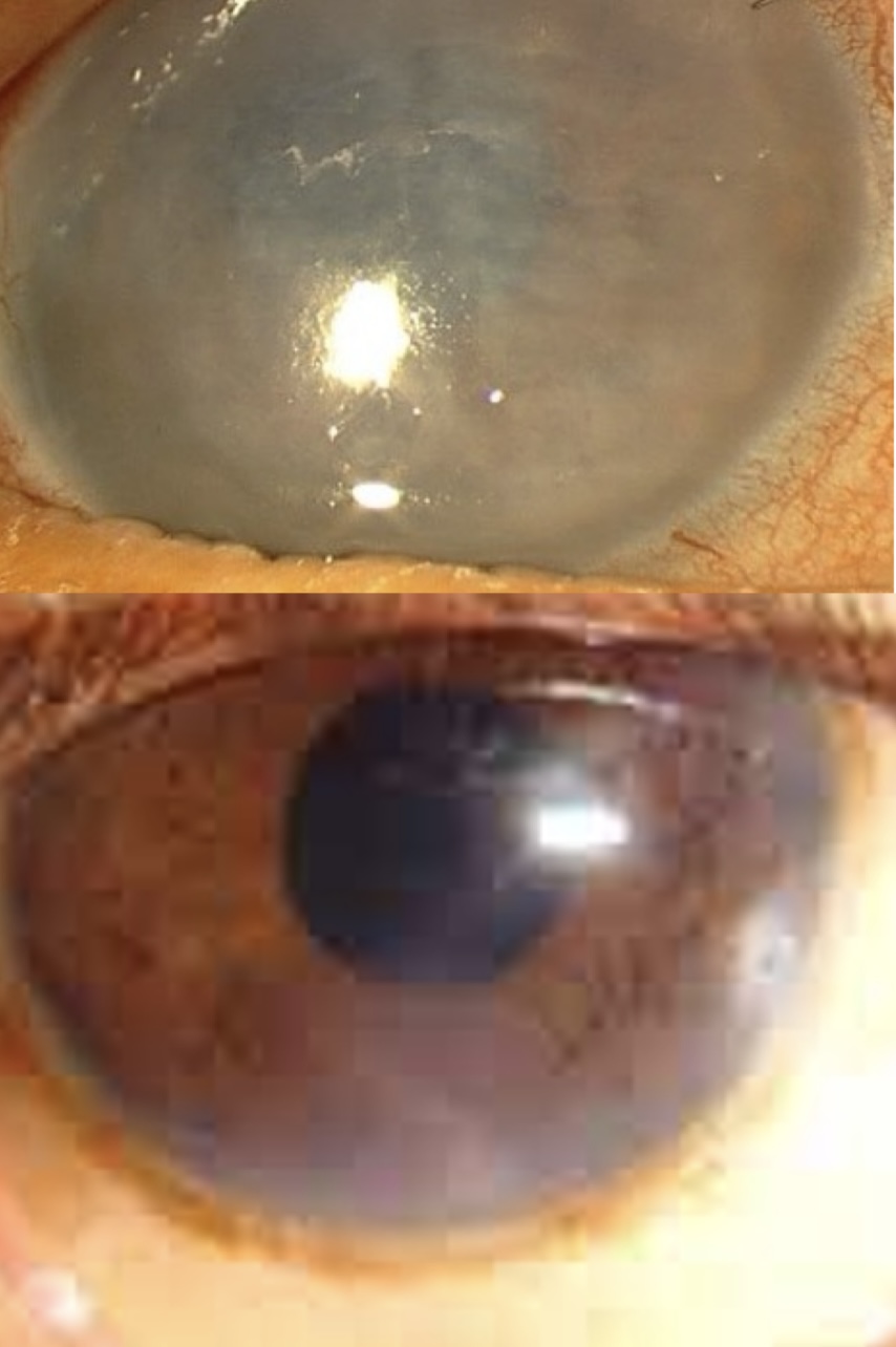
corneal edema
what is shown in the pic?
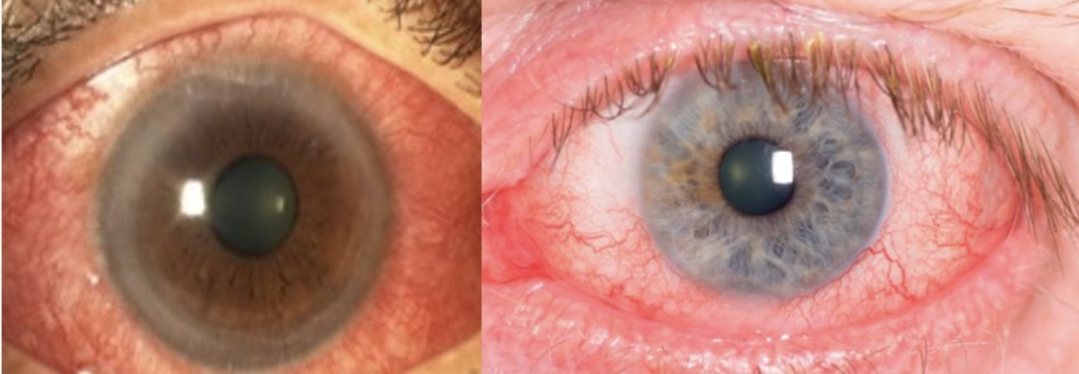
focal redness
what is shown in the pic?
corneal infiltrate
what is shown in the pic?
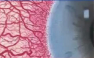
circumlimbal injection
what is shown in the pic?
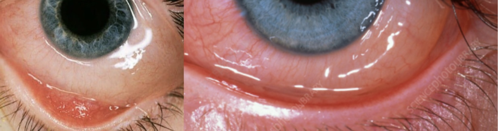
conjunctival edema
what is shown in the pic?
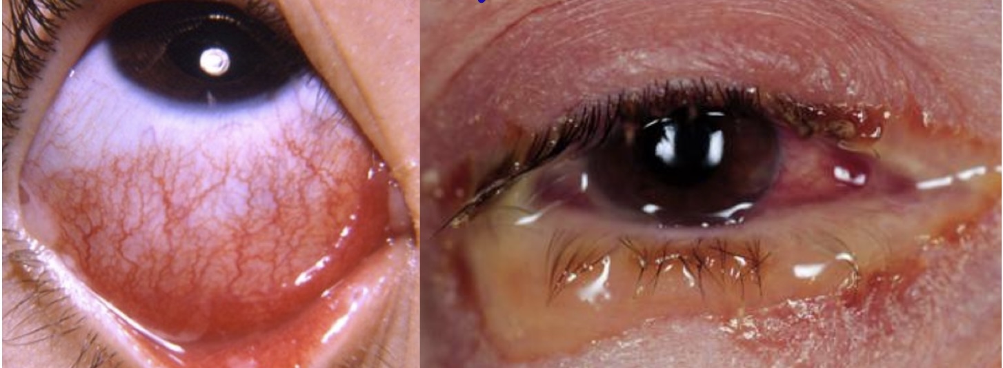
bacterial conjunctivitis
what is shown in the pic?
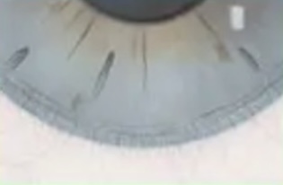
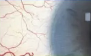
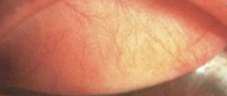
0
grade the injection
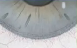
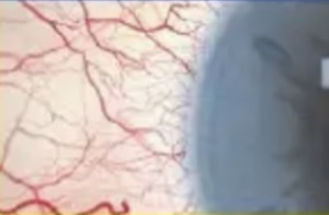
1
grade the injection
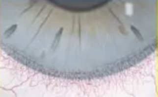
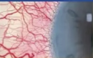
2
grade the injection
3
grade the injection

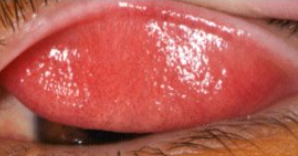
4
grade the injection
0
grade the injection

1
grade the injection
2
grade the injection
3
grade the injection
4
grade the injection
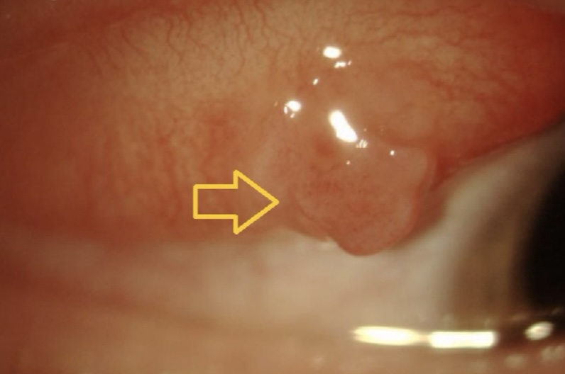
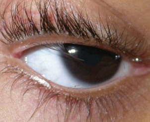
nevus
what is shown in the pic?
nevus
what is shown in the pic?
nevus
what is shown in the pic?
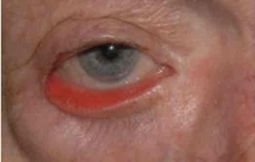
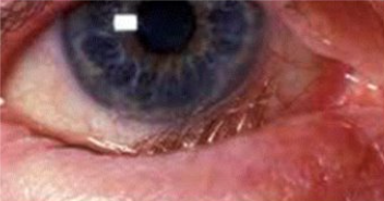
ectropion
what is shown in the pic?
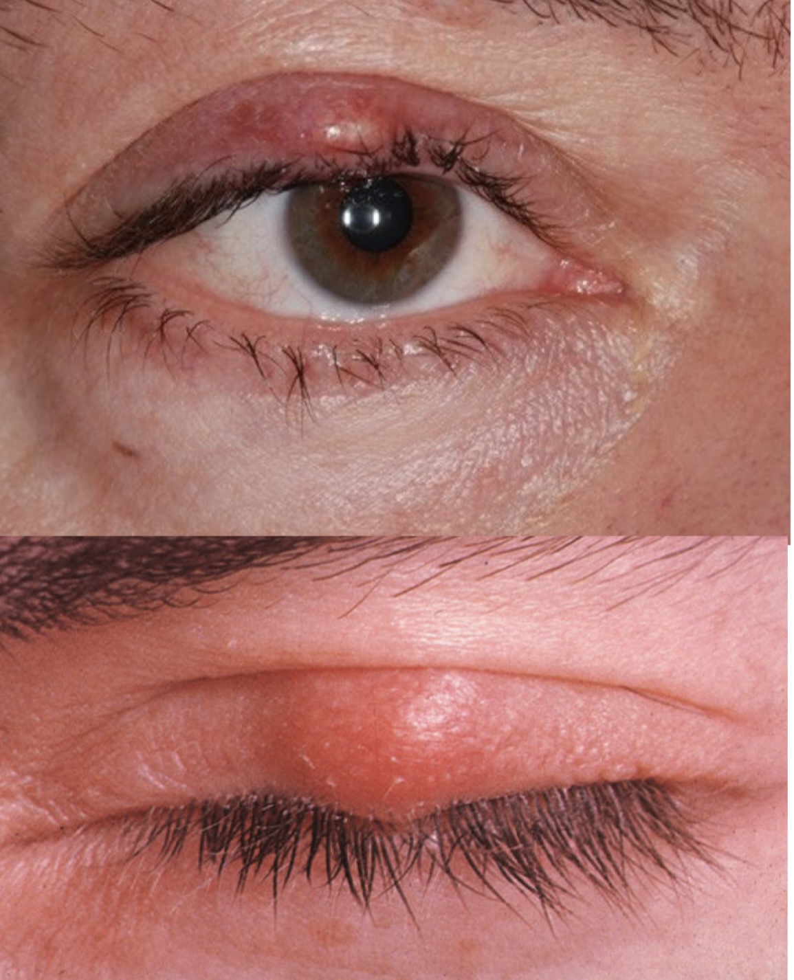
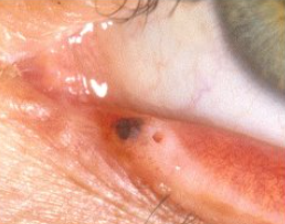
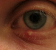
hordeolum
what is shown in the pic?
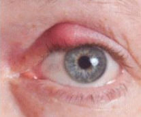
hordeolum
what is shown in the pic?
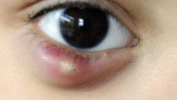
hordeolum
what is shown in the pic?
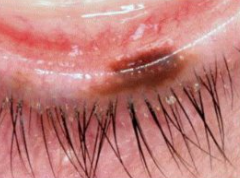
chalazion
what is shown in the pic?
chalazion
what is shown in the pic?
entropion
what is shown in the pic?
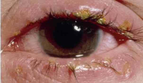
demodex blepharitis
what is shown in the pic?
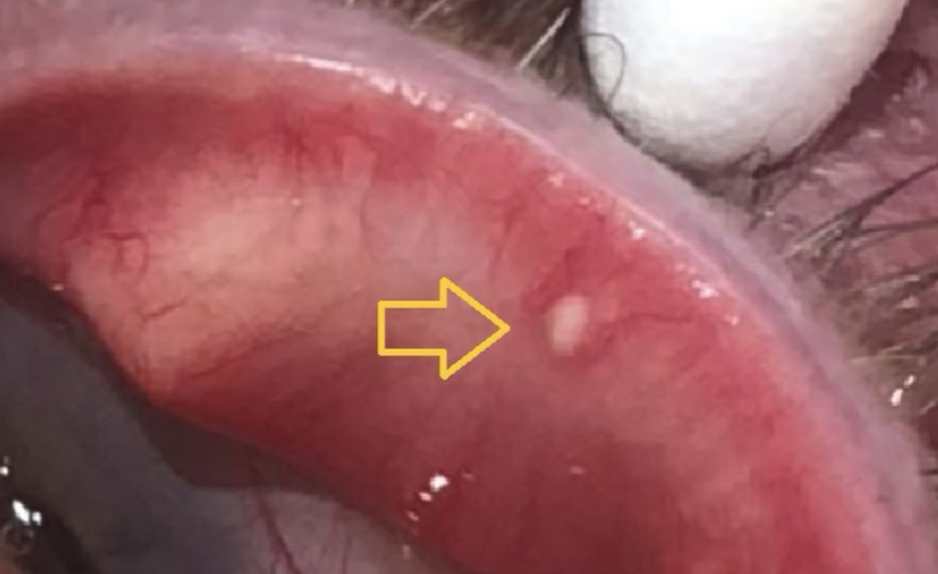
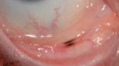
concretion
what is shown in the pic?
papillae
what is shown in the pic?

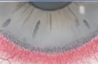
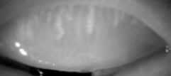
3: severe gland loss
grade the meibomian glands
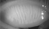
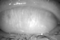
0: normal
grade the meibomian glands
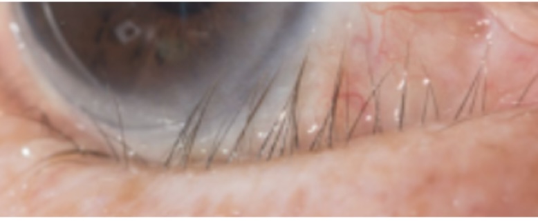
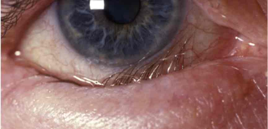
distichiasis & trichiasis
what is shown in the pic?
0
grade the papillae
1
grade the papillae
3
grade the papillae
3
grade the papillae

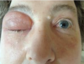
orbital cellulitis (inflammation is restricted to the orbit)
what is shown in the pic?
miebomitis/posterior blepharitis
what is shown in the pic?
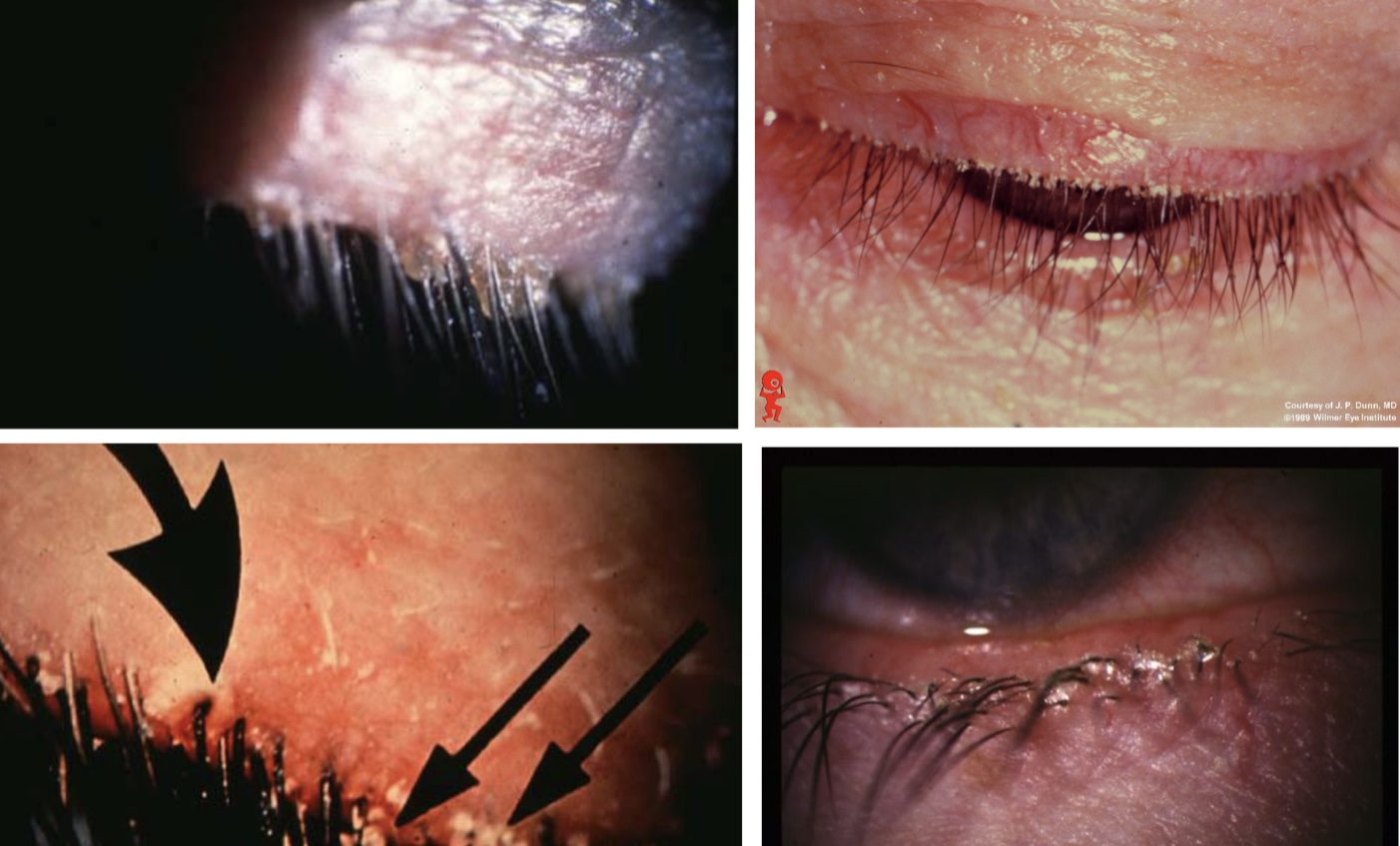
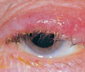
staphylococcal blepharitis
what is shown in the pic?
1: mild gland loss
grade the meibomian glands
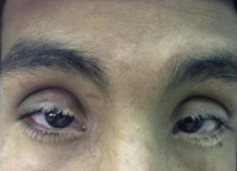
poliosis
what is shown in the pic?