Cardio Week 1
 Call Kai
 Learn
Learn Practice Test
 Spaced Repetition
 Match
Match Flashcards
 Knowt Play
1/479

There's no tags or description
Looks like no tags are added yet.
Name | Mastery | Learn | Test | Matching | Spaced | Call with Kai | Chat |
|---|
No analytics yet
Send a link to your students to track their progress
480 Terms
Which ventricle holds more blood at end‑diastole?
RV EDV > LV EDV (RV holds slightly more blood).

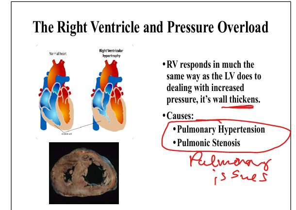
Causes of RV pressure overload
Pulmonary hypertension, pulmonic stenosis.
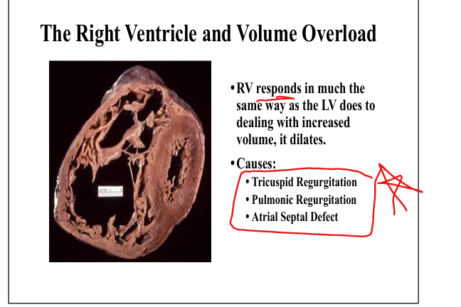
Causes of RV volume overload
Tricuspid regurgitation, pulmonic regurgitation, atrial septal defect.
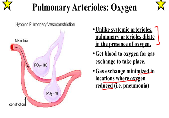
How do pulmonary arterioles respond to oxygen?
They dilate in the presence of oxygen
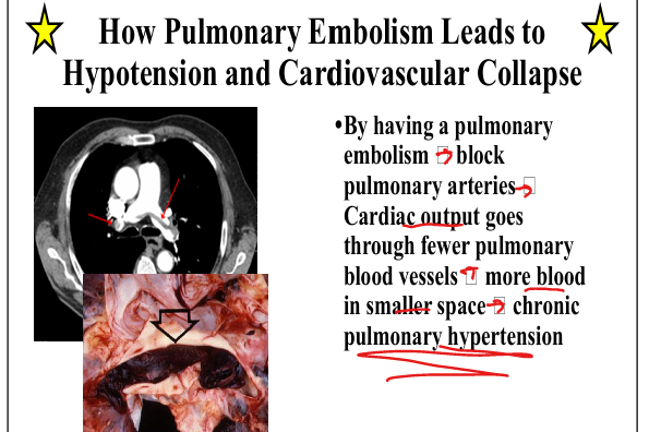
How does pulmonary embolism cause hypotension?
Blocked pulmonary arteries → CO forced through fewer vessels → ↑PVR → RV failure → hypotension.
Effect of left‑to‑right shunt on pulmonary vasculature
Systemic pressure damages pulmonary arteries → hypertrophy → ↑PVR → impaired oxygenation.


What causes reversal to right‑to‑left shunt?
PVR > SVR → deoxygenated blood enters systemic circulation.

Signs of Eisenmenger syndrome
Clubbing, polycythemia, hemoptysis, heart failure.

Is Eisenmenger reversible?
No — irreversible pulmonary hypertension

What is Tetralogy of Fallot?
VSD + Pulmonic stenosis + Overriding aorta + RV hypertrophy. PROV

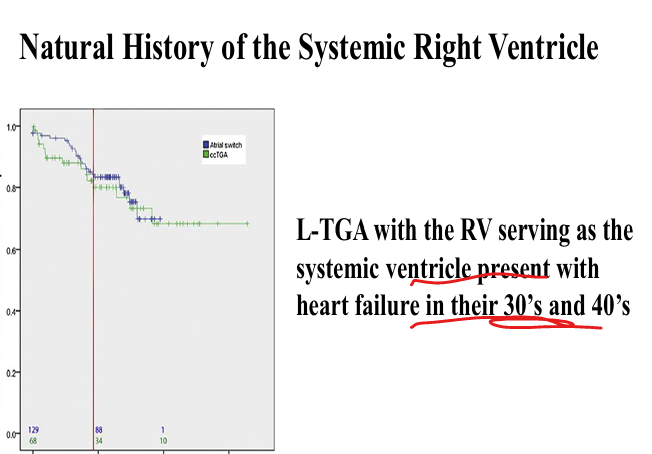
What is L‑TGA (Congenital Corrected Transposition of the Great Arteries)?
TGA=Transposition of Great Arteries=>Congenital corrected transposition where RV becomes systemic ventricle.

Natural history of systemic RV (L‑TGA)
RV fails in 30s–40s due to chronic systemic workload.
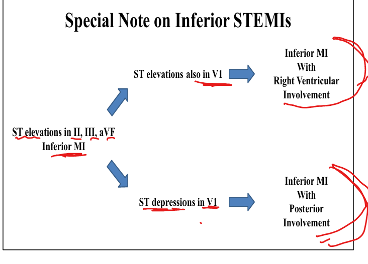
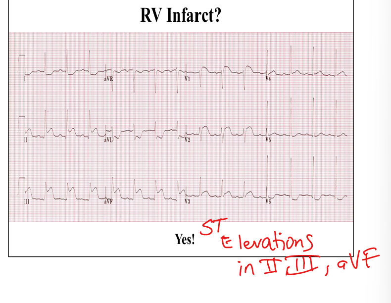
ECG finding suggesting RV infarct in inferior MI
ST elevation in II, III, aVF plus ST elevation in V1
Q: What anatomical fact allows the jugular venous pulse to directly reflect right atrial pressure?
A: There is no valve between the SVC and right atrium, so JVP directly transmits RA pressure.


Q: What does the “a wave” represent on the JVP waveform?
A: Atrial contraction.


Q: What does the “x descent” represent on the JVP waveform?
A: Atrial relaXation.
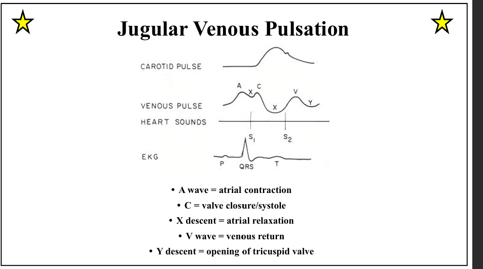
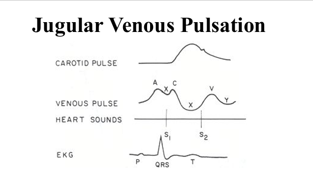
Q: What does the “v wave” represent on the JVP waveform?
A: Venous return into the right atrium.
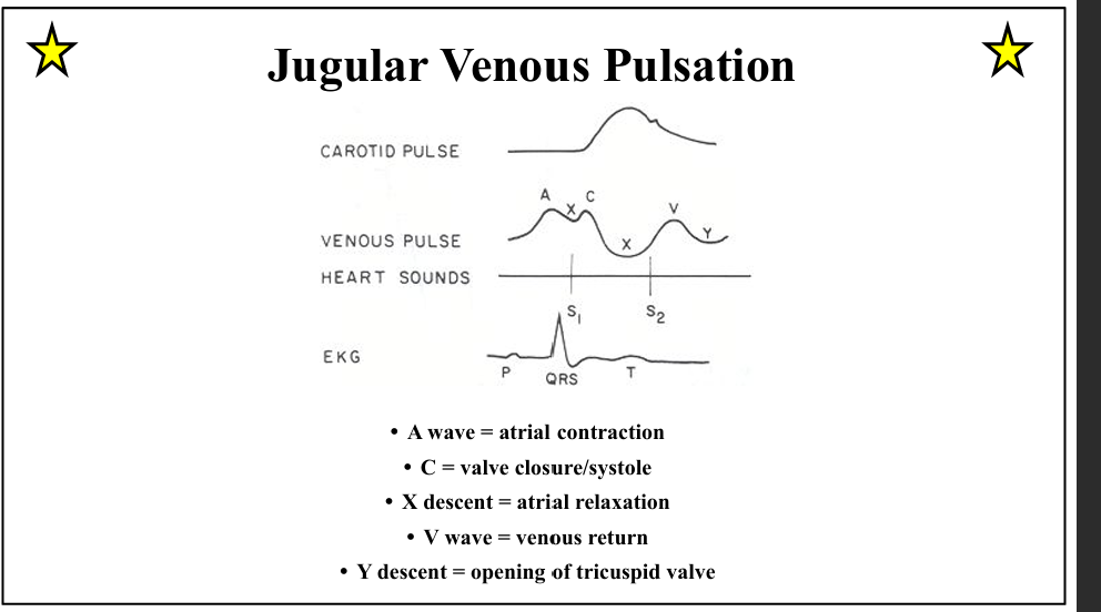

Q: What does the “y descent” represent on the JVP waveform?
A: Opening of the tricuspid valve and early RV filling.

Q: How do you differentiate JVP from carotid pulse?
A: JVP has two pulsations per cardiac cycle

Q: What are the two major layers of the pericardium?
A: Visceral and parietal layers separated by a thin fluid layer.
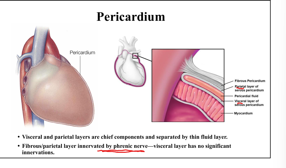
Q: Which pericardial layer is innervated by the phrenic nerve?
A: The fibrous/parietal pericardium. The visceral layer has no significant innervation.

Q: Where do the coronary arteries run relative to the pericardium?
A: Within epicardial fat inside the visceral pericardium.

Q: What are the functional roles of the pericardium?
A: Maintains anatomic orientation, prevents acute LV dilation, and limits spread of infection.
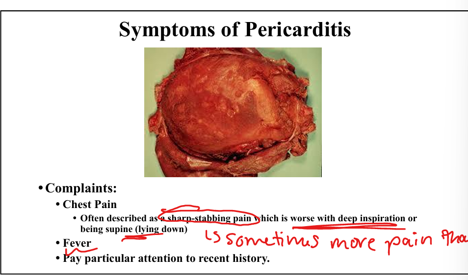
Q: What is the classic chest pain description in acute pericarditis?
A: Sharp, stabbing pain worse with inspiration and lying supine.

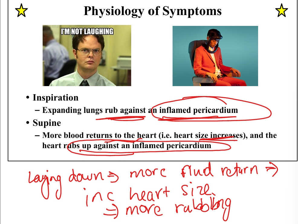
Q: Why does inspiration worsen pericarditis pain?
A: Expanding lungs rub against the inflamed pericardium.

Q: Why does lying supine worsen pericarditis pain?
A: Increased venous return enlarges the heart, increasing friction against the inflamed pericardium.
Q: What physical exam finding is characteristic of pericarditis?
A: A pericardial friction rub with components from ventricular contraction, ventricular relaxation, and atrial contraction.

Q: What EKG findings suggest acute pericarditis?
A: Diffuse ST elevation and PR depression.

Q: What medications treat pericarditis?
A: NSAIDs (aspirin, ibuprofen) + colchicine

Causes of pericarditis
Infectious: HIV, Coxsackie, TB, bacterial
Non-infectious: MI, Post CABG, Renal failure, Radiation, Lupus/RA

Q: What three factors determine symptoms in pericardial effusion?
A: Total volume, rate of accumulation, and pericardial stiffness.

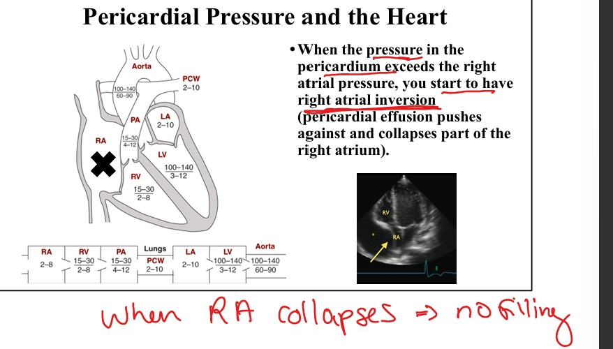
Q: What happens when pericardial pressure exceeds right atrial pressure?
A: Right atrial inversion/collapse.
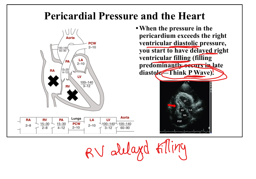
Q: What happens when pericardial pressure exceeds RV diastolic pressure?
A: Delayed RV filling, occurring mainly in late diastole (P‑wave timing).
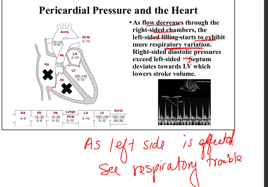
Q: Why does respiratory variation in LV filling increase in tamponade?
A: High right‑sided pressures push the septum toward the LV, reducing LV stroke volume(back flow to pulmonary veins)
Q: What ultimately causes hypotension in cardiac tamponade?
A: Reduced LV filling → decreased cardiac output → reflex tachycardia.

Q: What is Beck’s triad for cardiac tamponade?
A: JVD, hypotension with pulsus paradoxus, muffled heart sounds.

Q: What JVP change is seen in tamponade?
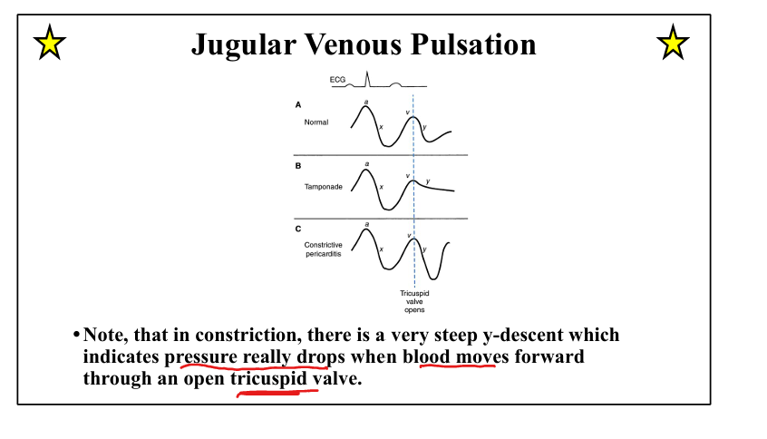
A: Preserved x‑descent(atrial relaxation) but absent y‑descent(atrial pressure with opening of tricuspid) due to impaired RV filling.

Q: What defines pulsus paradoxus?
A: >10 mmHg drop in systolic BP during inspiration.

Q: Why does pulsus paradoxus occur in tamponade?
A: Inspiration increases venous return, but RV diastolic pressure > LV diastolic pressure → septal bowing → reduced LV output.

Q: What EKG finding suggests tamponade?
A: Electrical alternans (beat‑to‑beat QRS amplitude variation).


Q: What is the definitive treatment for tamponade?
A: Echo‑guided pericardiocentesis with drain placement.

Causes of Cardiac Tamponade
Any cause of acute pericarditis, acute hemorrhage into pericardium, Acute aortic dissection, LV Free wall rupture STEMI

Q: What causes pericardial constriction?
A: Chronic scarring/fibrosis ± calcification from recurrent pericarditis, radiation, or prior TB.

Q: What exam finding is pathognomonic for constriction?
A: Kussmaul’s sign—JVD increases with inspiration. Not seen in tamponade.

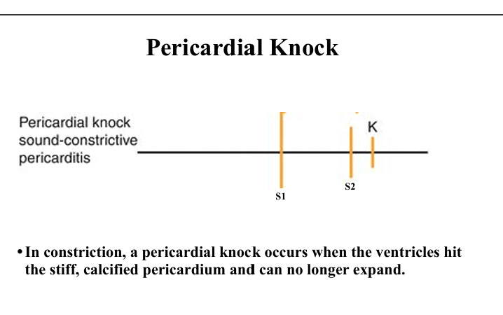
Q: What heart sound is associated with constriction?
A: Pericardial knock—early diastolic sound when ventricles hit the rigid pericardium.
Q: What JVP change is seen in constriction?
A: Exaggerated y‑descent due to rapid early filling followed by abrupt cessation.
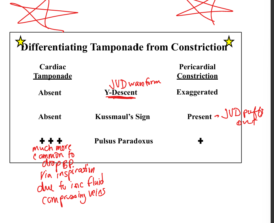
How to differentiate Pericardial constriction vs Cardiac Tamponade
Pericardial constriction: has Kussmaul’s sign(when you inhale the JV distends because the stiff pericardium will not allow more blood to enter the right side of the heart), exaggerated Y-descent
Cardiac tamponade: no Y descent, no kussmauls, much more pulsus paradoxus(drop BP due to inspiration)


Q: What is the treatment for severe constriction?
A: Pericardial stripping.

Q: How does pulsus paradoxus differ between tamponade and constriction?
A: Tamponade: prominent

Q: How does the y‑descent differ between tamponade and constriction?
A: Tamponade: absent
Present in Pericardial constriction
Q: How does Kussmaul’s sign differentiate tamponade vs constriction?
A: Present in constriction

Define the physiologic definition of CHF.
The inability of the heart to maintain circulatory demands without a rise in LV filling pressure
Clinical syndrome that can result from any structural or functional cardiac disorder that impairs the ability of the heart to fill with or eject blood


List the most specific sign and symptom of CHF.
Most specific sign: S3 gallop
Most specific symptom: orthopnea/Paroxysmal Nocturnal Dyspnea.

Explain why dyspnea occurs in left-sided HF
Increased LA pressure is transmitted to pulmonary veins → pulmonary capillary pressure rises → pulmonary edema → dyspnea


Explain orthopnea physiologically.
Supine position increases venous return from legs → increased LV filling pressures → pulmonary congestion → SOB.
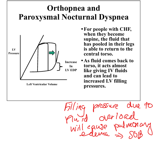
Explain paroxysmal nocturnal dyspnea.
Fluid redistribution during sleep increases LV filling pressures patient awakens gasping and improves when upright.

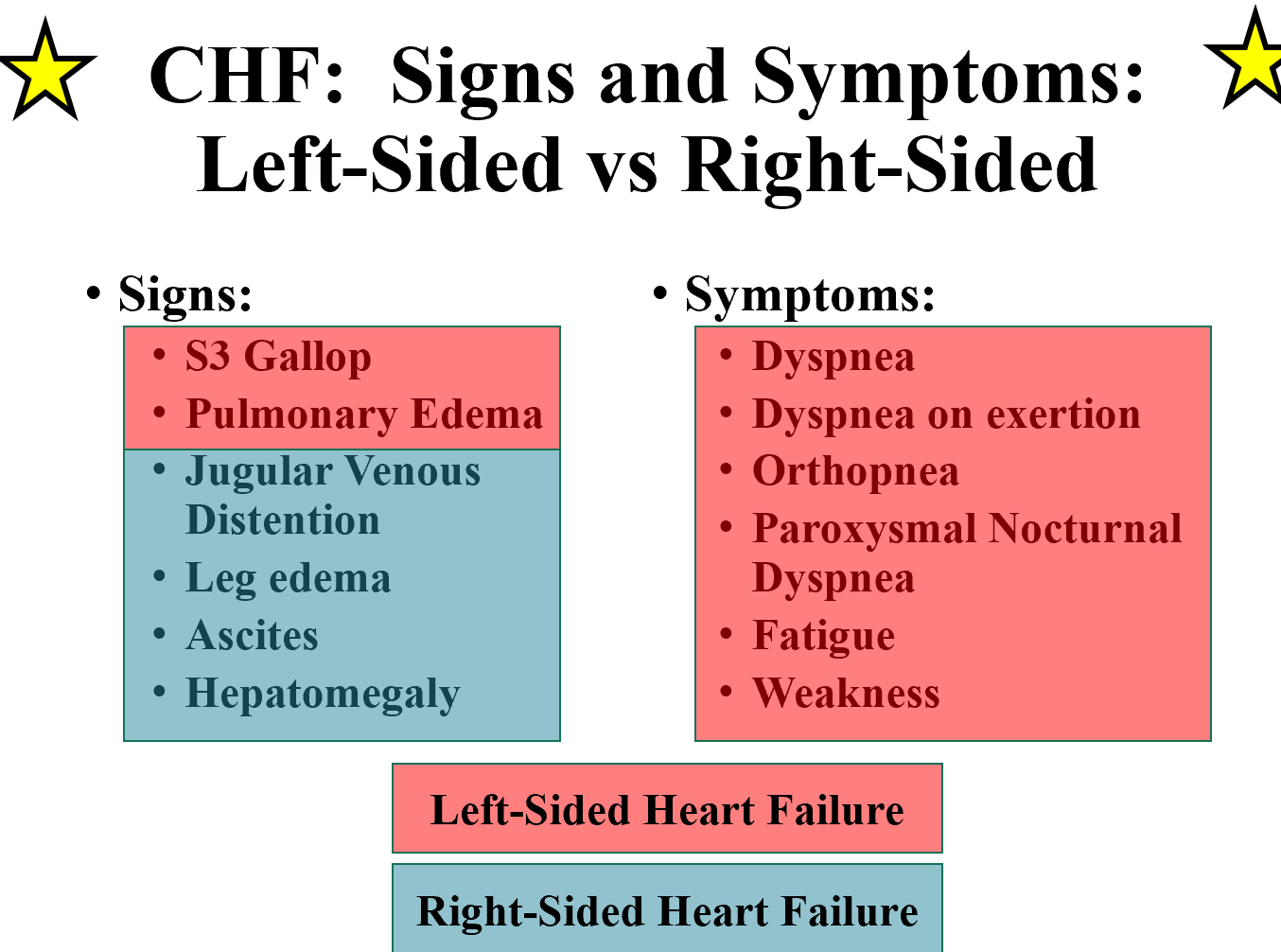
List signs of right-sided HF
HF. JVD, hepatomegaly, ascites, lower extremity edema
“By far, the greatest cause of right-sided heart failure is left-sided heart failure.”


Differentiate left- vs right-sided HF symptoms.
Left: dyspnea, orthopnea, PND, pulmonary edema.
Right: JVD, leg edema, ascites, hepatomegaly.

Define high-output vs low-output HF.__Discard
Low-output: AS, HTN, MI.
High-output: anemia, thyrotoxicosis, AVM, pregnancy. Vast majority is low-output.

Describe the “wet vs dry” and “warm vs cold” HF profiles.
Wet = congestion (orthopnea, PND, rales, JVD). Cold = low perfusion (cool extremities, narrow pulse pressure, renal dysfunction).

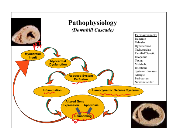
Explain LV remodeling after injury.
Reduced EF → LV dilates to maintain SV (↑EDV).

List causes of remodeling.
Myocardial insult → dysfunction → reduced perfusion → sympathetic/RAAS activation → altered gene expression → apoptosis → remodeling.
Describe sympathetic system effects in HF.
↑Automaticity, ↑Contractility, ↑Afterload, ↑Preload, chronic stimulation → myocyte death + arrhythmia.
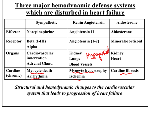
Describe RAAS effects in HF.
Ang II → ↑Afterload, ↑Preload chronic → hypertrophy + ischemia.

Describe aldosterone effects in HF.
↑Afterload, ↑Preload, chronic → cardiac fibrosis.
Explain beta-1 receptor changes in HF.
Downregulated and desensitized due to chronic epinephrine stimulation (“less likely to stimulate cAMP production”).

Explain ANP release.
Atrial stretch from ↑LV filling pressures → ANP release → natriuresis, vasodilation.

Explain BNP release.
Ventricular tension/stretch → BNP release → natriuresis, ↓renin, ↓aldosterone, ↓preload/afterload.
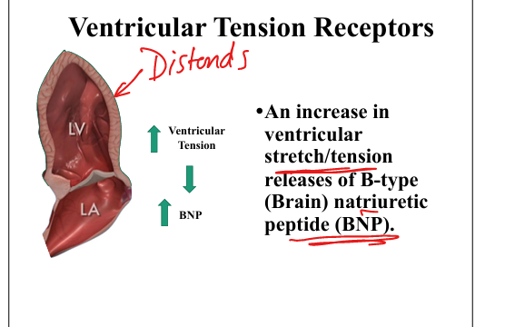
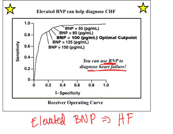
State BNP’s diagnostic utility.
BNP elevation helps diagnose CHF

Define HFpEF, HFmrEF, HFrEF.
HFpEF ≥50%, HFmrEF 41–49%, HFrEF <40%

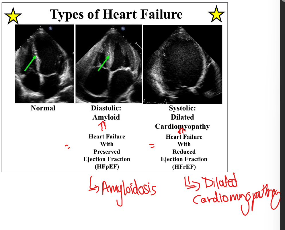
Describe HFpEF remodeling pattern.
Concentric hypertrophy: ↑mass, normal volume, ↓diastolic function, preserved systolic function. (think hypertrophy)
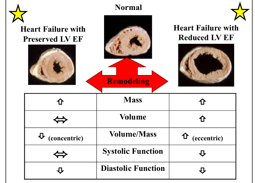

Describe HFrEF remodeling pattern.
Eccentric dilation: ↑volume, ↑mass, ↓systolic function, ↓diastolic function. (think dilated cardiomyopathy)


List causes of HFpEF(pressure overload) vs HFrEF(volume overload).
HFpEF: HTN, DM, Aortic stenosis (pressure overload). HFrEF: MI, alcohol, idiopathic (volume overload).

Define pulsus alternans and what it is a strong indicator of
Alternating strong/weak pulses; good indicator of HFrEF.


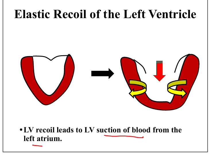
Define LV suction.
Elastic recoil during early diastole generates negative pressure → pulls blood from LA.
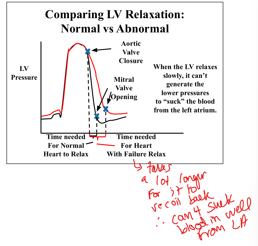
Explain impaired LV relaxation.
Slow relaxation prevents generation of low LV pressure → impaired suction → ↑LA pressure → exertional dyspnea.

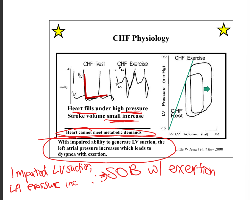
Describe normal vs CHF filling pressures.
Normal: LV fills under low pressure. CHF: LV fills under high pressure → small SV increase with exercise.
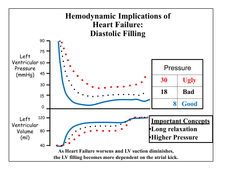
Explain dependence on atrial kick.
As suction worsens, LV filling becomes more dependent on atrial contraction.
Does everyone with HF have diastolic dysfunction.
Yes! Everyone with HF—preserved or reduced EF—has abnormal diastolic filling.

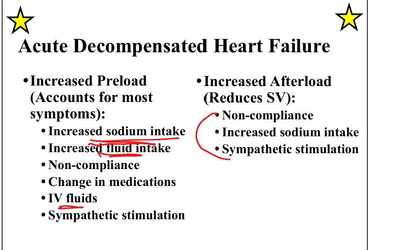
List acute HF causes of increased preload.
Na/fluid intake, non-compliance, IV fluids, sympathetic stimulation.
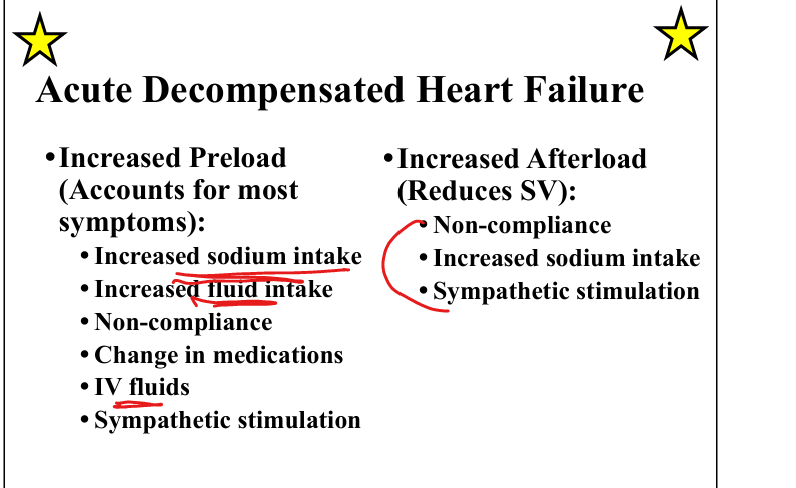
List acute HF causes of increased afterload.
Na intake, non-compliance, sympathetic stimulation.
Explain acute HF treatment goals.
Decrease preload (diuretics-dec venous congestion, nitrates-dec pulm edema); decrease afterload/inc contractility(ACEi/ARB/hydralazine/nitroprusside)

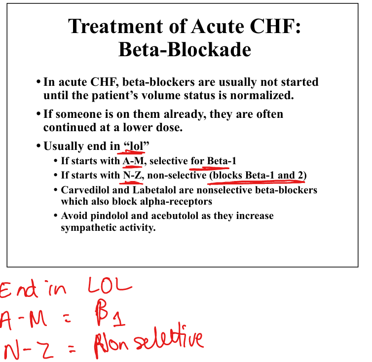
State when beta-blockers are used in acute HF.
Not started until euvolemic, if already on them, continue at lower dose.
List chronic HF life-prolonging therapies.
ACEi/ARB/ARNI(ARB+ Neprilysin inhibitor), beta-blockers (carvedilol, metoprolol XL, bisoprolol), aldosterone antagonists(spironalactone)

Explain ARNI(ARB + Neprilysin Inhibitor) role.
Used in symptomatic HFrEF (Class II–III) improves outcomes.

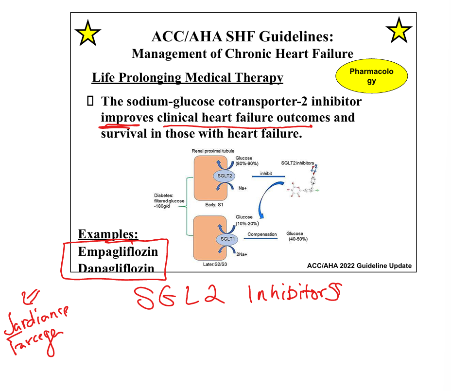
Explain SGLT2 inhibitor role + Ex
Improves HF outcomes and survival (empagliflozin, dapagliflozin) by dec fluid and glucose
State ICD indications
LVEF EF ≤35% and no recovery after being on guideline directed therapy

Left sided HF sx
S3 gallop, Pulmonary edema, Dyspnea w exertion, orthopnea, Paroxysmal Nocturnal Dyspnea, fatigue, weakness
Treatment of Acute CHF meds
Diuresis(use furosemide(lasix)), Nitroglycerin to vasodilate and improve sx, and ACEi/ARBs, Hydralazine, Nitroprusside to inc stroke volume, and Dobutamine/milrinone to inc SV
Should you use Ca Channel Blockers in HF?
NO! Avoid Verapamil/diltiazem because they limit contractility

Treatment tree for HF
Initially start Beta blocker, ACEi/ARB, SGLT-2 inhibitor
Class III/IV: start aldosterone antagonist(spironalactone), maybe hydralazine nitrates
If EF <35% on meds»Use ICD + CRT

What does blood pressure depend on (components of equation)
BP= (LVEDV-LVESV) x HR x SVR: depends on CO(SVxHR) and SVR, so drugs target LVEDV (preload), LVESV (contractility/afterload), HR (chronotropy), or SVR (vasoconstriction) to correct hypotension.

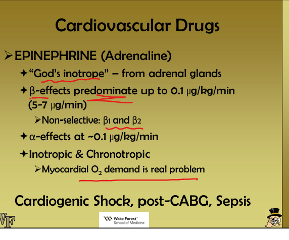
Which hemodynamic variable does epinephrine primarily modify, and how does this help differentiate shock types?
Epinephrine increases CO (β1/β2) and SVR (α1 at higher doses), improving hypotension in distributive(low SVR) or cardiogenic shock depending on the dominant deficit.
How does norepinephrine’s receptor profile help identify distributive shock physiology?
Norepinephrine’s strong α1 vasoconstriction corrects low SVR, the hallmark of distributive shock (e.g., sepsis), while modest β1 support maintains CO.

Why is phenylephrine inappropriate for cardiogenic shock when analyzing pump function?
Phenylephrine is a pure α1 vasoconstrictor that increases SVR/afterload without improving contractility, worsening CO in cardiogenic shock.

How does dobutamine’s effect on LVESV help differentiate cardiogenic shock from hypovolemic shock?
Dobutamine(b1 agonist) reduces LVESV by increasing contractility (β1) and decreasing afterload (β2), improving CO in cardiogenic shock but ineffective if preload is low (hypovolemia).

How does isoproterenol’s physiology help identify obstructive vs cardiogenic shock?
Isoproterenol increases HR/contractility (beta agonist) and causes pulmonary vasodilation, improving RV output—useful in pulmonary hypertension or RV failure but not in tamponade or PE obstruction.

How does angiotensin II help differentiate shock states with preserved CO but severe vasoplegia?
AT1 vasoconstriction rapidly increases SVR and BP, identifying distributive shock with intact cardiac function.
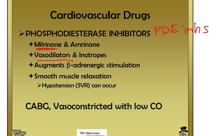
How do PDE inhibitors (milrinone) help evaluate afterload vs contractility problems?
Milrinone increases contractility and decreases SVR
Why does norepinephrine improve MAP more predictably than epinephrine in septic shock?
Norepinephrine’s dominant α1 vasoconstriction increases SVR without β2 vasodilation, producing a more stable MAP rise in vasoplegic states.
