Anatomy and Physiology Midterm Lab Practical/Exam Study Guide (edited 9/26/24)
1/99
There's no tags or description
Looks like no tags are added yet.
Name | Mastery | Learn | Test | Matching | Spaced |
|---|
No study sessions yet.
100 Terms
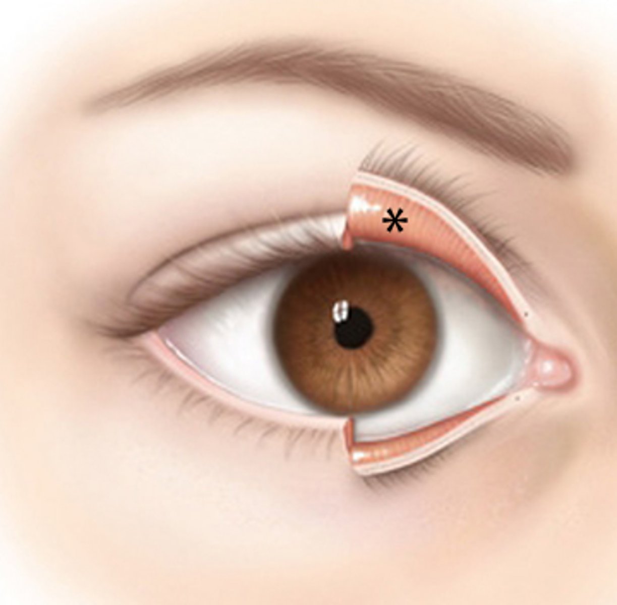
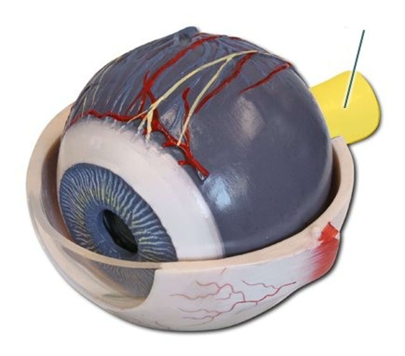
Upper/lower eyelid
External feature of the eye
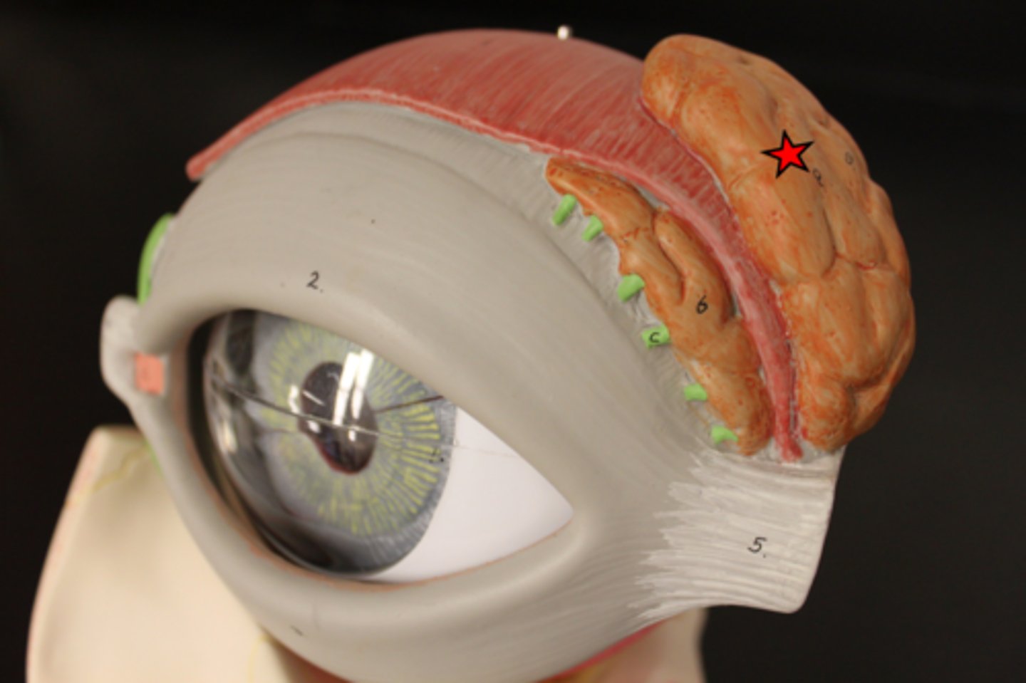
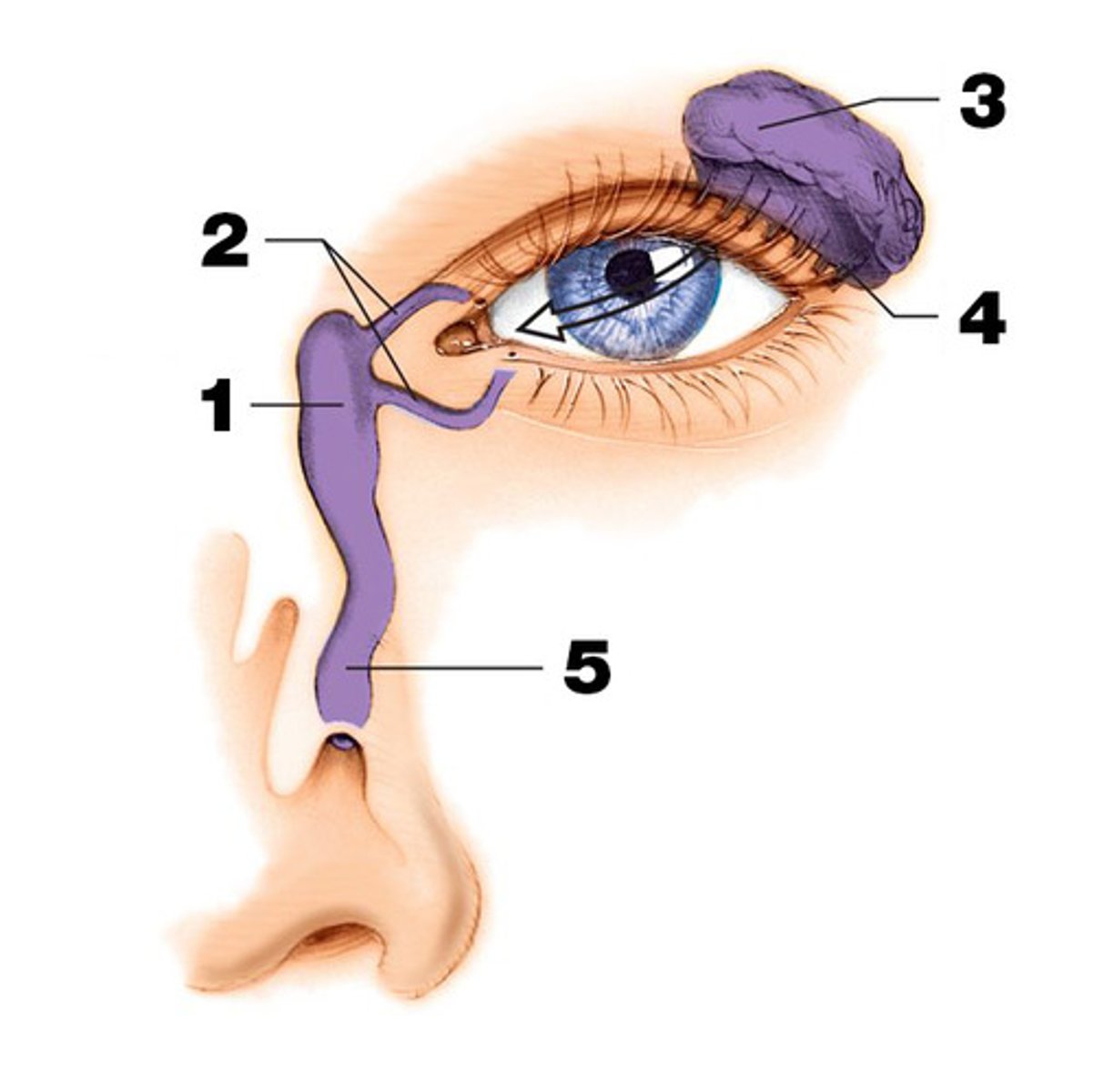
Lacrimal gland
gland located in the upper outer region above the eyeball that secretes tears
excretory lacrimal ducts
empty tears onto the surface of the conjunctiva of the upper lid
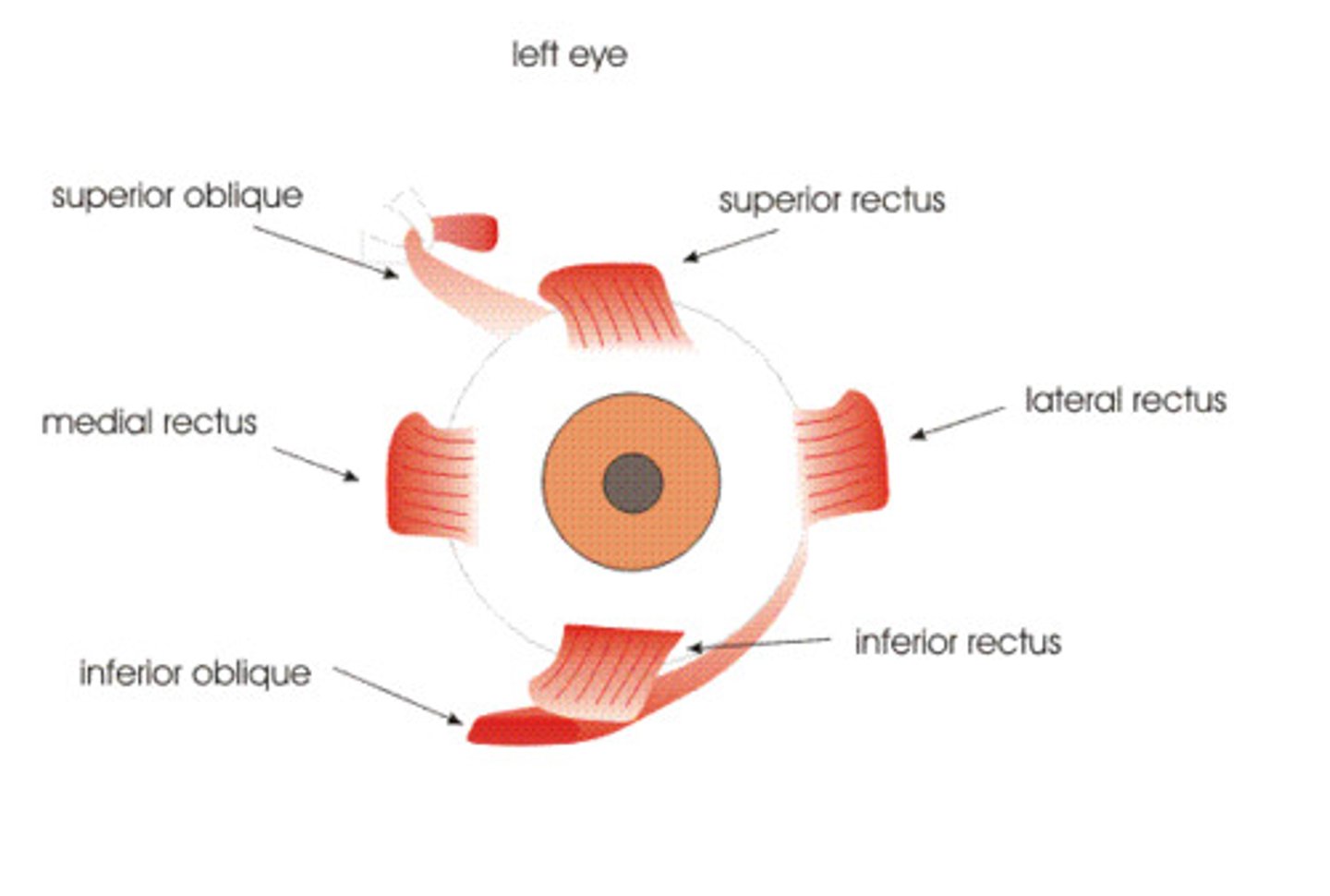
Lateral/Medial rectus
Moves eye left and right

Superior/Inferior rectus
move eye up and down
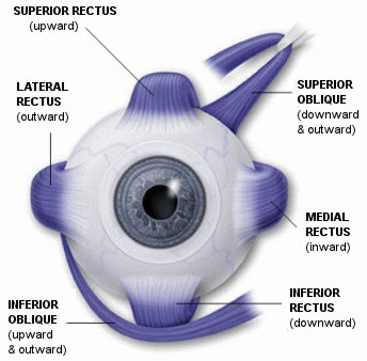
Superior/Inferior oblique
rotate eye opposite their name and also laterally
Pupil
The opening through which light enters the eye
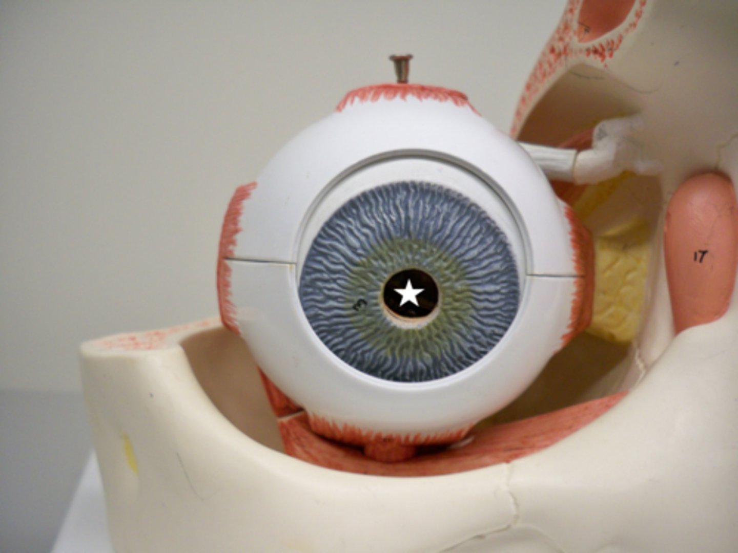
Iris
a ring of muscle tissue that forms the colored portion of the eye around the pupil and controls the size of the pupil opening
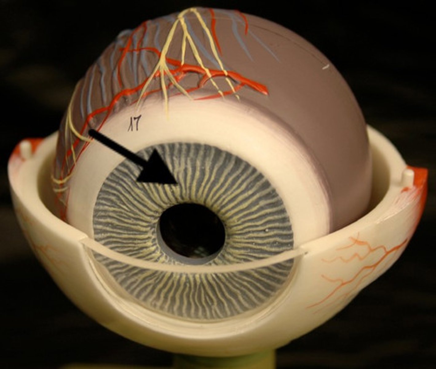
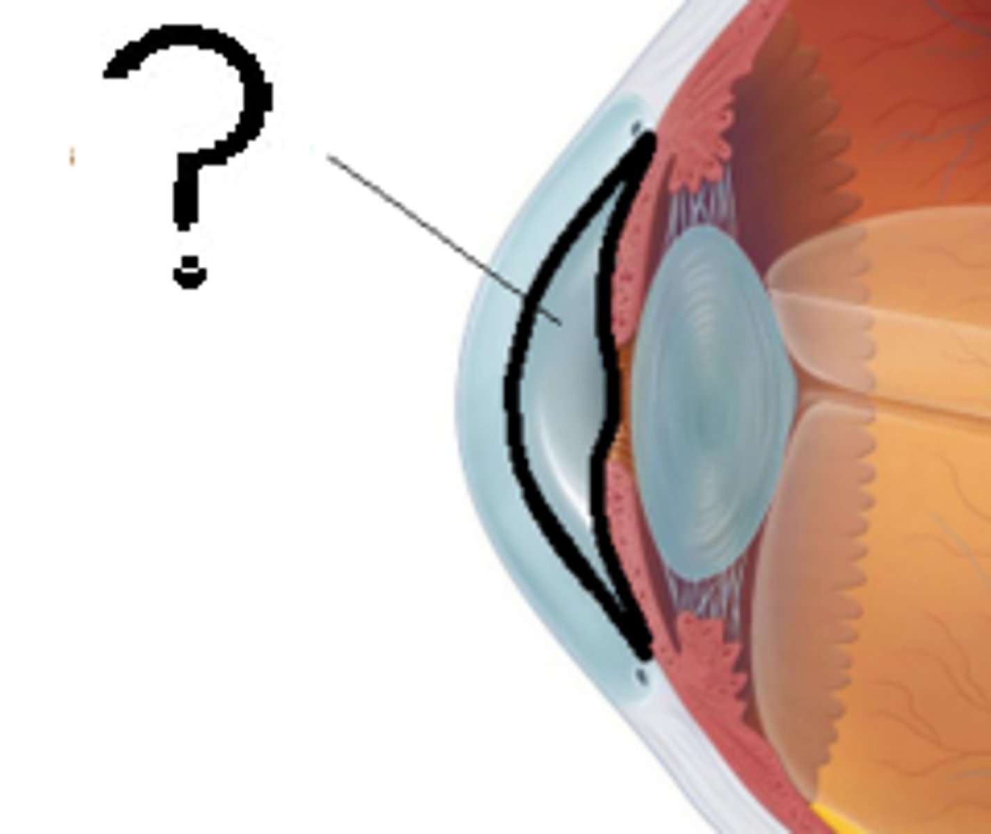
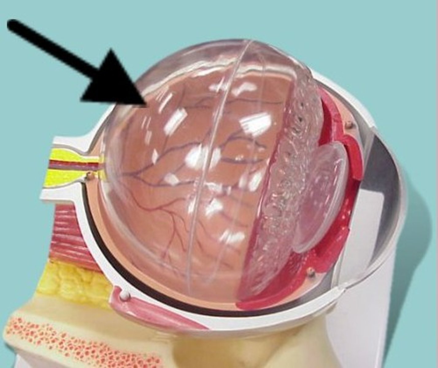
Sclera
white of the eye; fibrous tunic (outer layer)
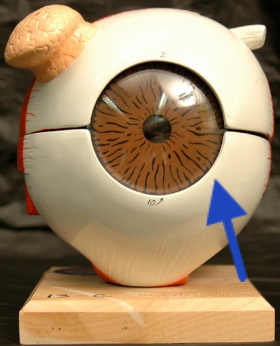
Cornea
The clear tissue that covers the front of the eye
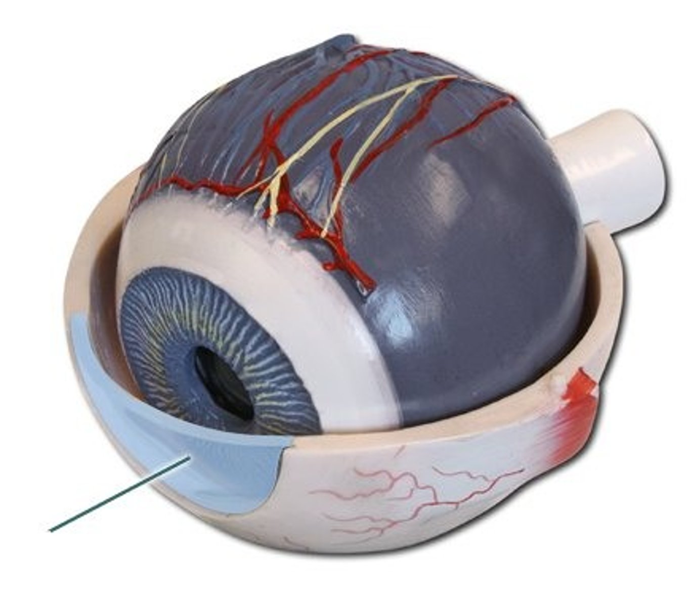
Conjunctiva
Delicate membrane lining the eyelids and covering the eyeball
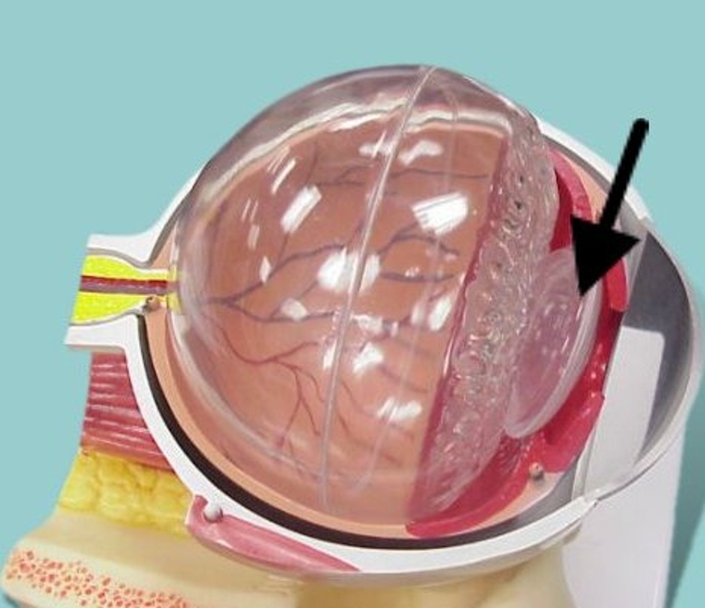
Lens
structure in the eye that focuses light rays on the retina
Optic nerve
the nerve that carries neural impulses from the eye to the brain
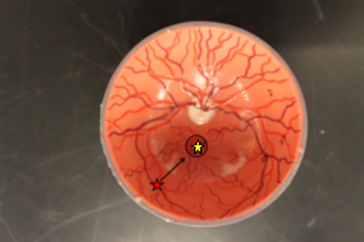
Optic disk
A hole in the retina where the optic nerve fibers exit the eye.
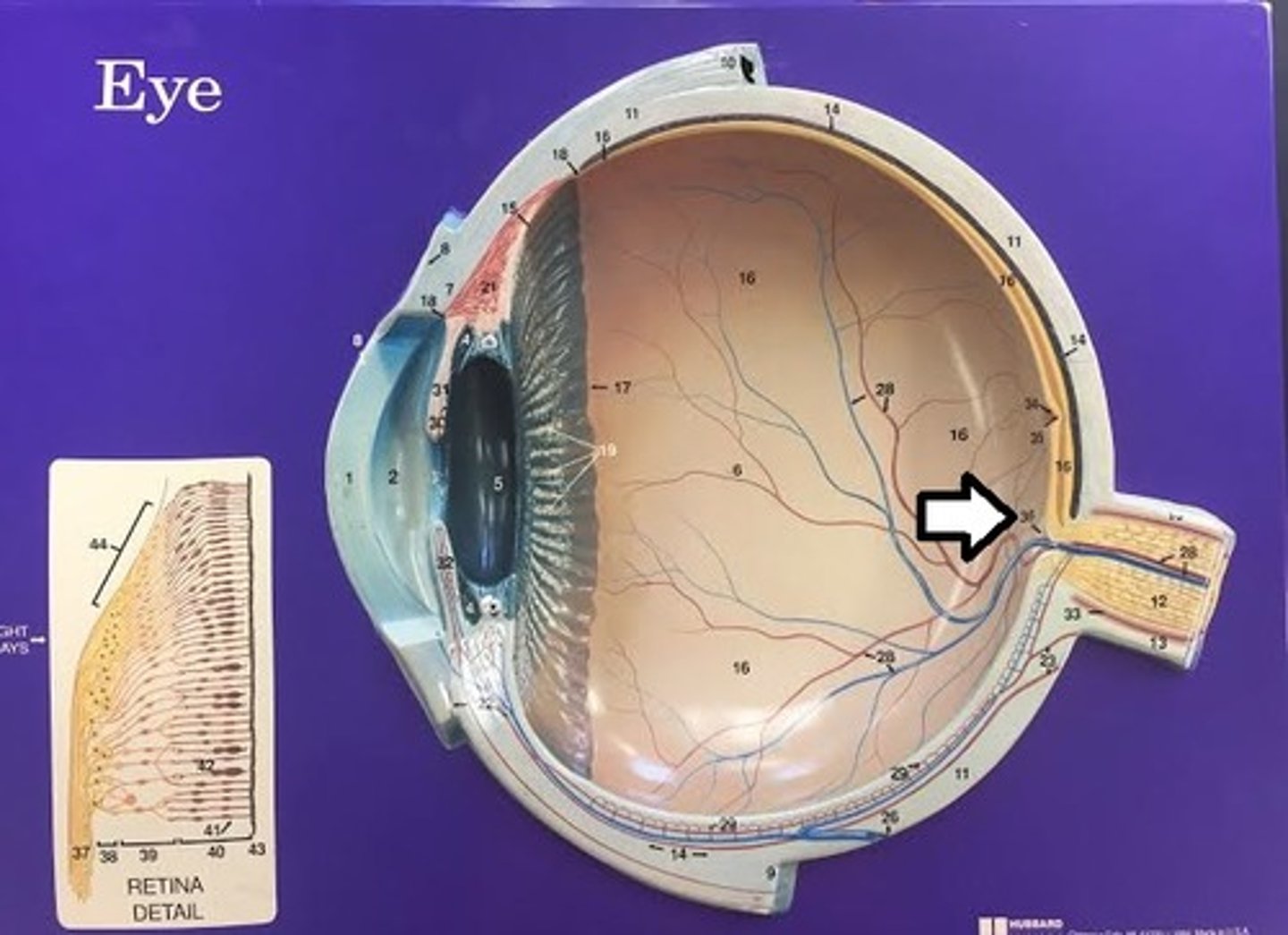
Macula
yellowish region on the retina lateral to and slightly below the optic disc that produces sharper vision
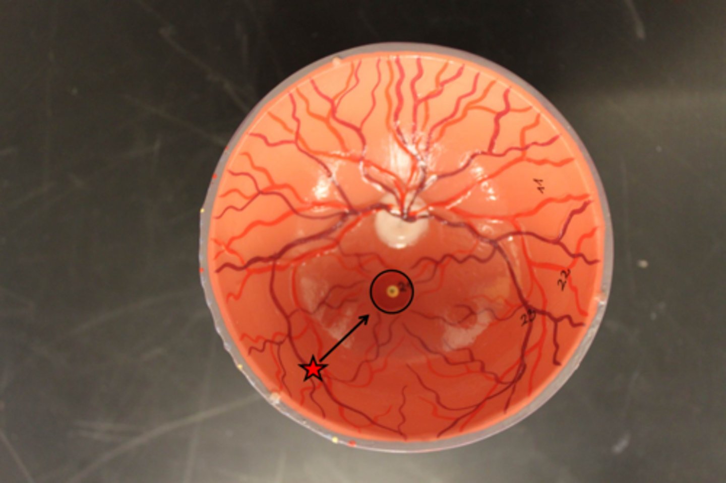
Fovea centralis
tiny pit or depression in the macula that is the region of clearest vision
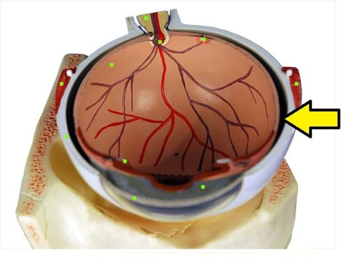
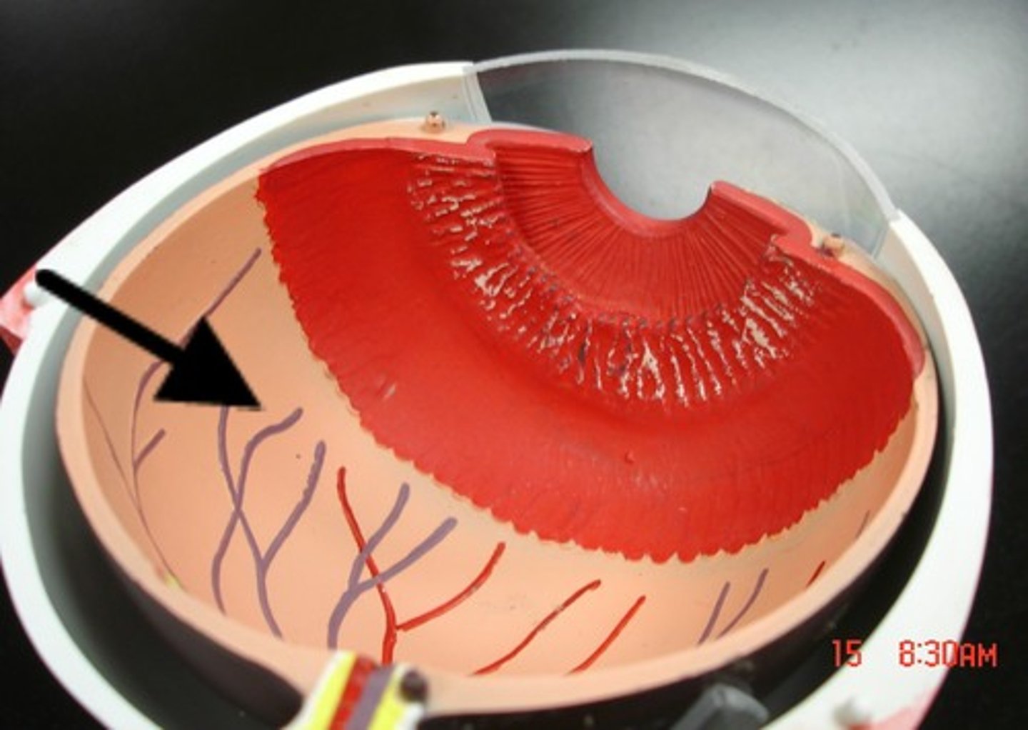
Choroid
Middle layer of the eye (vascular tunic) that includes the iris, pupil, and ciliary body/muscle
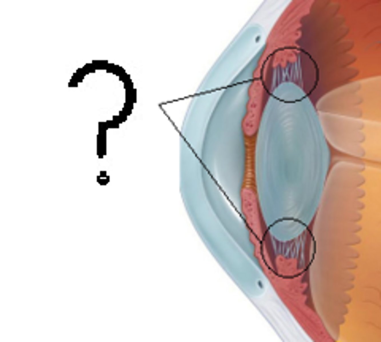
Ciliary body and muscle
Body: controls the shape of the lens, and the ciliary epithelium, which produces the aqueous humor. Muscle: controls accommodation for viewing objects
Retina
Inner "nervous" layer of the eye
Anterior cavity
contains aqueous humor that helps maintain shape of eyeball and supplies oxygen and nutrients to lens and cornea
Posterior cavity
Contains vitreous humor/body
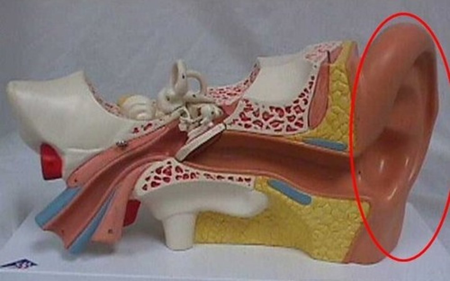
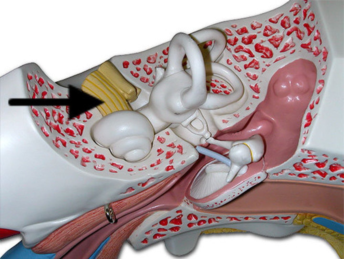
Auricle
external ear
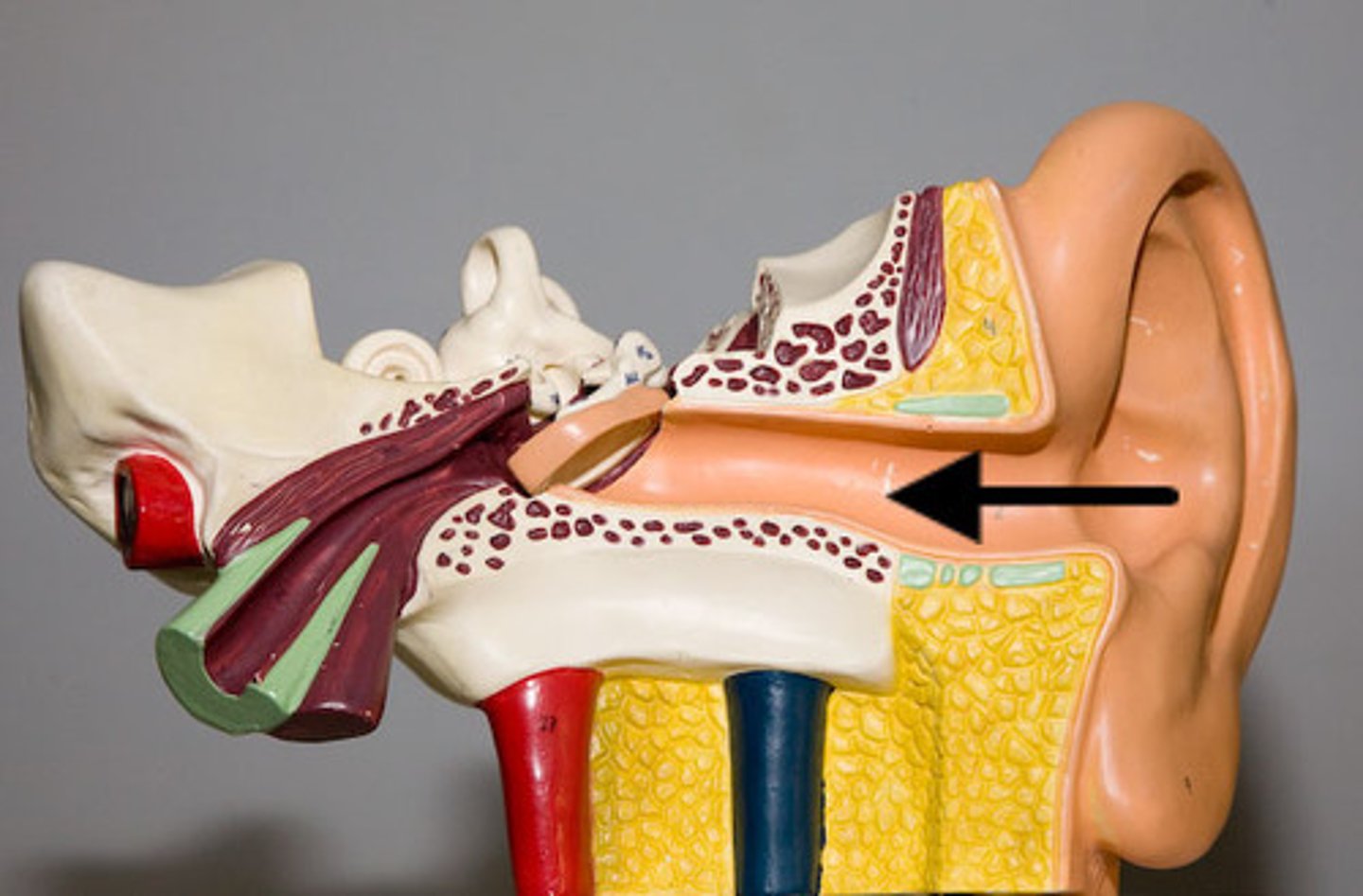
External acoustic meatus
ear canal
Tympanic membrane
eardrum
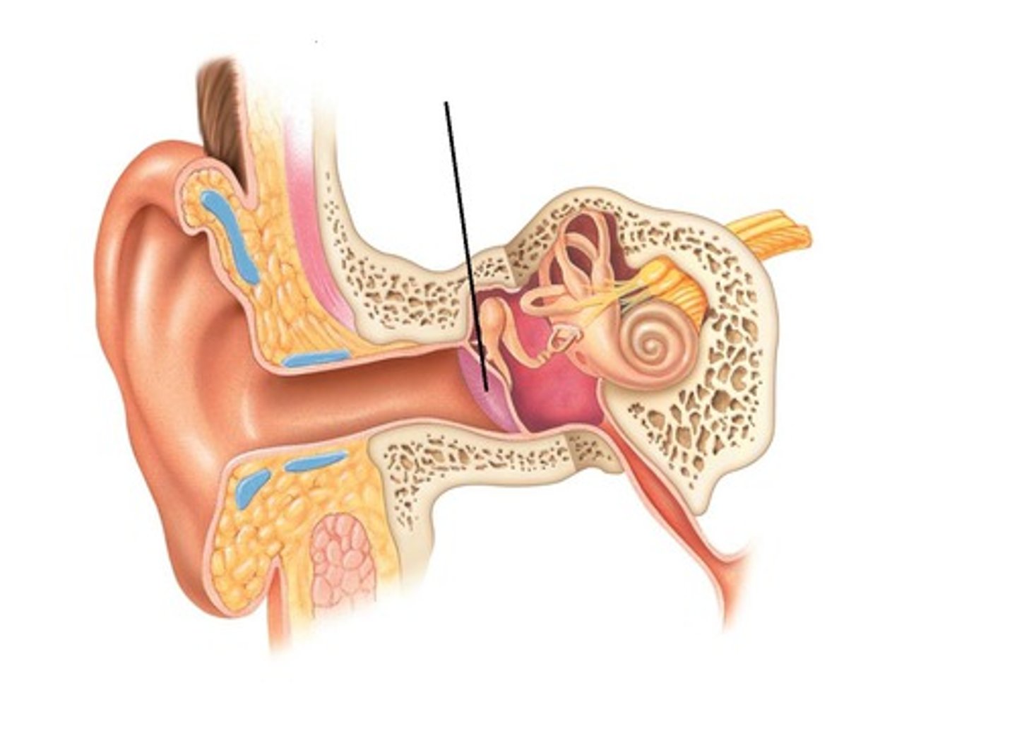
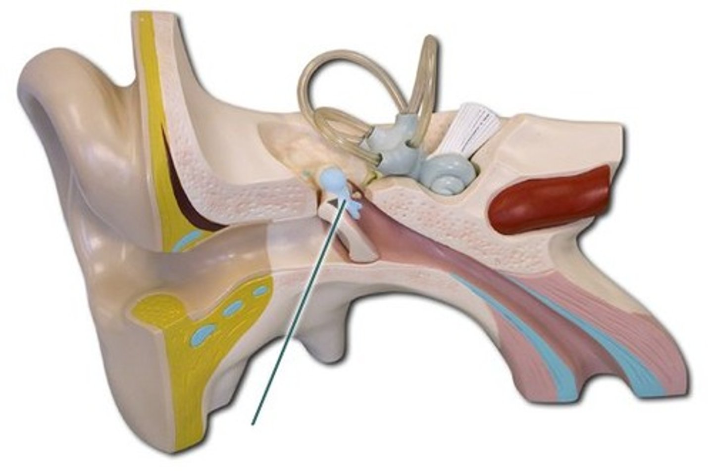
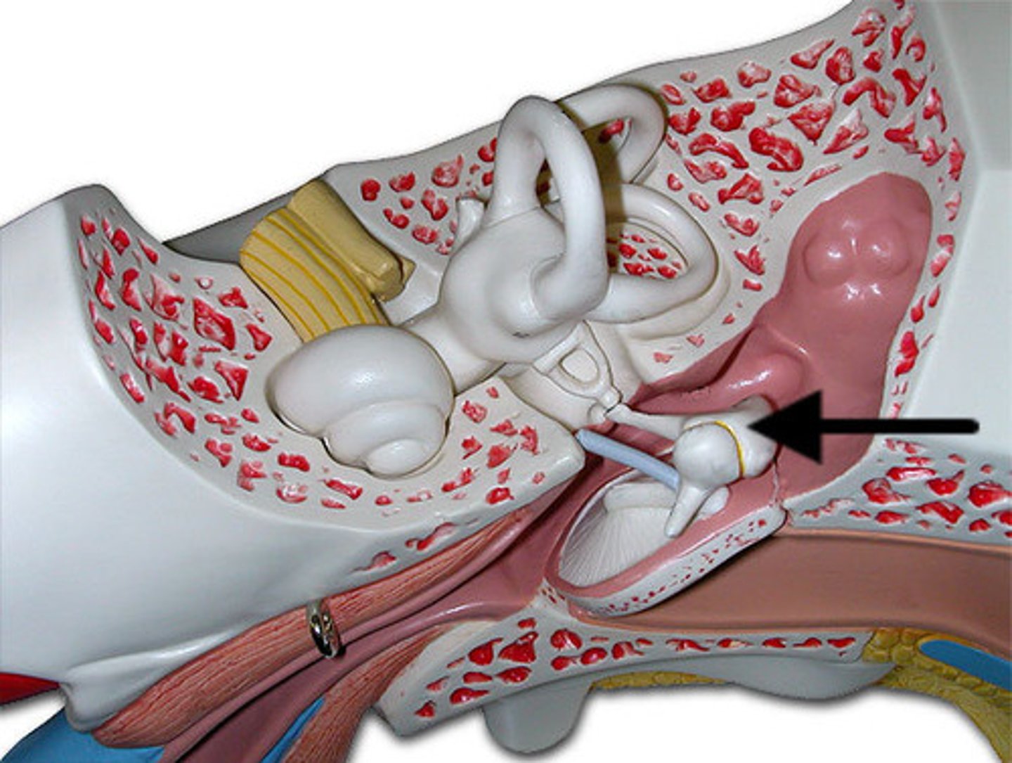
Oval window
membrane at the enterance to the cochlea through which the ossicles transmit vibrations
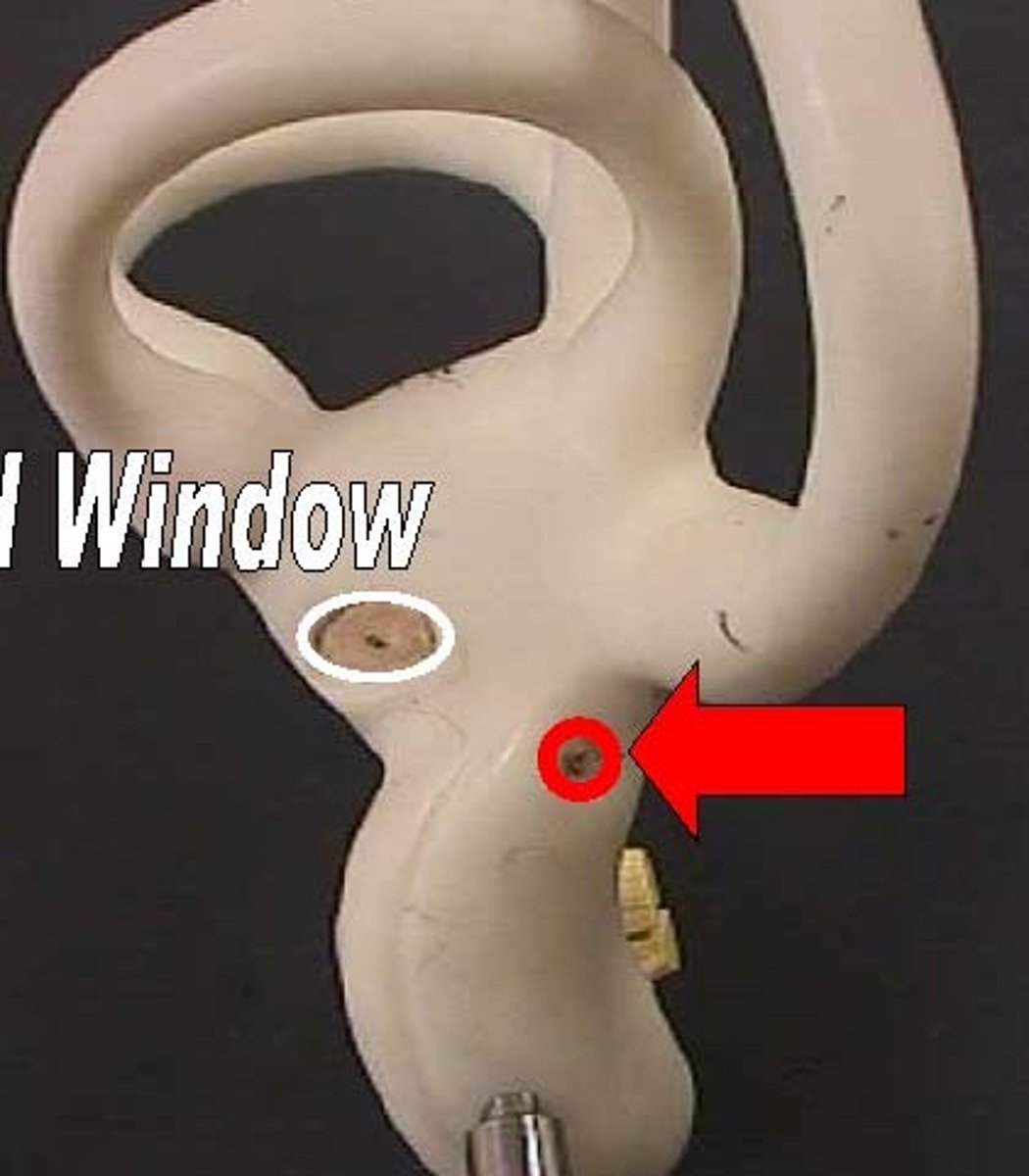
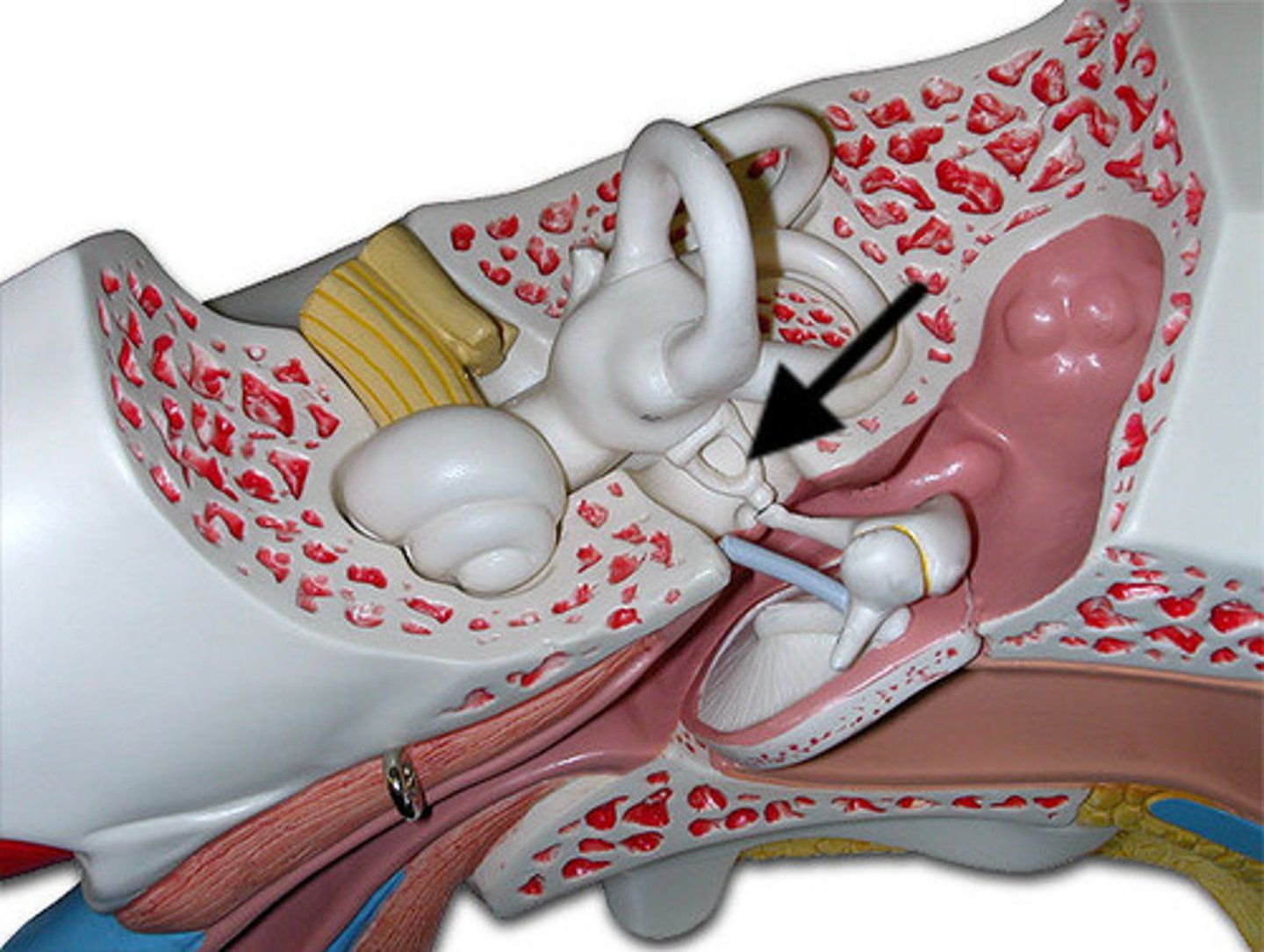
Round window
The membrane that relieves pressure from the vibrating waves in the cochlear fluid.
Malleus
hammer; first of the three auditory ossicles of the middle ear
Incus
anvil; middle of the three auditory ossicles of the middle ear
Stapes
stirrup; last of the three auditory ossicles of the middle ear
auditory (eustachian) tube
channel between the middle ear and the nasopharynx
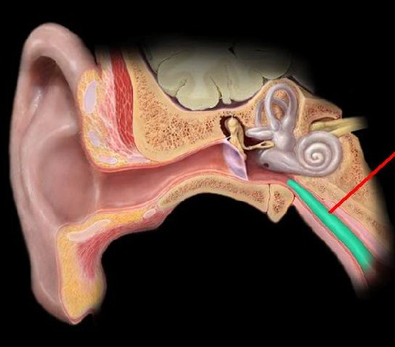
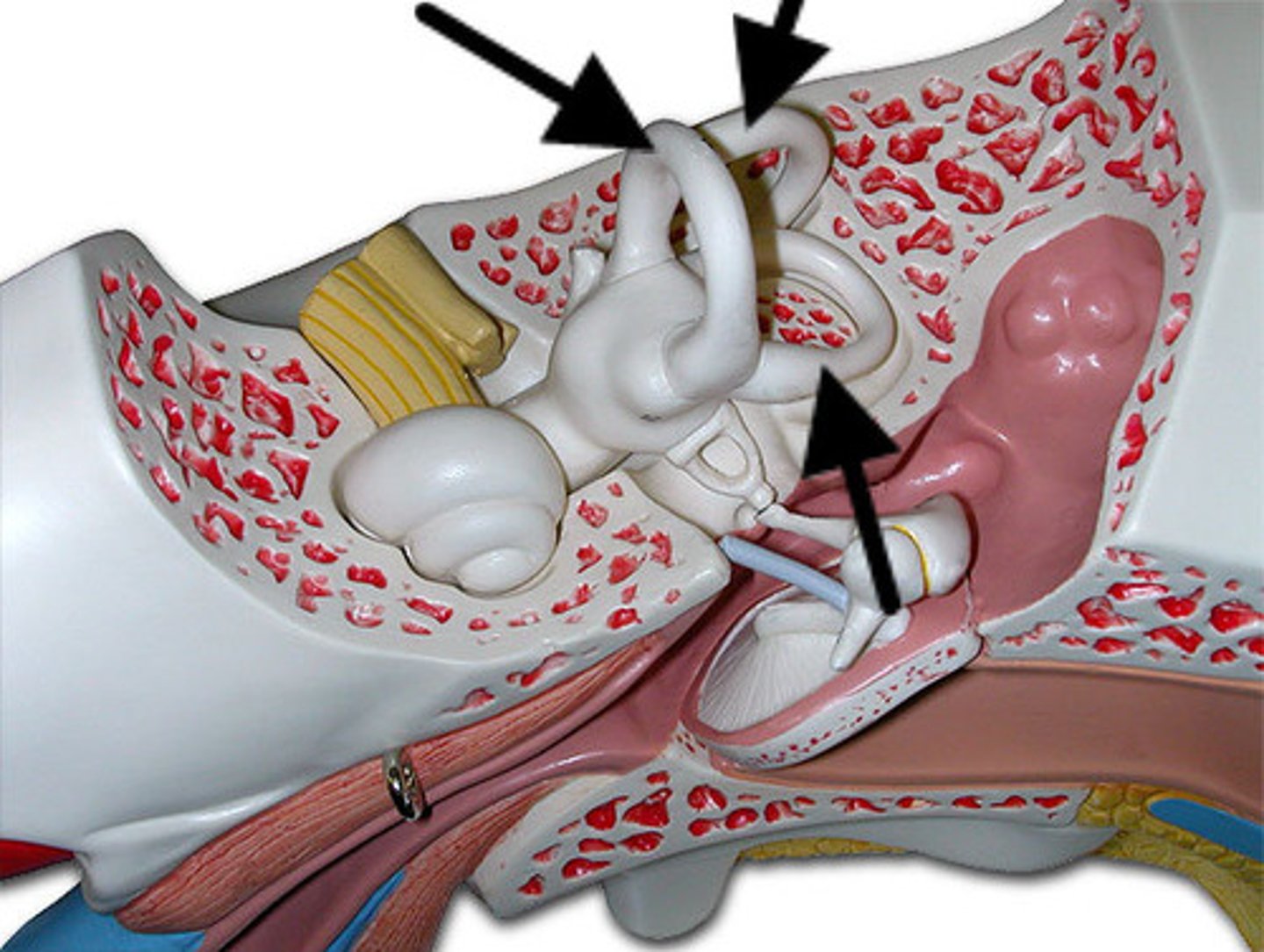
Vestibule
The portion of the inner ear that senses the position of the head. Its sensory epithelium is contained in two saclike spaces: the utricle and the saccule.
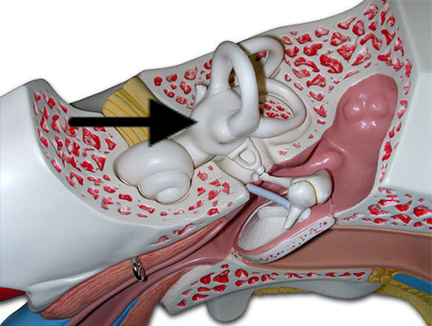
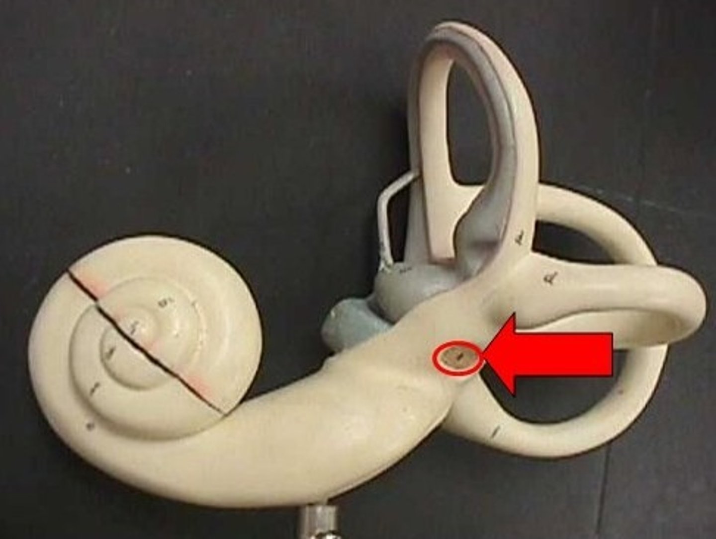
Cochlea
a coiled, bony, fluid-filled tube in the inner ear through which sound waves trigger nerve impulses
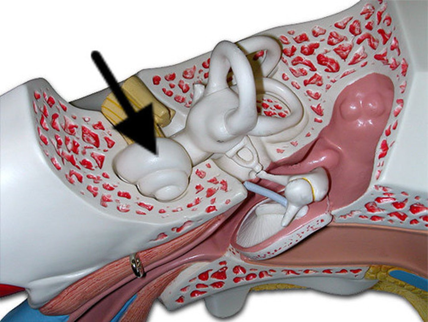
Semicircular canals
three canals within the inner ear that contain specialized receptor cells that generate nerve impulses with body movement
vestibulocochlear nerve
transmits hearing and balance impulses to the brain
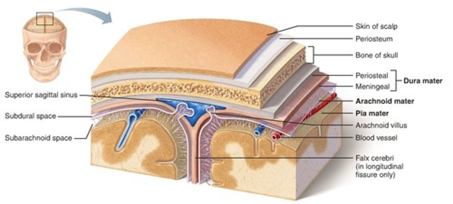
Meninges
Protective membranes surrounding the brain and spinal cord
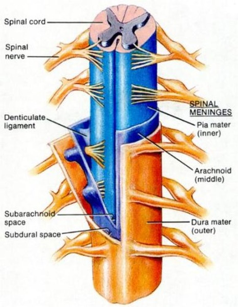
Dura Mater
Outermost and toughest layer of the meninges

Arachnoid mater
Middle layer of the meninges, filled with cerebrospinal fluid

Pia Mater
Innermost layer of the meninges, directly attached to the brain and spinal cord
Cerebrum
Largest part of the brain responsible for higher functions such as intellect, creativity, awareness, and language
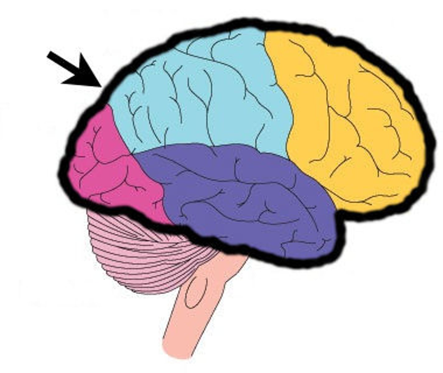
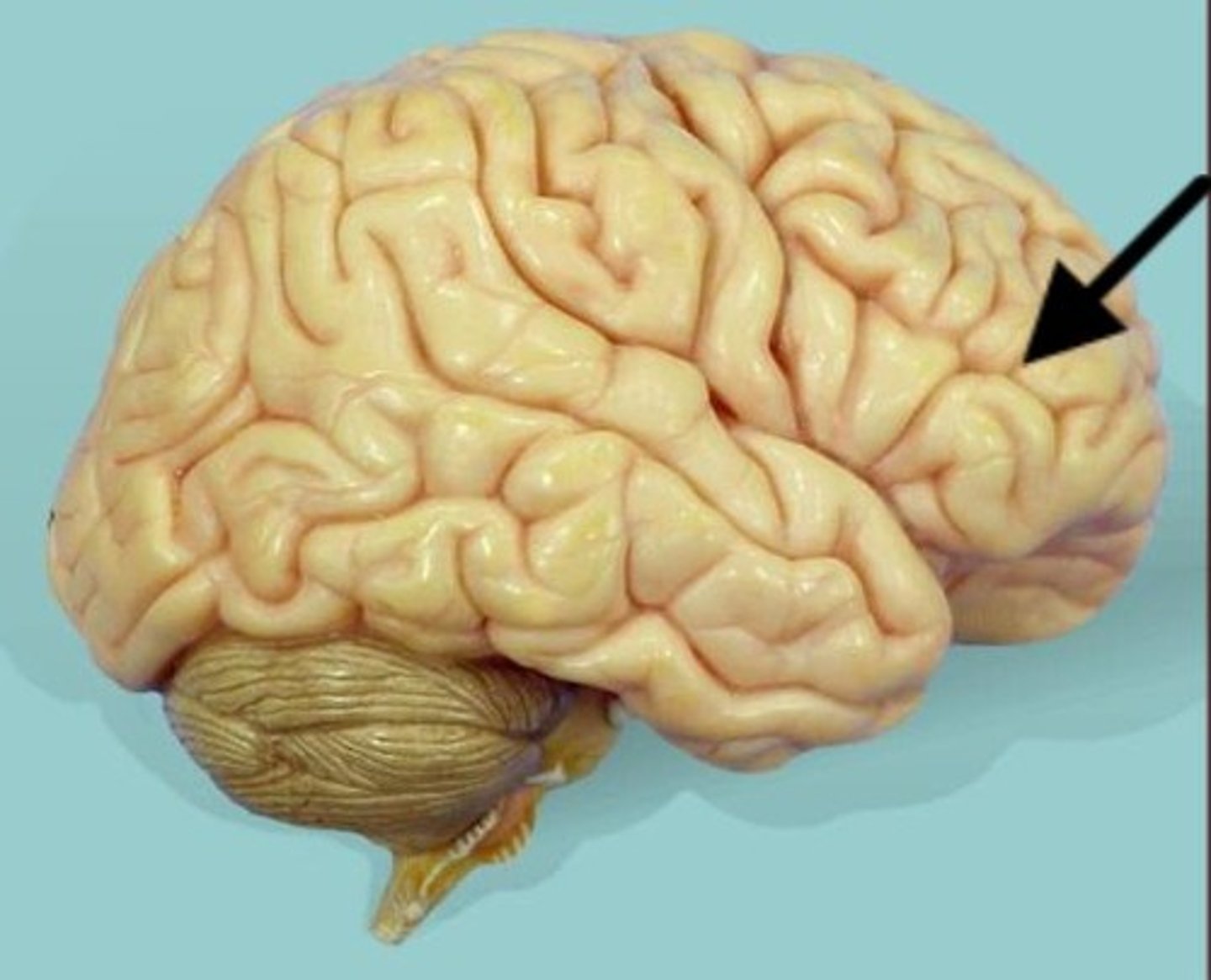
Frontal Lobe
Part of the cerebrum responsible for higher functions such as intellect, creativity, awareness, and language
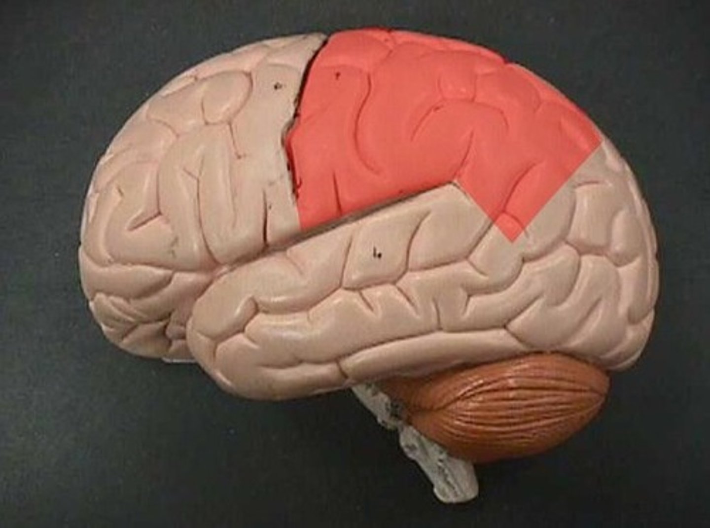
Parietal Lobes
Part of the cerebrum responsible for receiving sensory information from the body
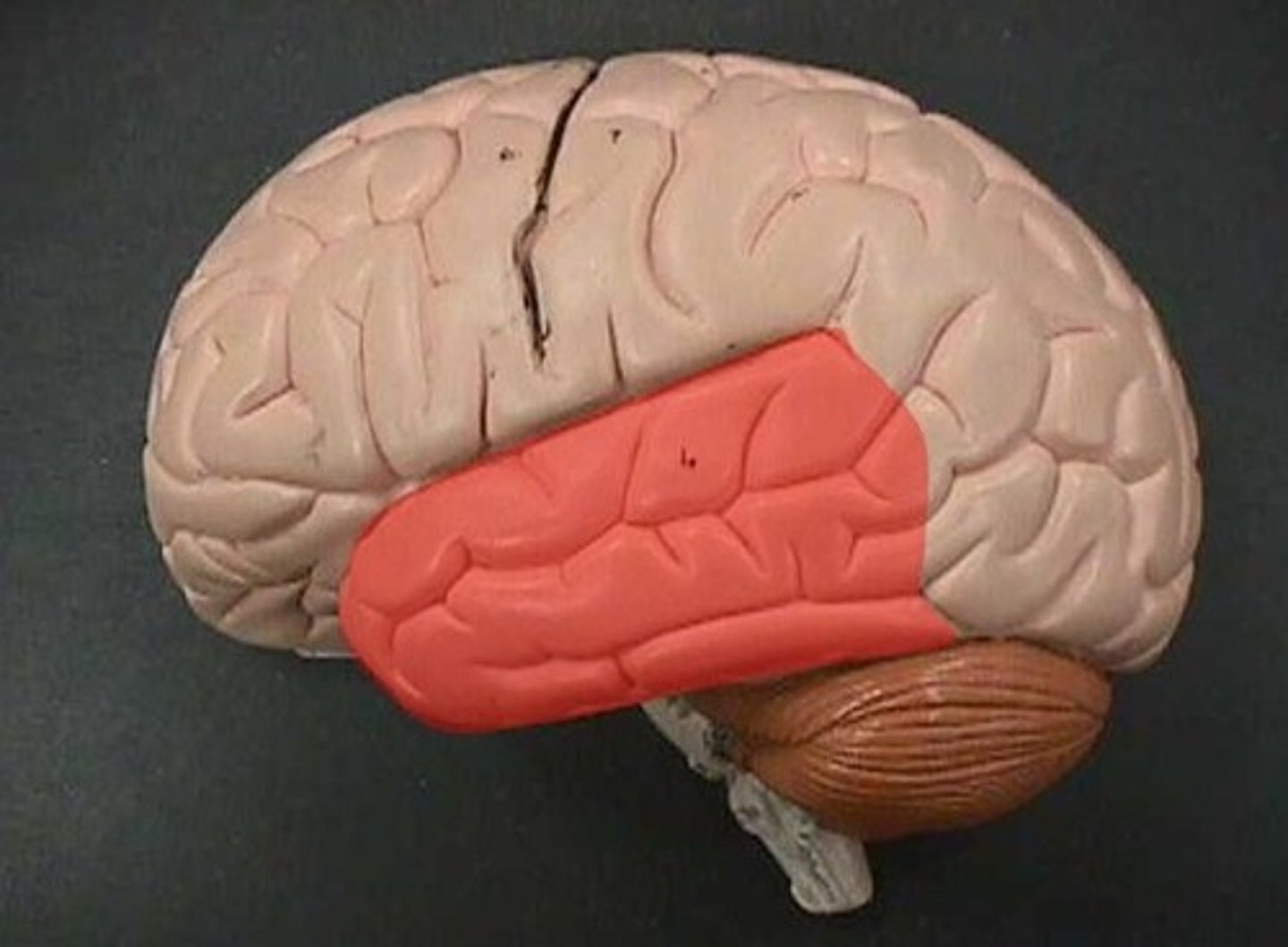
Temporal Lobes
Part of the cerebrum containing the primary auditory cortex and responsible for hearing
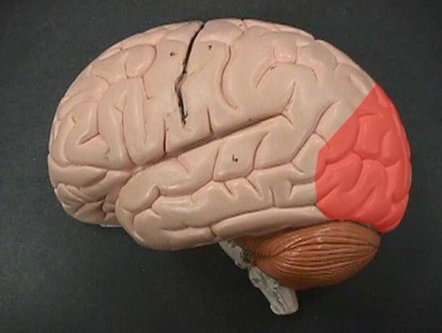
Occipital Lobe
Part of the cerebrum responsible for vision
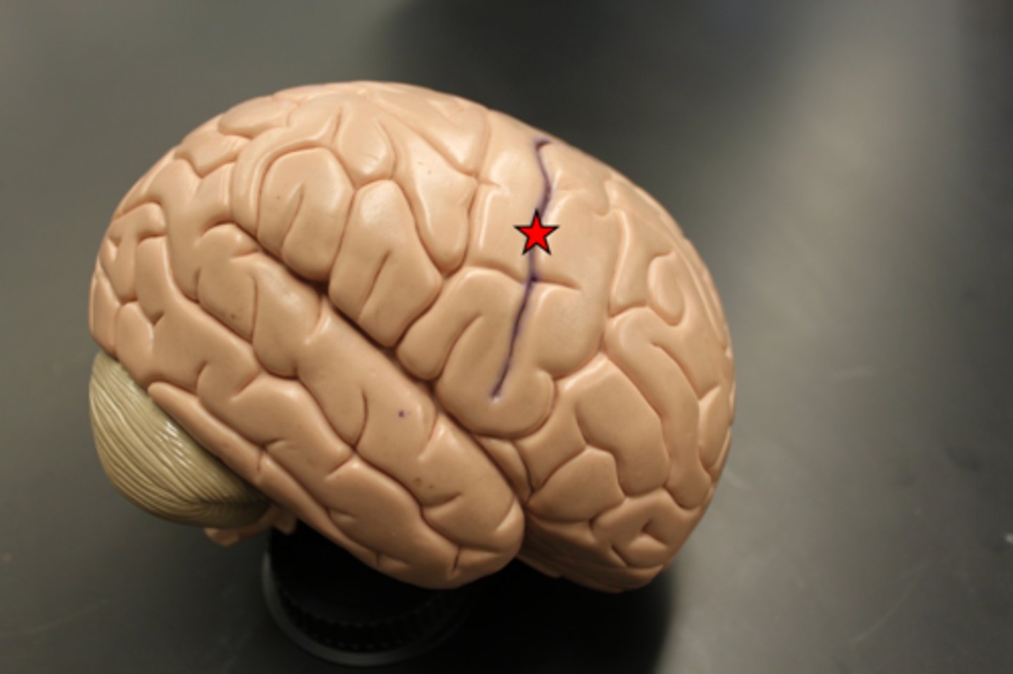
Central sulcus
A groove that divides the frontal and parietal lobes
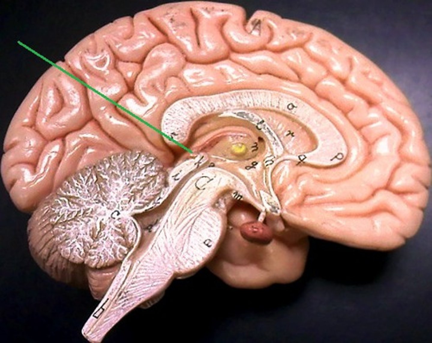
Diencephalon
Contains thalamus and hypothalamus
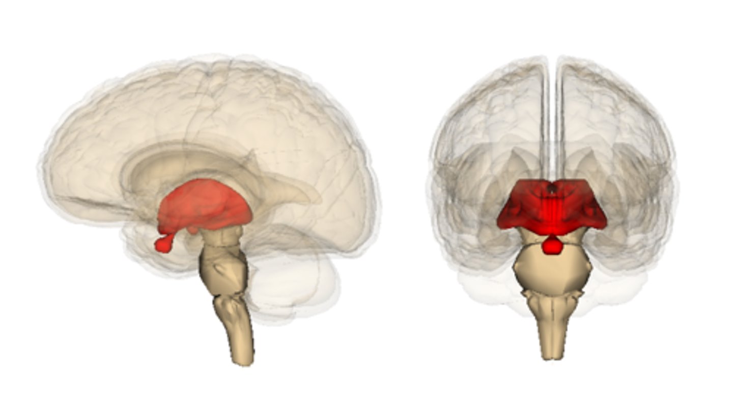
Thalamus
Part of the diencephalon that receives almost all sensory information from the body and sends it to the cerebral cortex
Hypothalamus
Part of the diencephalon involved with the autonomic nervous system and pituitary gland, and responsible for functions such as thirst, hunger, temperature regulation, pleasure, sexual desire, and aggression
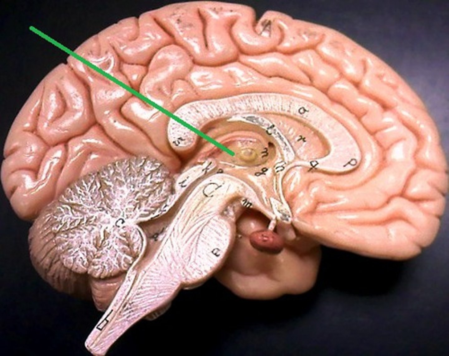
Pituitary Gland
Gland in the brain that controls many hormones in the body
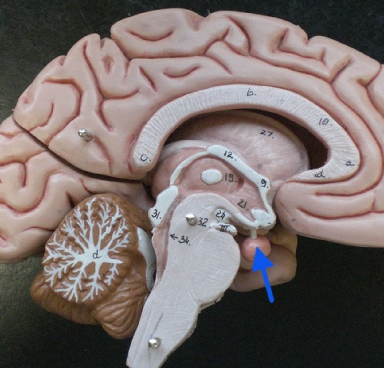
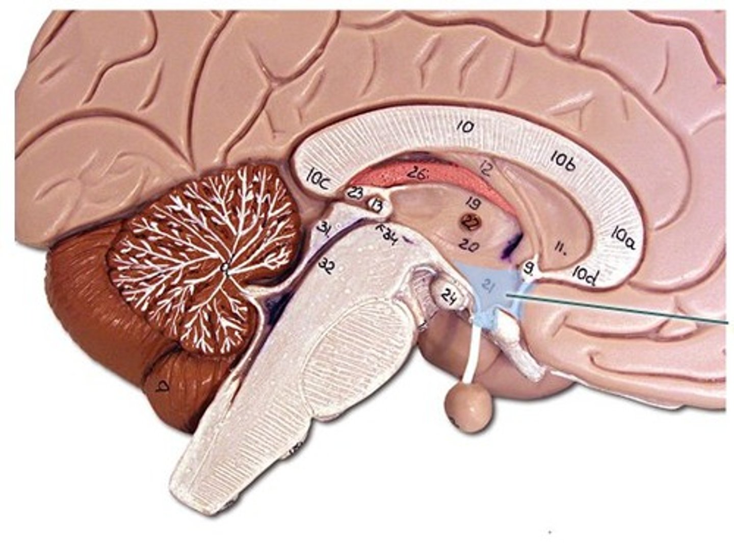
Pineal Gland
Gland in the brain that secretes melatonin, which regulates daily sleep/wake rhythms
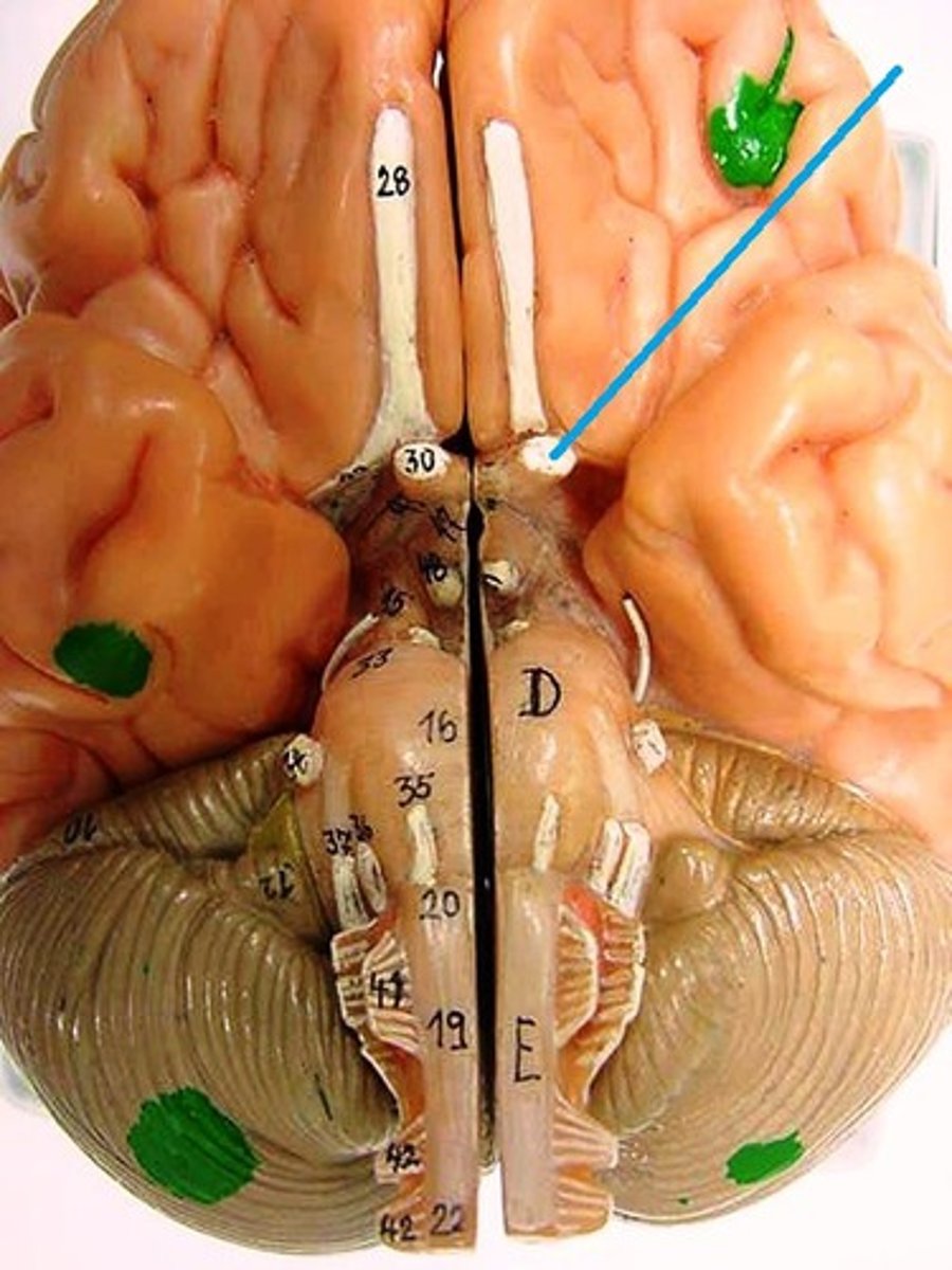
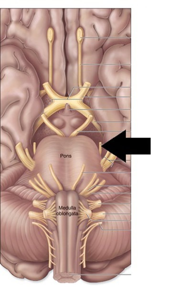
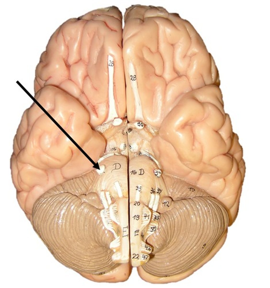
Midbrain
Part of the brainstem involved in sensory and motor functions
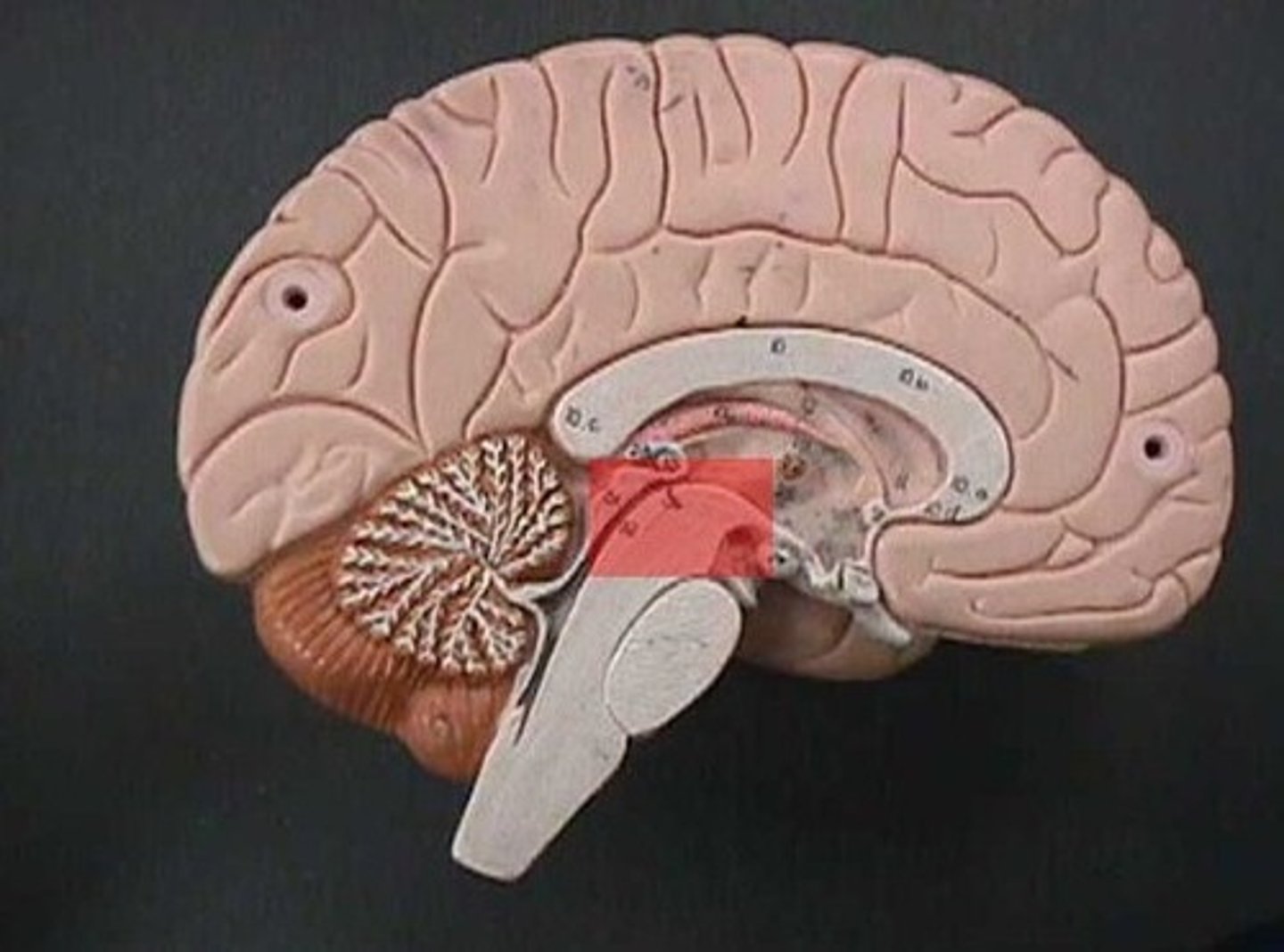
Pons
Part of the brainstem involved in relaying signals between the cerebrum and cerebellum and handles some unconscious processes
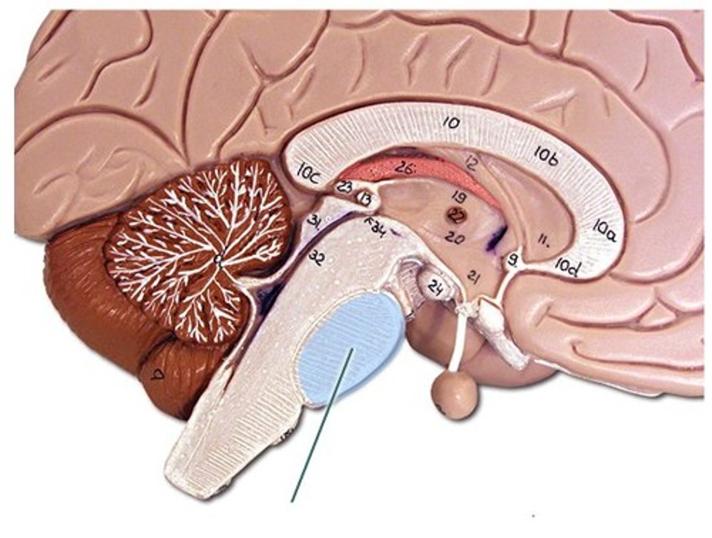
Medulla Oblongata
Part of the brainstem responsible for vital functions such as breathing and heart rate
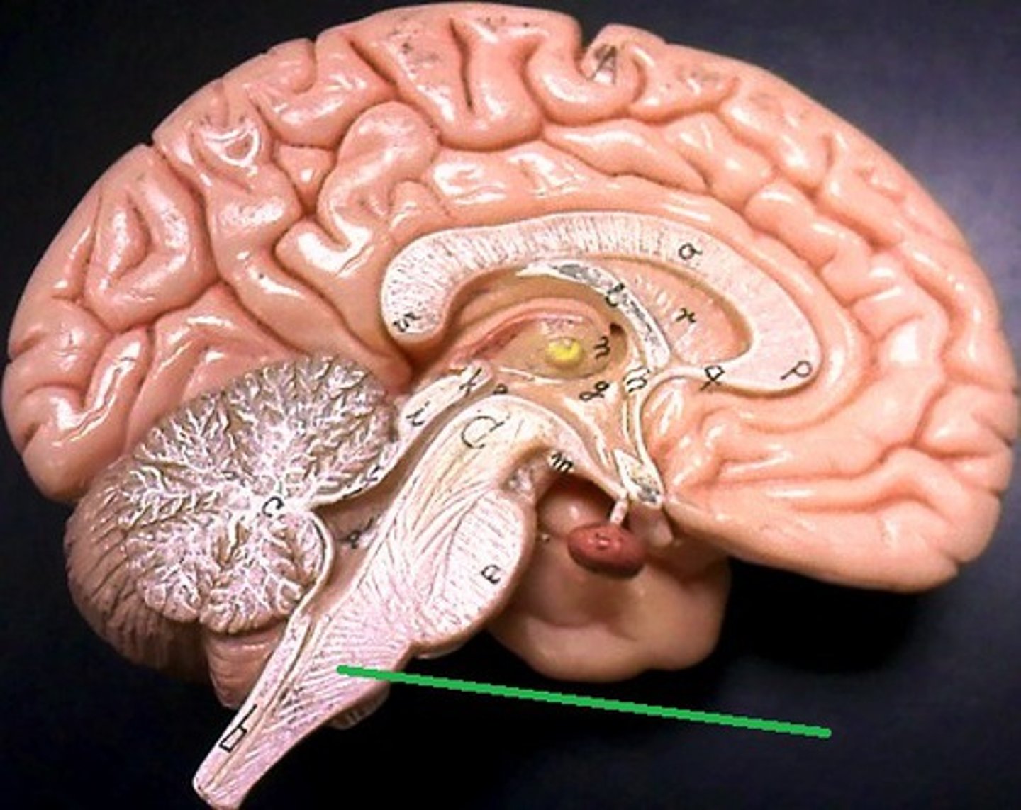
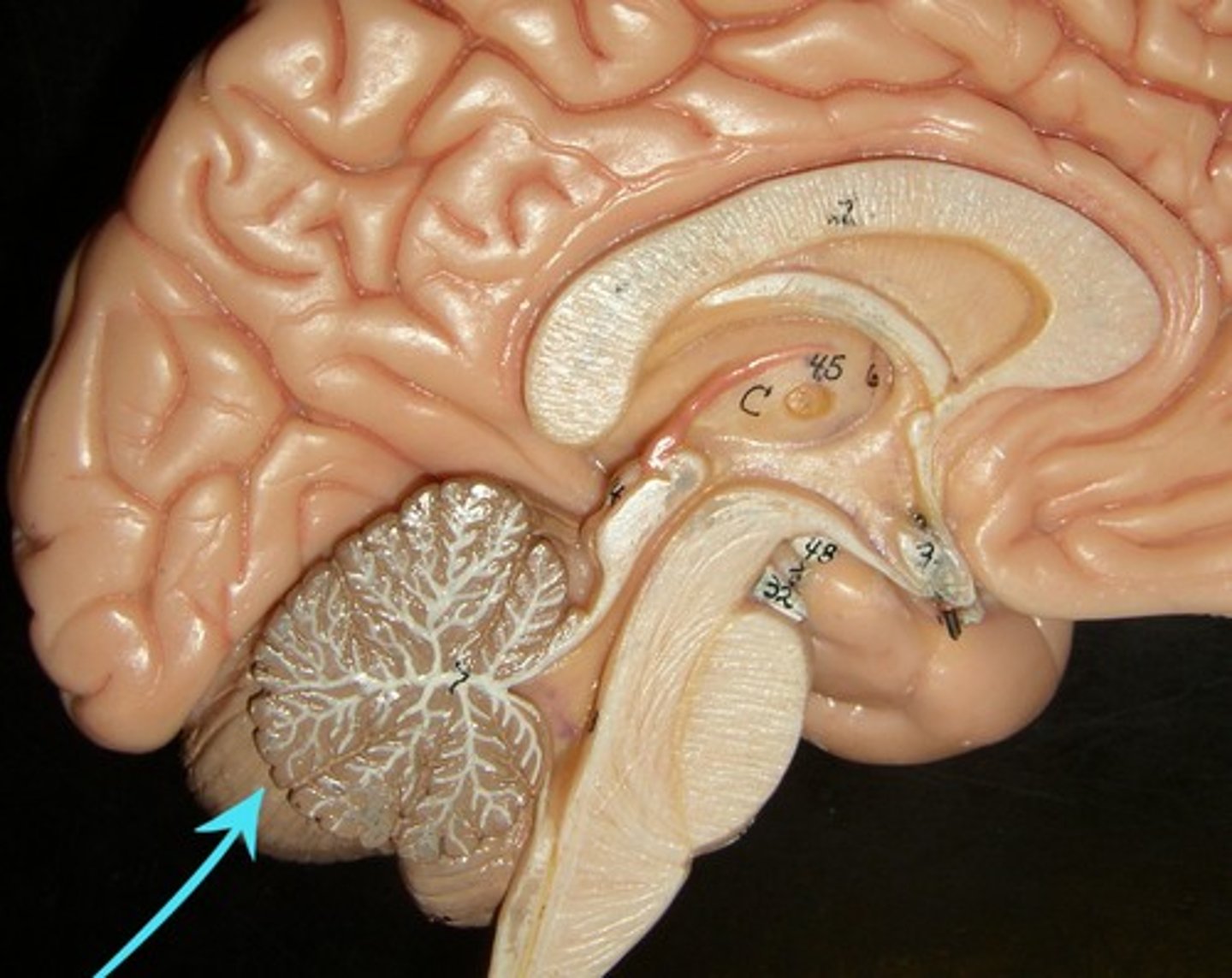
Cerebellum
Part of the brain responsible for balance, coordination, and planning
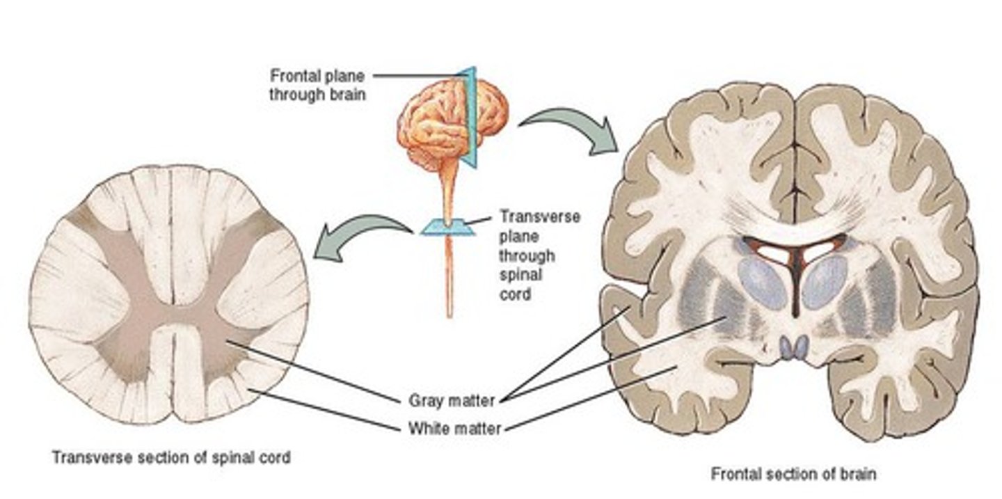
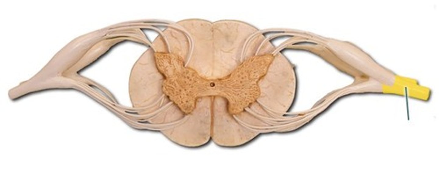
White and Grey matter
Tissues that make up the spinal cord, with white matter containing nerve fibers and grey matter containing cell bodies
Ventral and Dorsal root
Nerve roots that emerge from the spinal cord, with ventral roots carrying motor signals and dorsal roots carrying sensory signals
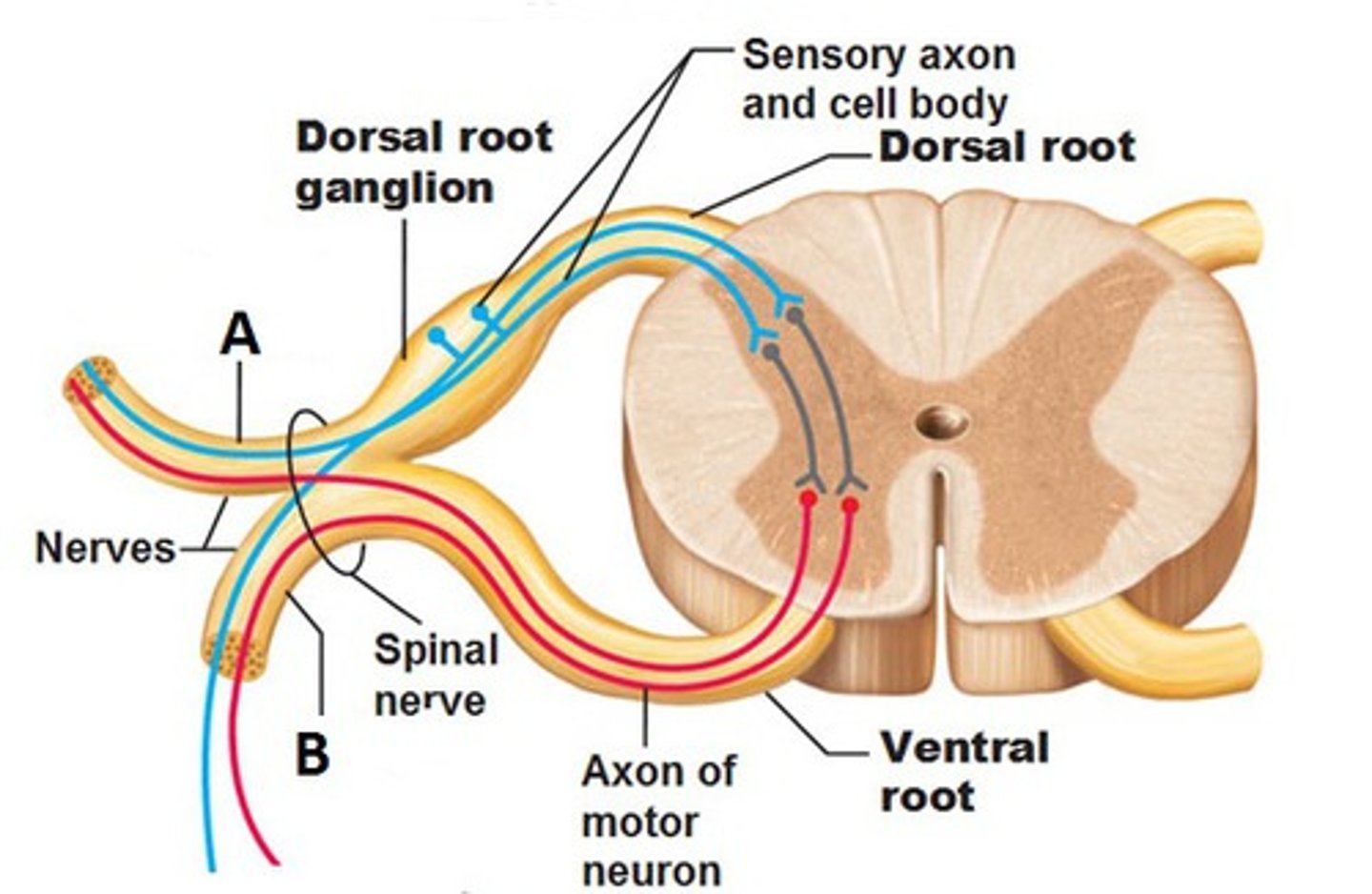
Dorsal horn
Part of the spinal cord grey matter that receives sensory information
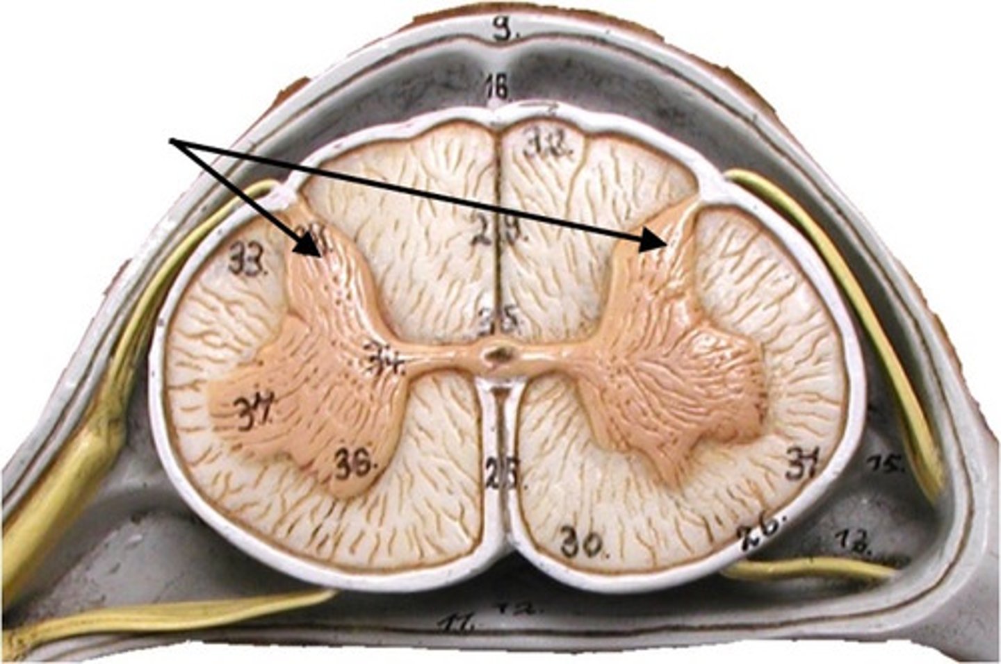
Dorsal root ganglion
Cluster of sensory neuron cell bodies located in the dorsal root
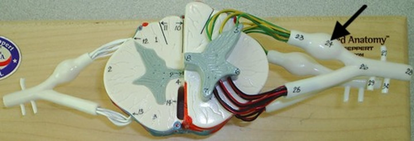
Spinal nerve
Nerve that emerges from the spinal cord and carries both sensory and motor signals
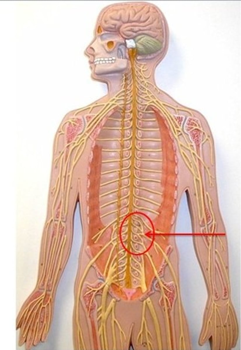
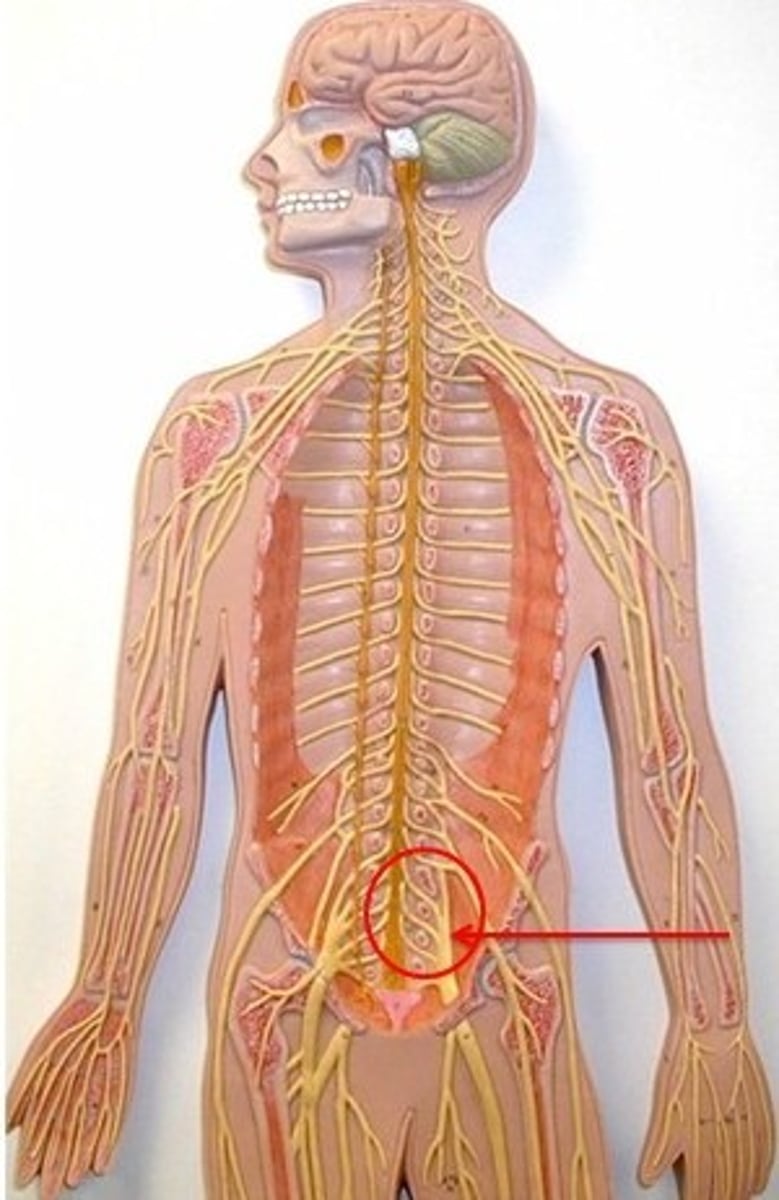
Conus medullaris
End of the spinal cord, usually at the level of the first or second lumbar vertebra
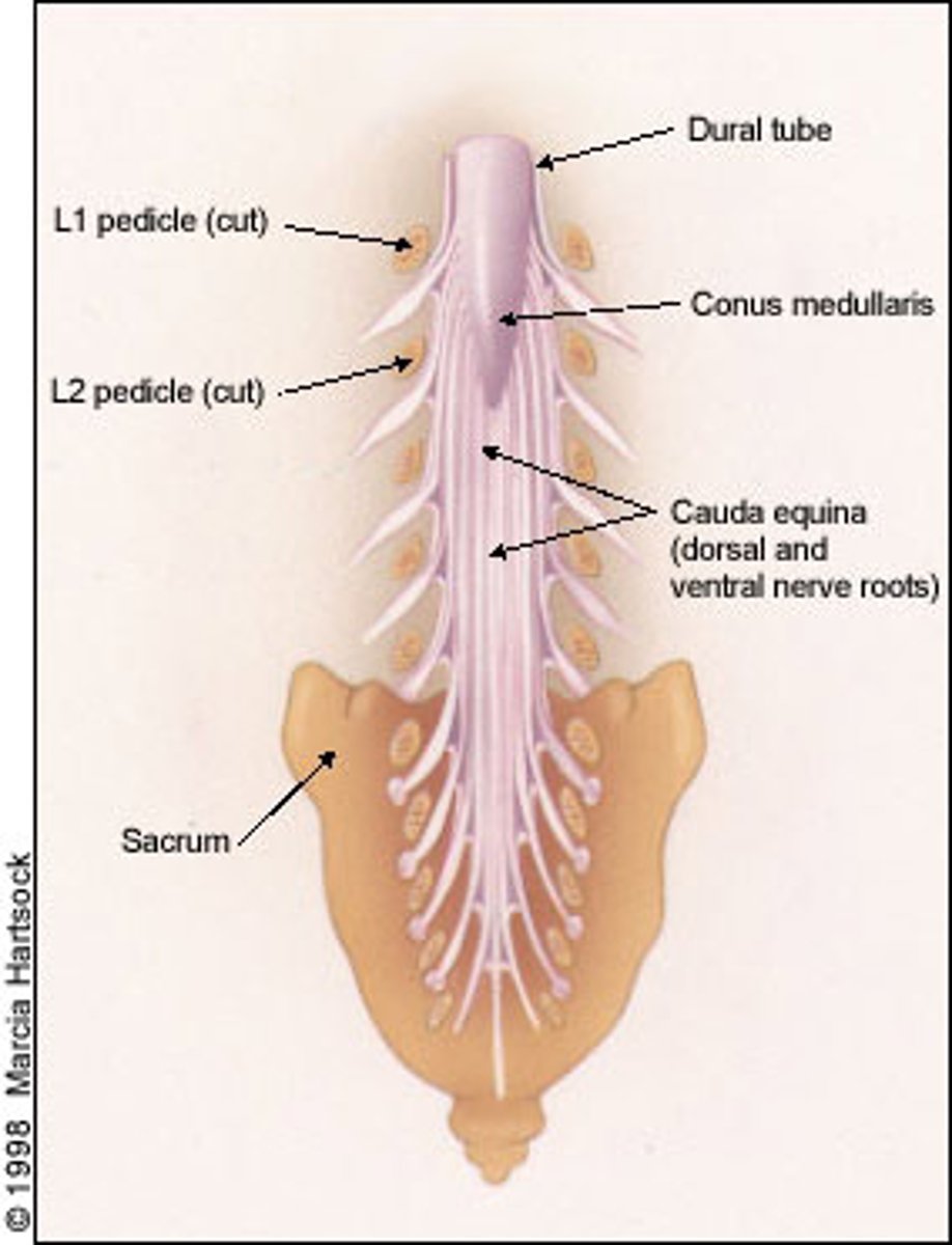
Cauda equina
Bundle of spinal nerves below the conus medullaris
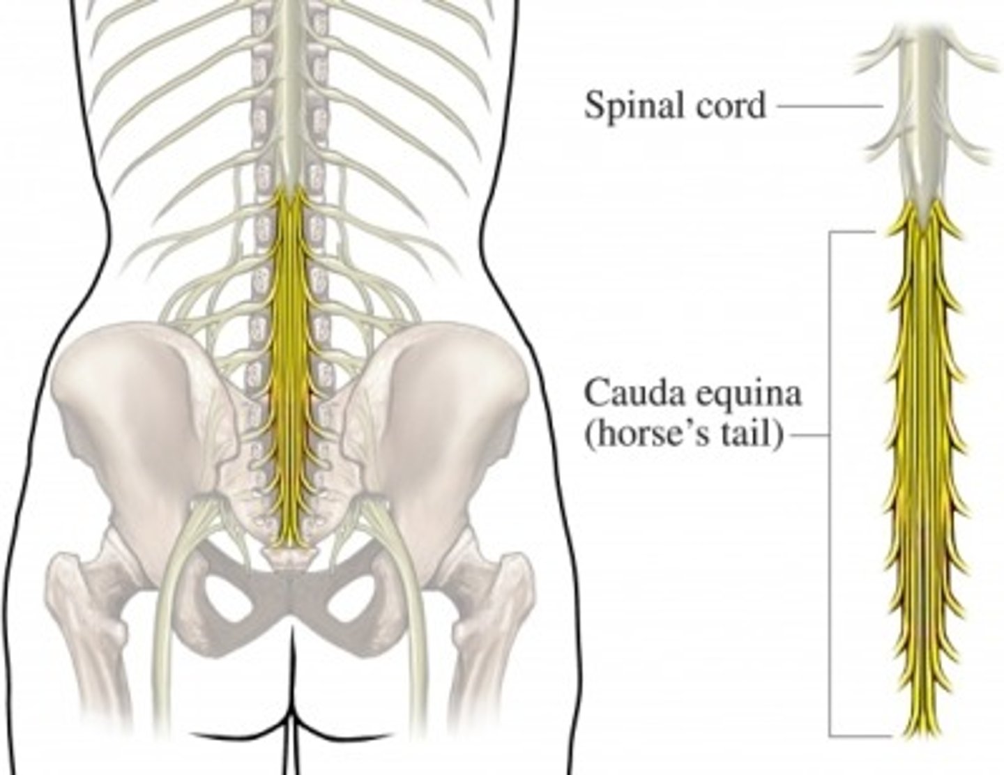
Filum terminal
Thin strand of fibrous tissue that extends from the conus medullaris and anchors the spinal cord
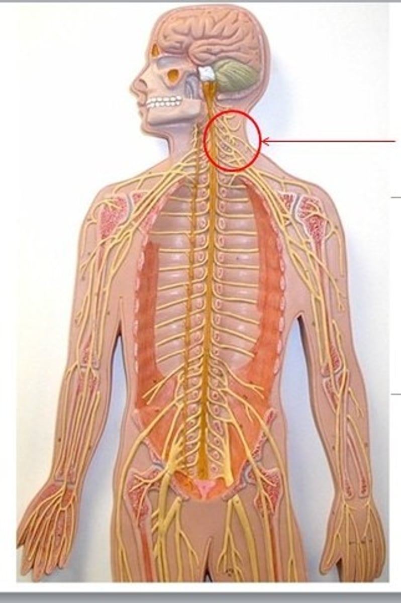
Cervical plexus
Plexus formed by spinal nerves C1-C4, serving the neck and upper shoulder
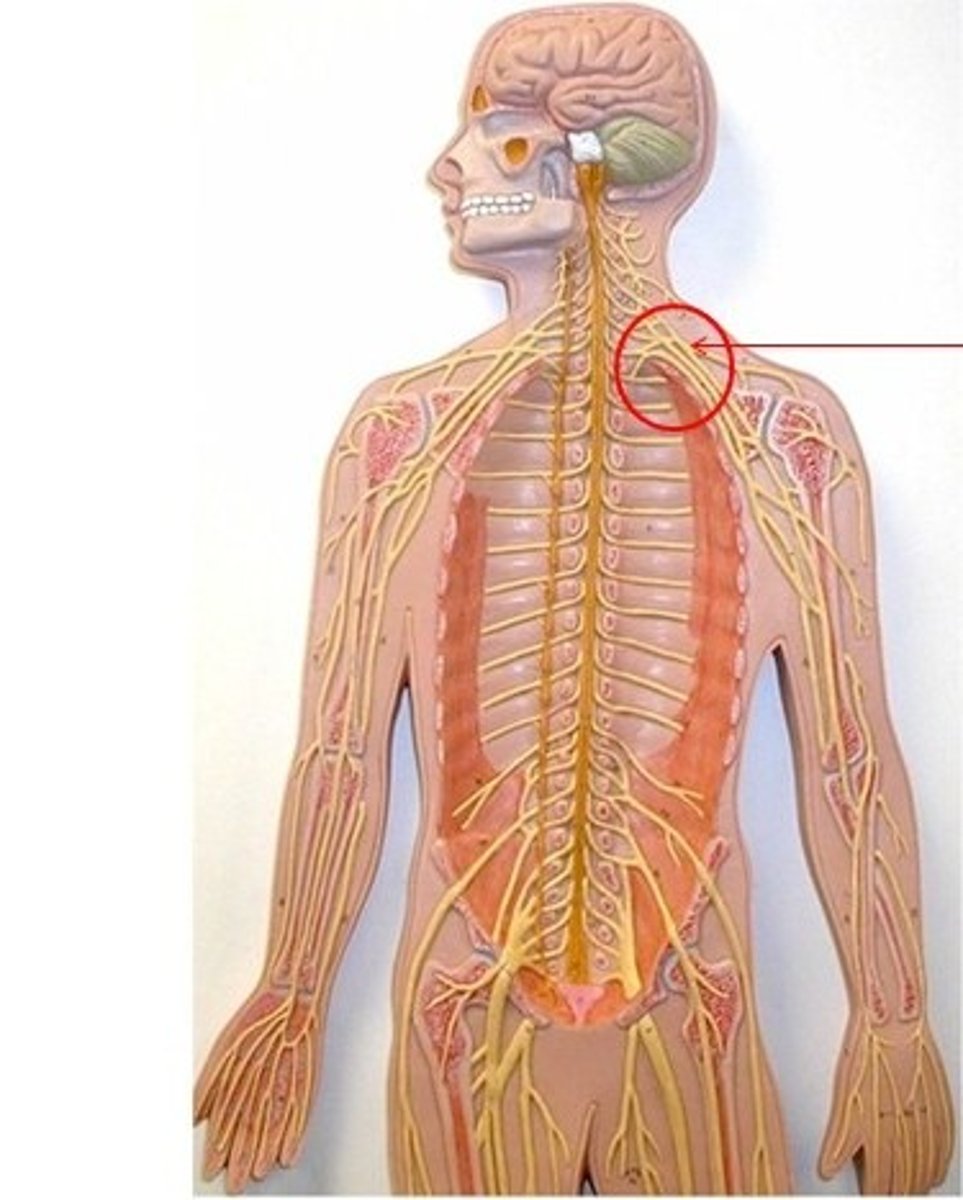
Brachial plexus
Plexus formed by spinal nerves C5-T1, serving the upper limb
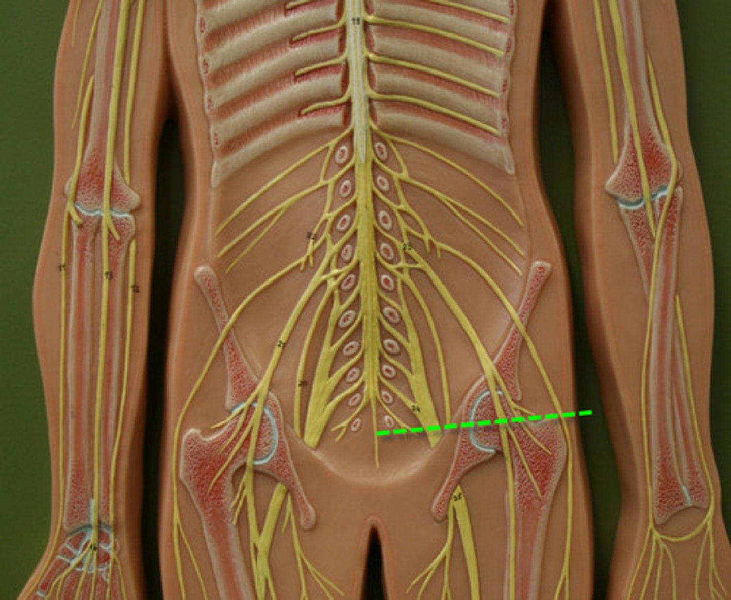
Lumbar plexus
Plexus formed by spinal nerves L1-L4, serving the lower abdomen and anterior thigh
Sacral plexus
Plexus formed by spinal nerves L4-S4, serving the posterior thigh, leg, and foot
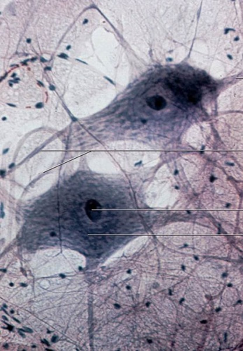
Neurons
Nerve cells responsible for transmitting information in the nervous system
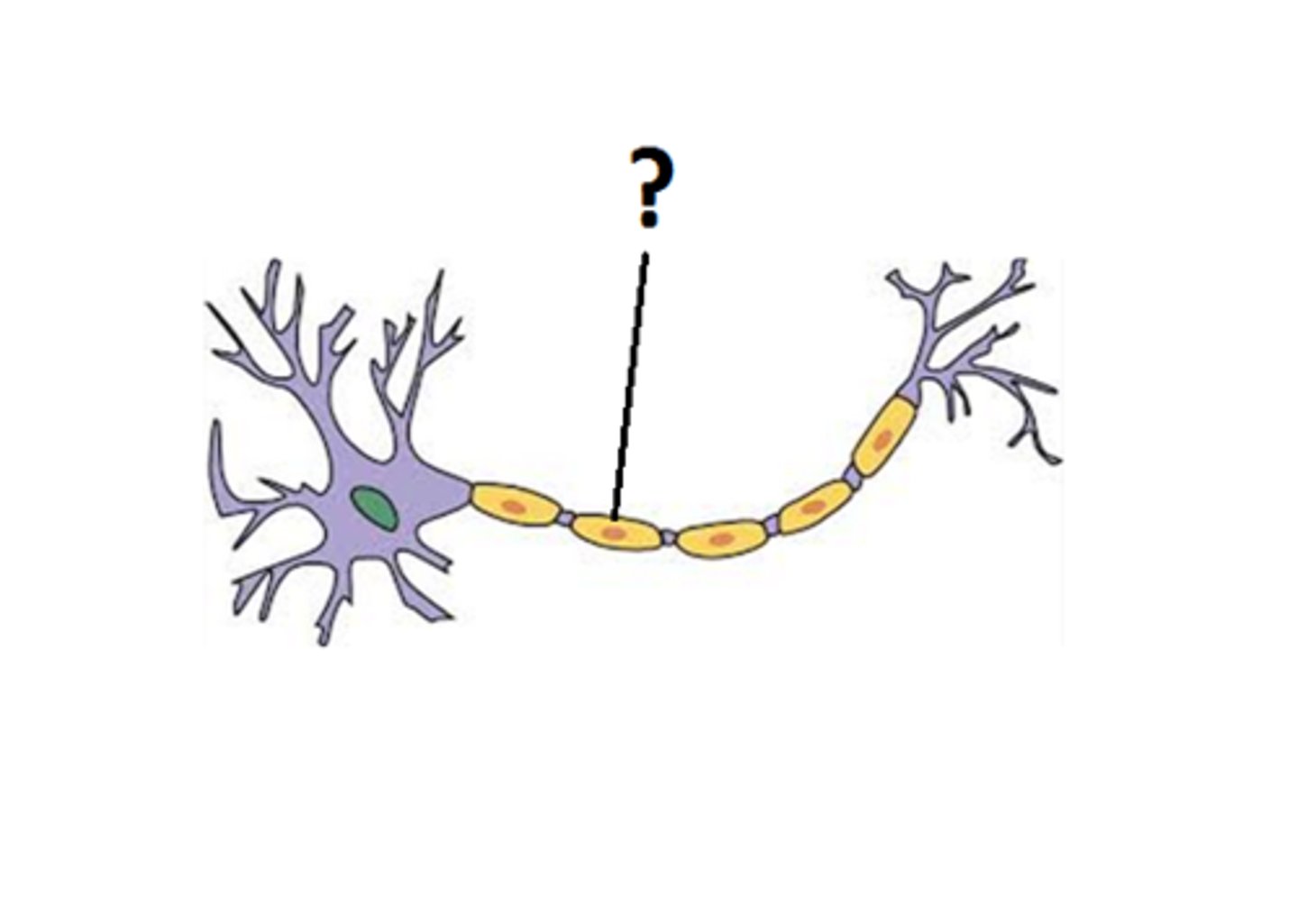
Axon
Long fiber of a neuron that carries nerve impulses away from the cell body
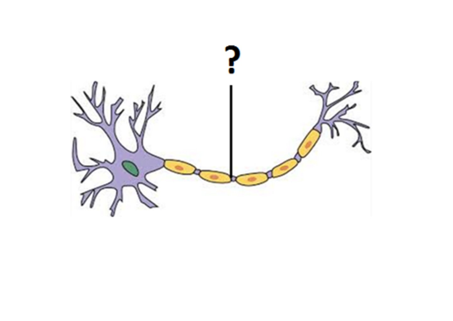
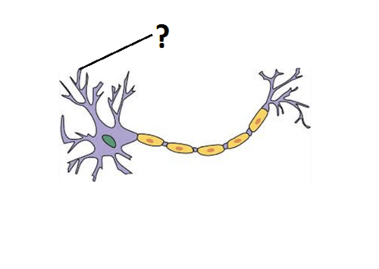
Dendrite
Branch-like extension of a neuron that receives signals from other neurons
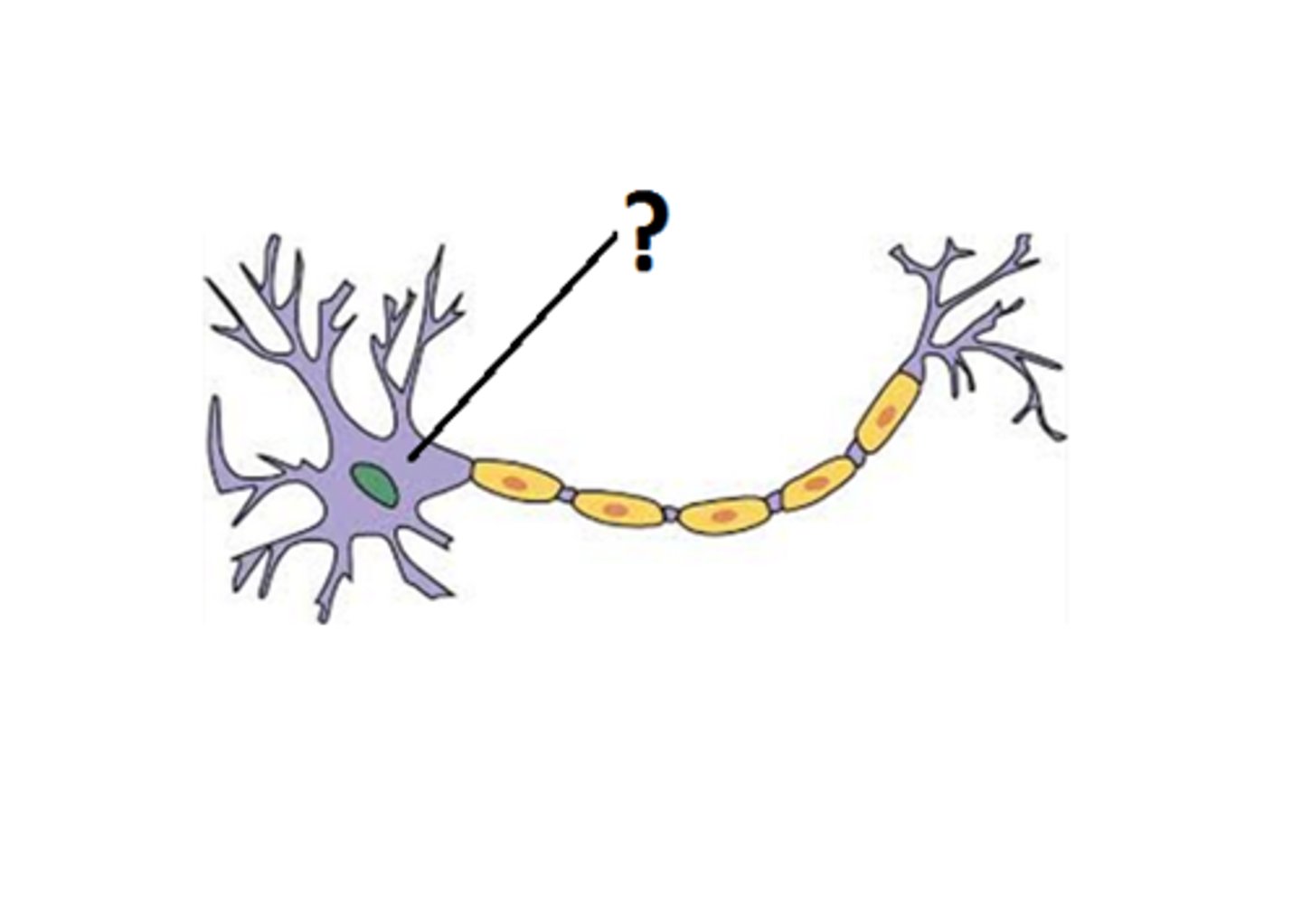
Neuron cell body
Part of a neuron that contains the nucleus and other organelles
Myelin
Fatty substance that surrounds and insulates axons, allowing for faster transmission of nerve impulses
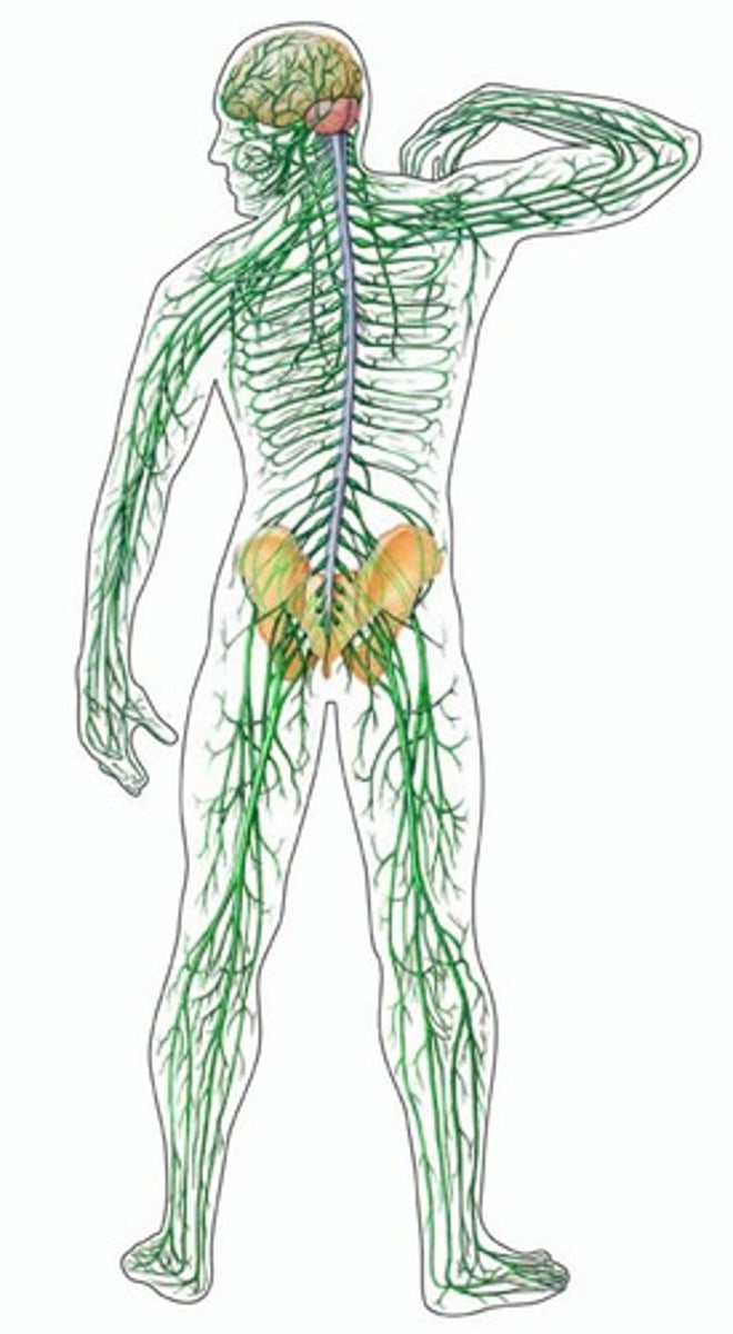
Peripheral nerves
Nerves that connect the central nervous system to the rest of the body
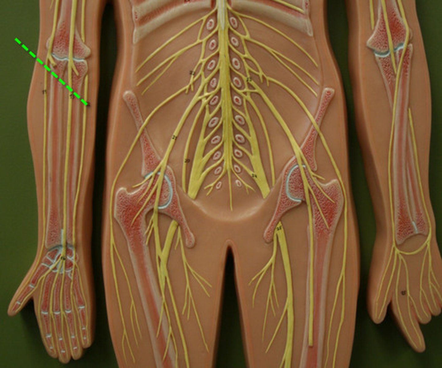
Median nerve
Nerve in the upper extremity responsible for sensation and movement in the thumb and first three fingers

Radial nerve
Nerve in the upper extremity responsible for sensation and movement in the back of the hand and forearm
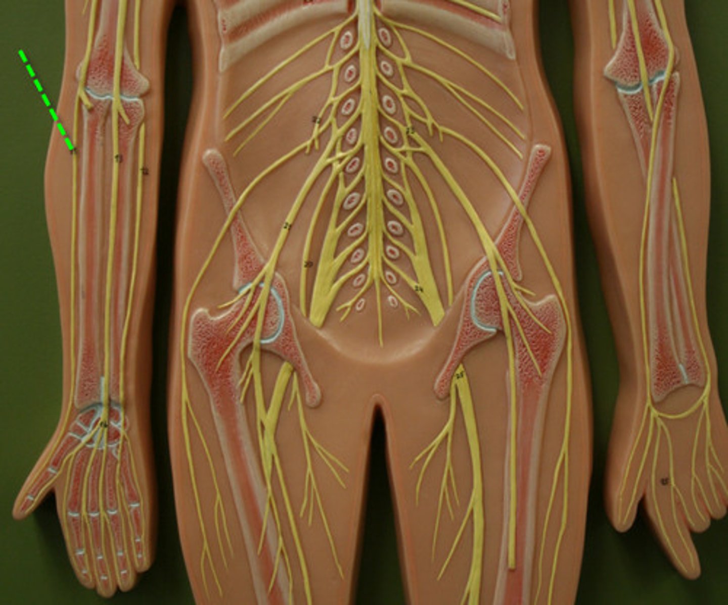
Ulnar nerve
Nerve in the upper extremity responsible for sensation and movement in the pinky finger and part of the ring finger
Femoral nerve
Nerve in the lower extremity responsible for sensation and movement in the front of the thigh and lower leg
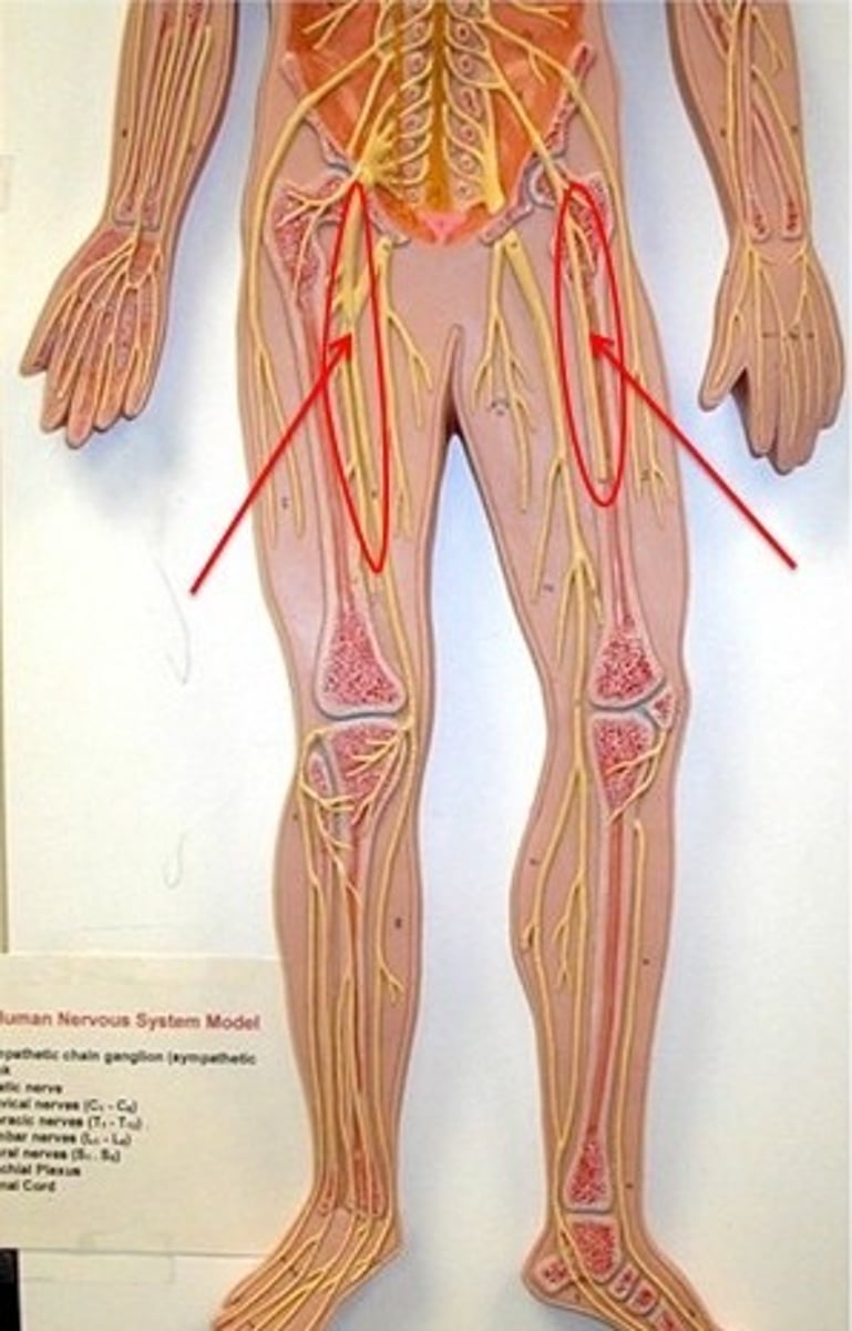
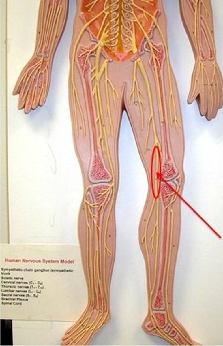
Sciatic nerve
Nerve in the lower extremity responsible for sensation and movement in the back of the thigh, leg, and foot
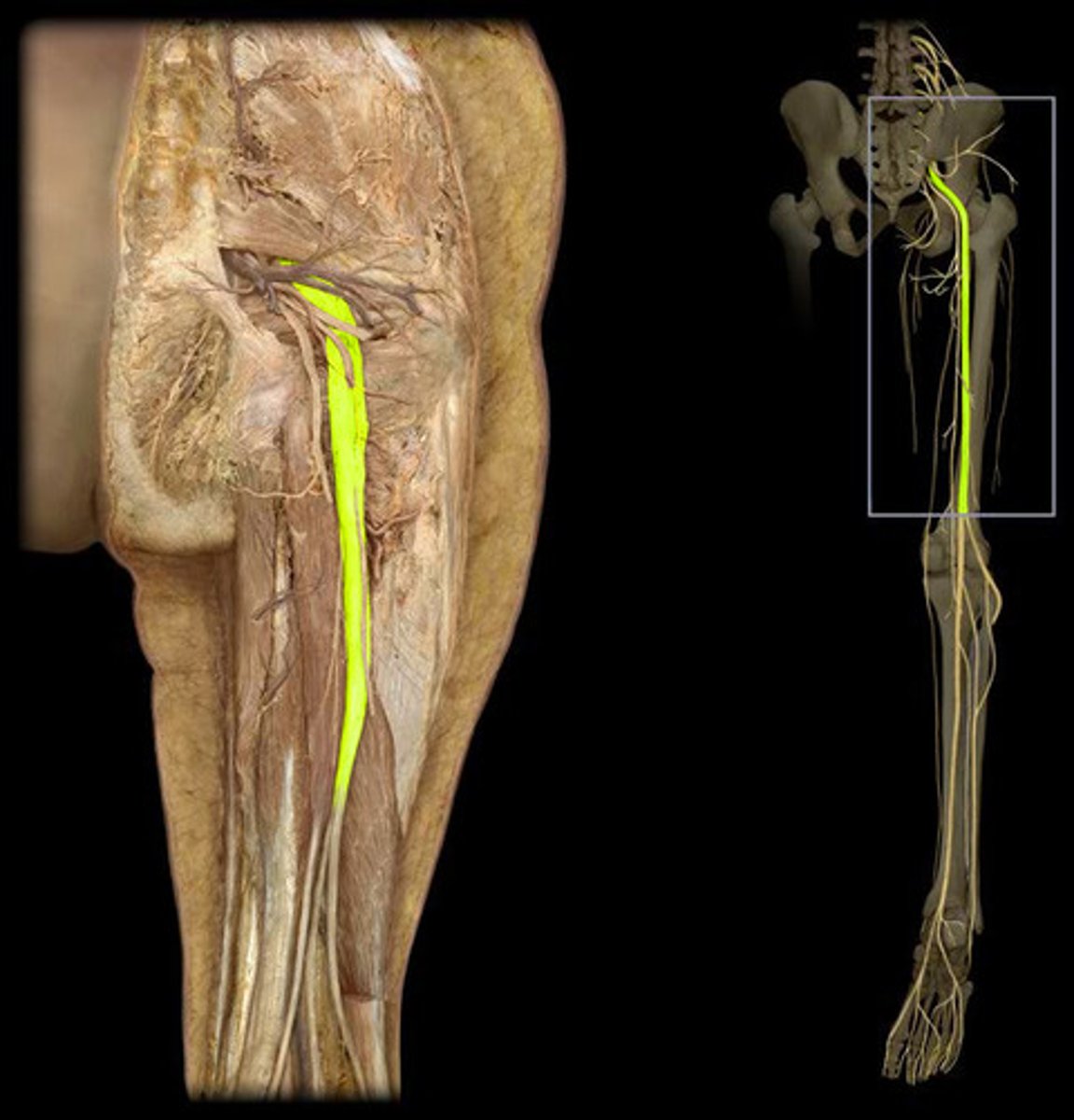
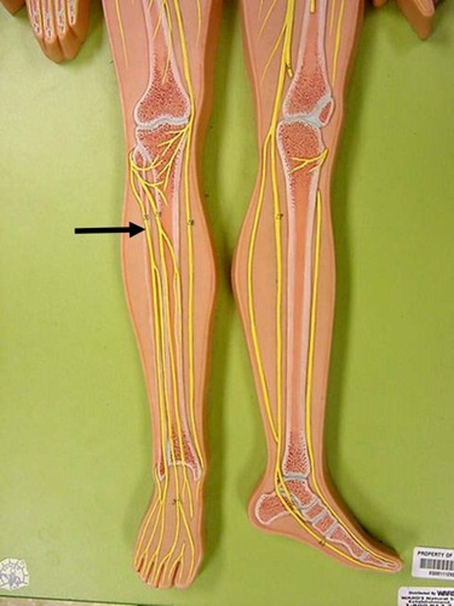
Tibial nerve
Nerve in the lower extremity responsible for sensation and movement in the back of the leg and sole of the foot
Fibular nerve
Nerve in the lower extremity responsible for sensation and movement in the front of the leg and top of the foot
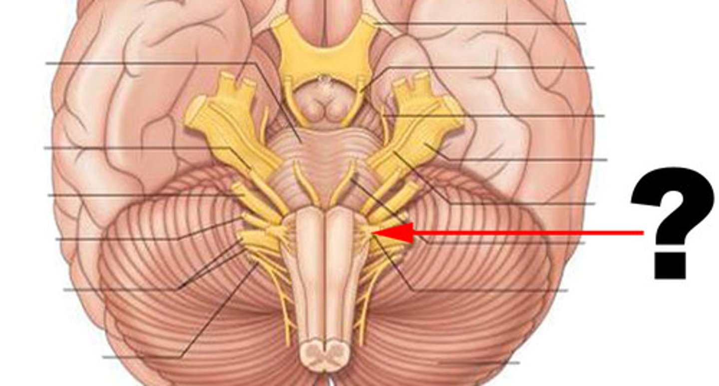
Cranial Nerves
Nerves that emerge directly from the brain and serve various functions
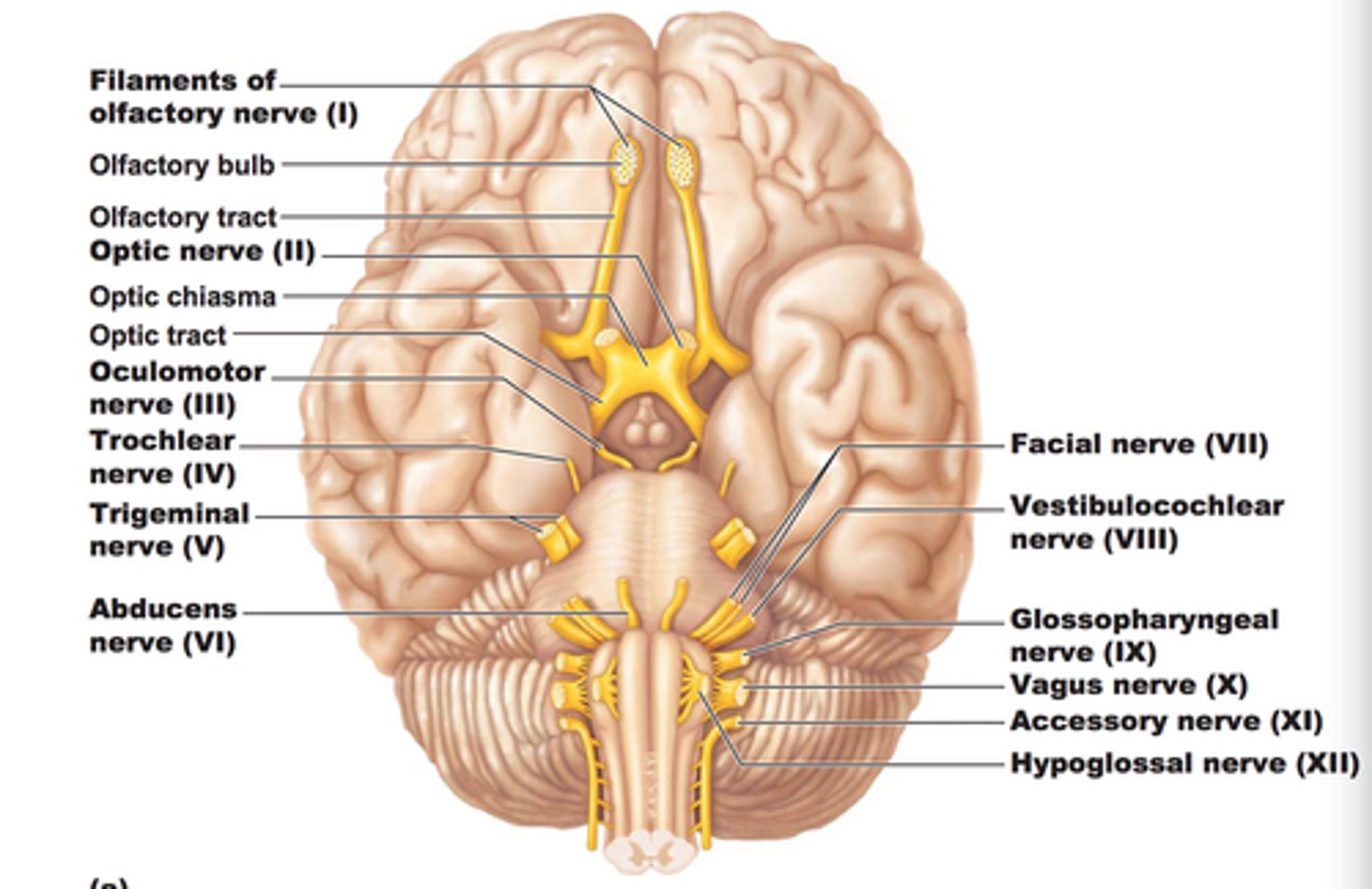
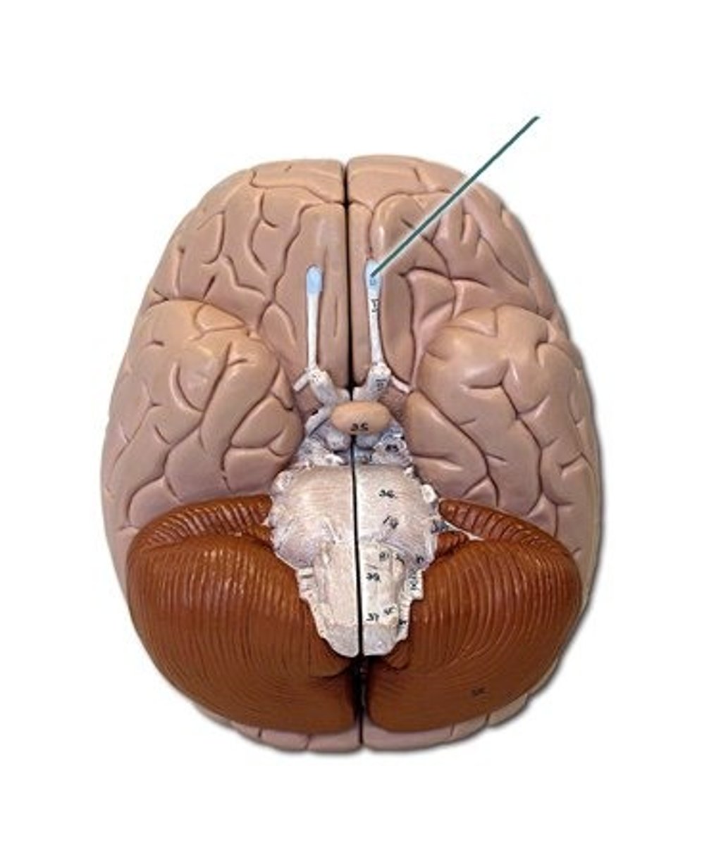
CN I
Olfactory nerve responsible for sensations for smelling
CN II
Optic nerve responsible for sensations for vision
CN III
Oculomotor nerve responsible for motor control of eye movements
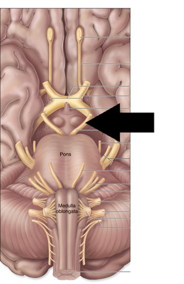
CN IV
Trochlear nerve responsible for motor control of the superior oblique eye muscle
CN V
Trigeminal nerve responsible for sensory input from the head and motor control to the head
CN VI
Abducens nerve responsible for motor control of lateral eye movements
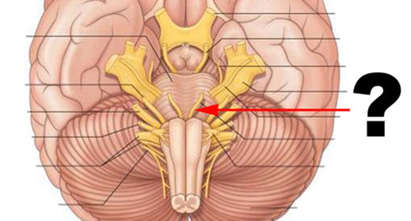
CN VII
Facial nerve responsible for sensory input from the anterior tongue and motor control to head muscles

CN VIII
Vestibulocochlear nerve responsible for sensory input for hearing and vestibular function for balance
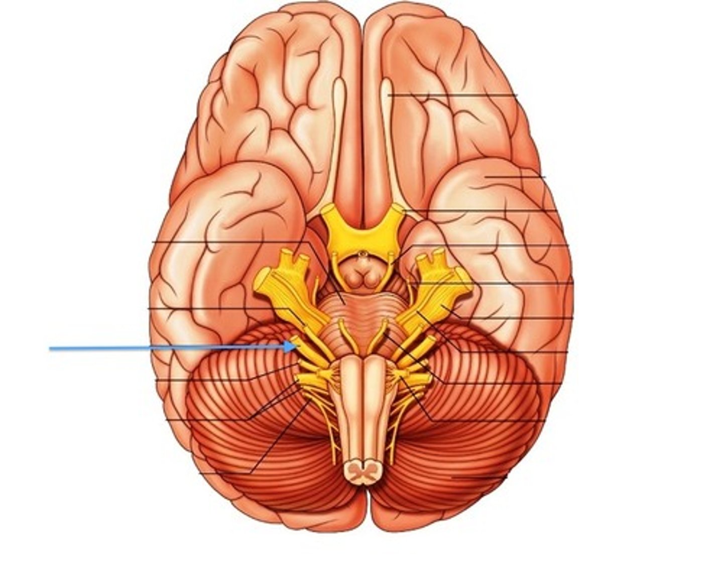
CN IX
Glossopharyngeal nerve responsible for mixed sensory and motor functions for the tongue and throat
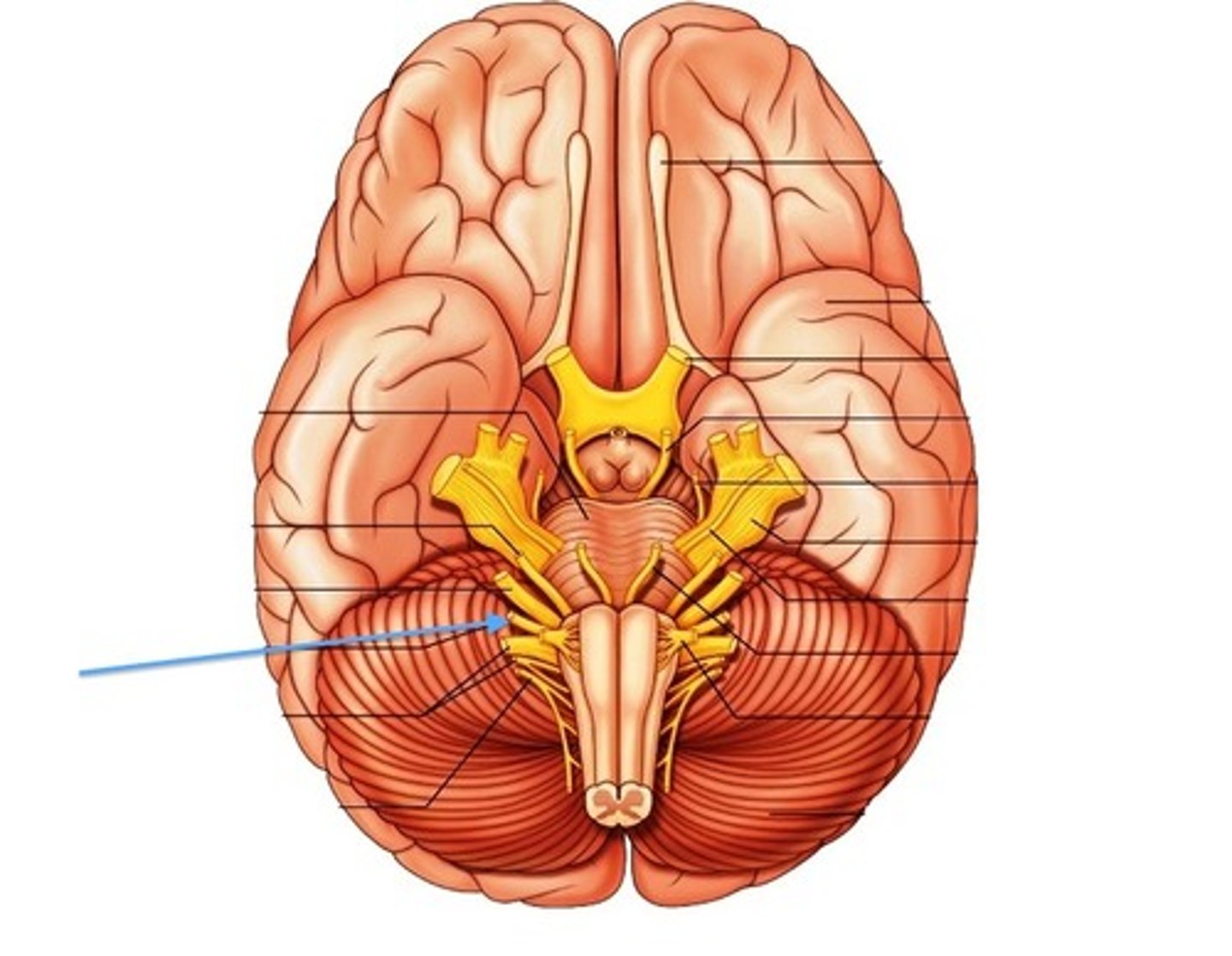
CN X
Vagus nerve responsible for sensory input from the abdomen, thorax, neck, and tongue, and motor control to the pharynx and larynx, as well as autonomic functions
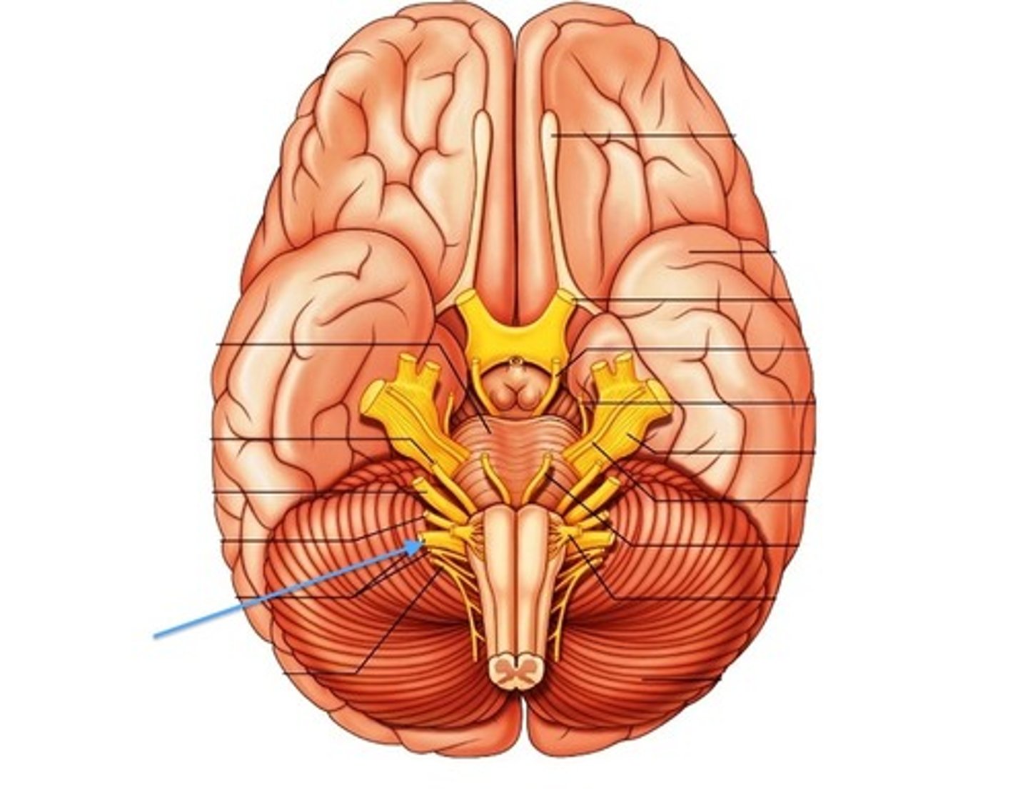
CN XI
Accessory nerve responsible for motor control of neck muscles
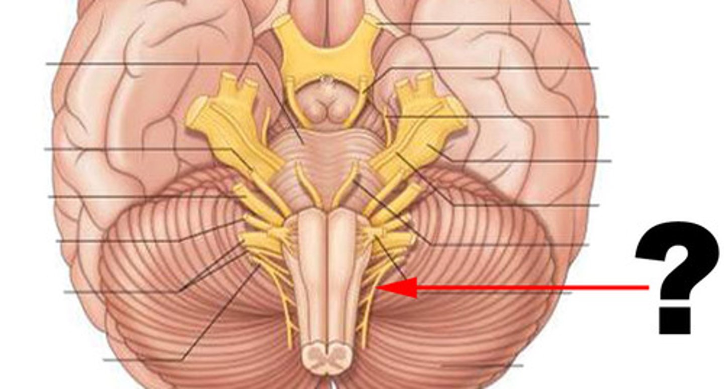
CN XII
Hypoglossal nerve responsible for motor control of the tongue
facial muscles
Temporalis, Masseter, Orbicularis Oculi, Orbicularis Oris, Zygomaticus Major, Buccinator
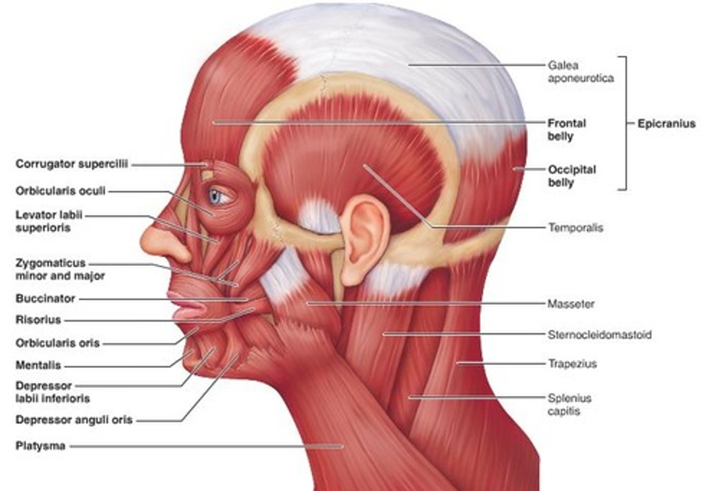
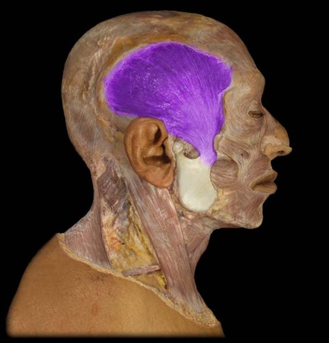
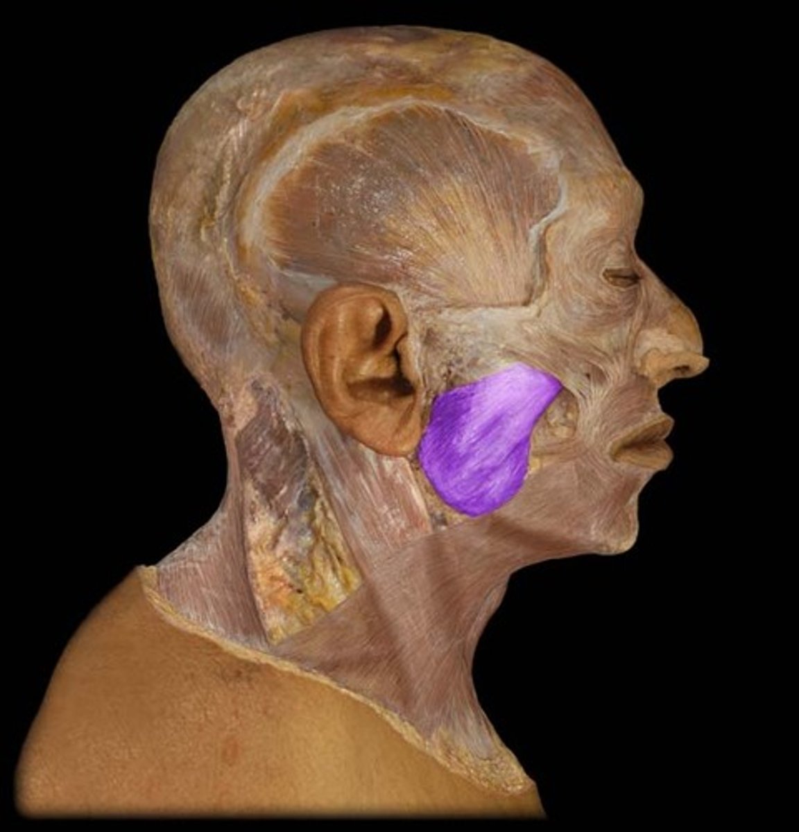
temporalis muscle
Mastication (chewing)
masseter muscle
Mastication (chewing)
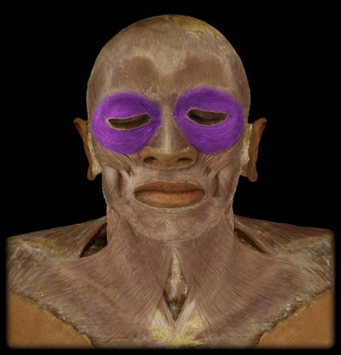
orbicularis oculi muscle
Winking, closing eye
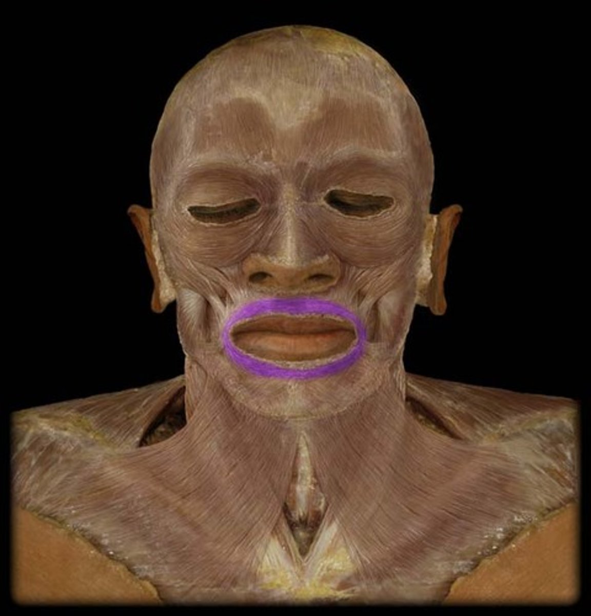
orbicularis oris muscle
Kissing, speech
zygomaticus major muscle
Smiling, laughing
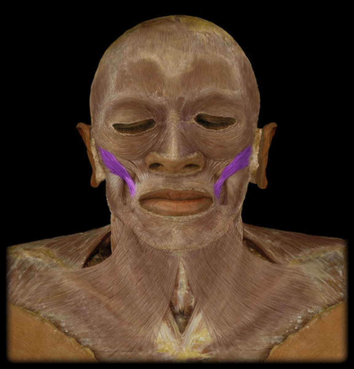
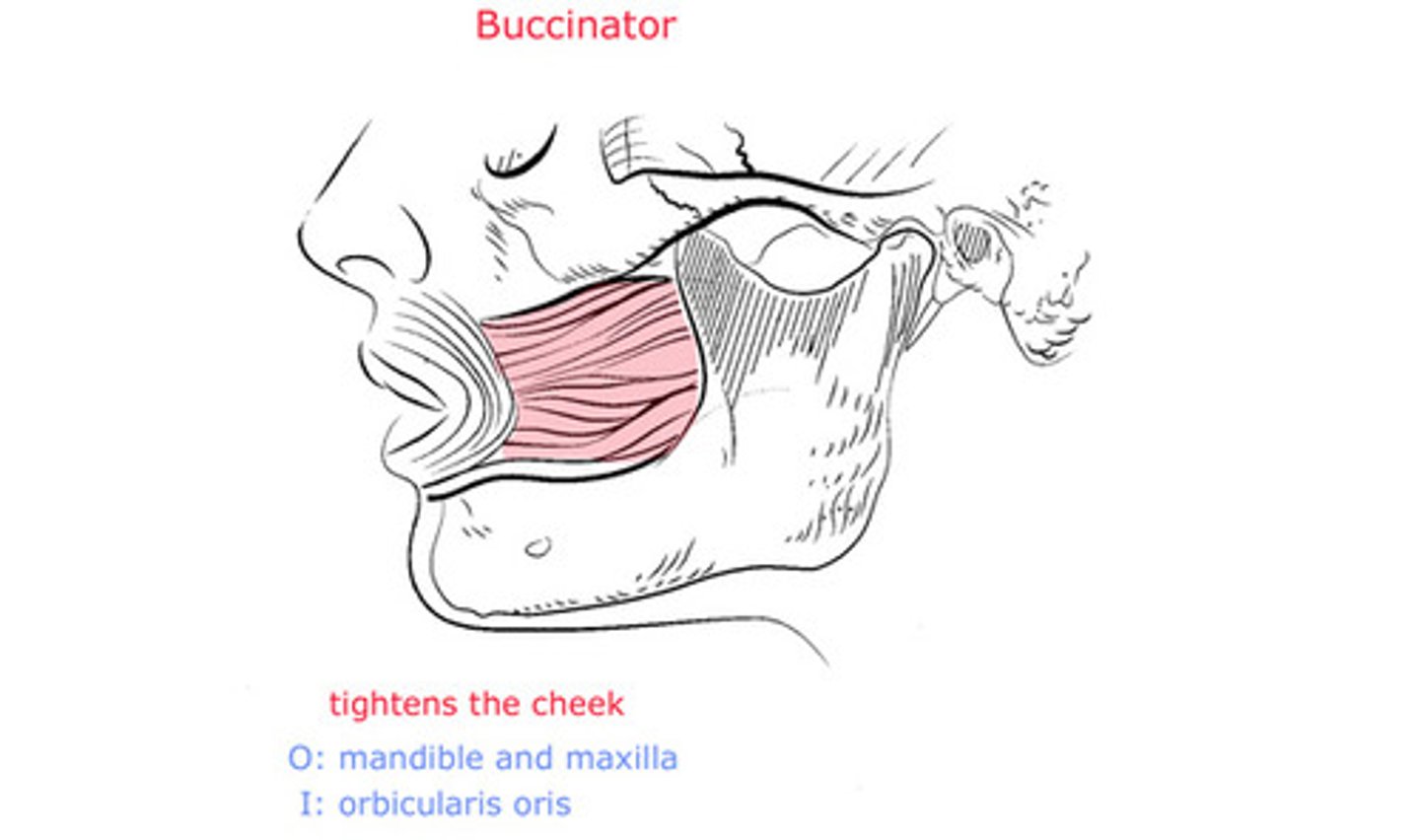
buccinator muscle
Whistling, blowing
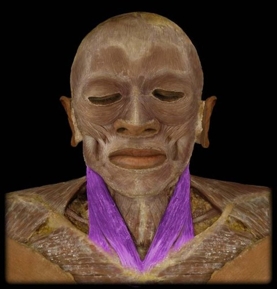
sternocleidomastoid muscle
Rotation of head to opposite side and flexion of neck
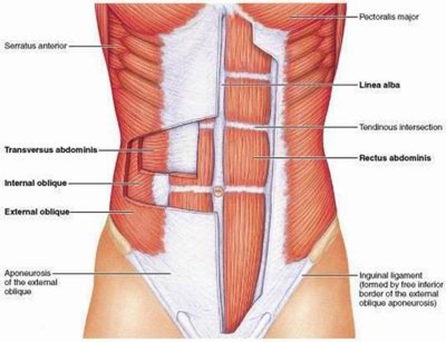
muscles of the abdomen
Intercostals (Internal and External), Diaphragm, Rectus Abdominis, Linea Alba, Obliques (Internal and External), Transverse Abdominis