A&P II Exam 3

0.0(0)
learnLearn
examPractice Test
spaced repetitionSpaced Repetition
heart puzzleMatch
flashcardsFlashcards
Card Sorting

1/108

encourage image

There's no tags or description

Looks like no tags are added yet.

Study Analytics
Name
Mastery
Learn
Test
Matching
Spaced

No study sessions yet.

109 Terms

1
New cards

Nasal cavity

The hollow space inside of the nose, containing structures such as the conchae.
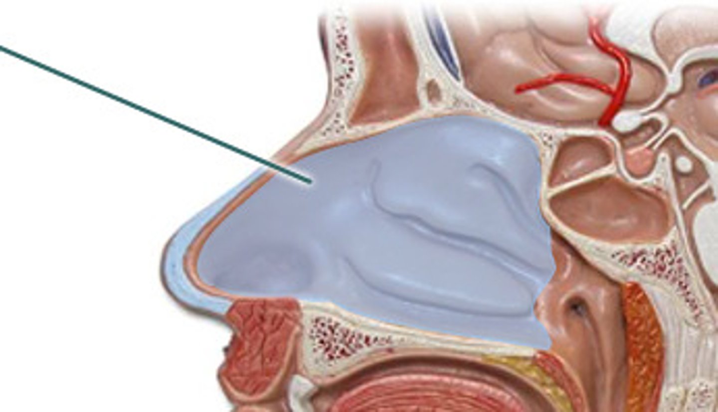
<p>The hollow space inside of the nose, containing structures such as the conchae.</p>
2
New cards

External nares

2 openings which open into the nasal cavity; where air enters the respiratory system.
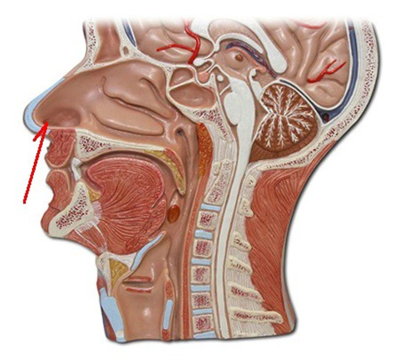
<p>2 openings which open into the nasal cavity; where air enters the respiratory system.</p>
3
New cards

Nasal septum

a wall of cartilage that divides the nose into two equal sections, spans the length of the entire nasal cavity
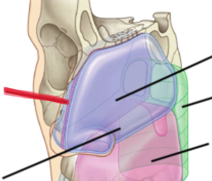
<p>a wall of cartilage that divides the nose into two equal sections, spans the length of the entire nasal cavity</p>
4
New cards

Inferior conchae

the thin, scroll-like bones that form part of the interior of the nose, functions to create turbulence within the nasal cavity and helps keep air in there longer to help clean and humidify air.
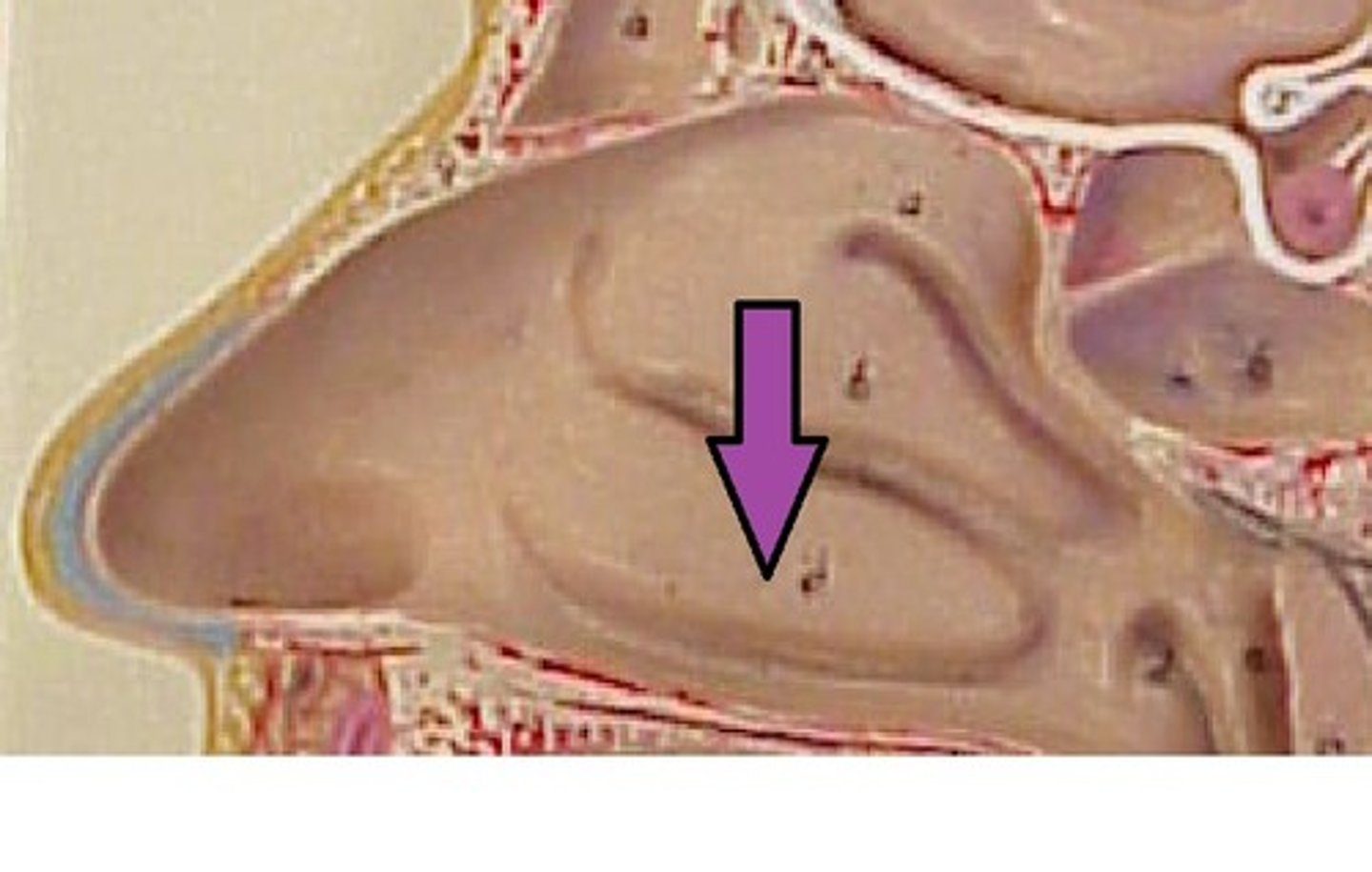
<p>the thin, scroll-like bones that form part of the interior of the nose, functions to create turbulence within the nasal cavity and helps keep air in there longer to help clean and humidify air.</p>
5
New cards

middle conchae

part of the ethmoid bone.

-on lateral walls.

-increase surface area of mucus membrane (moistens air).
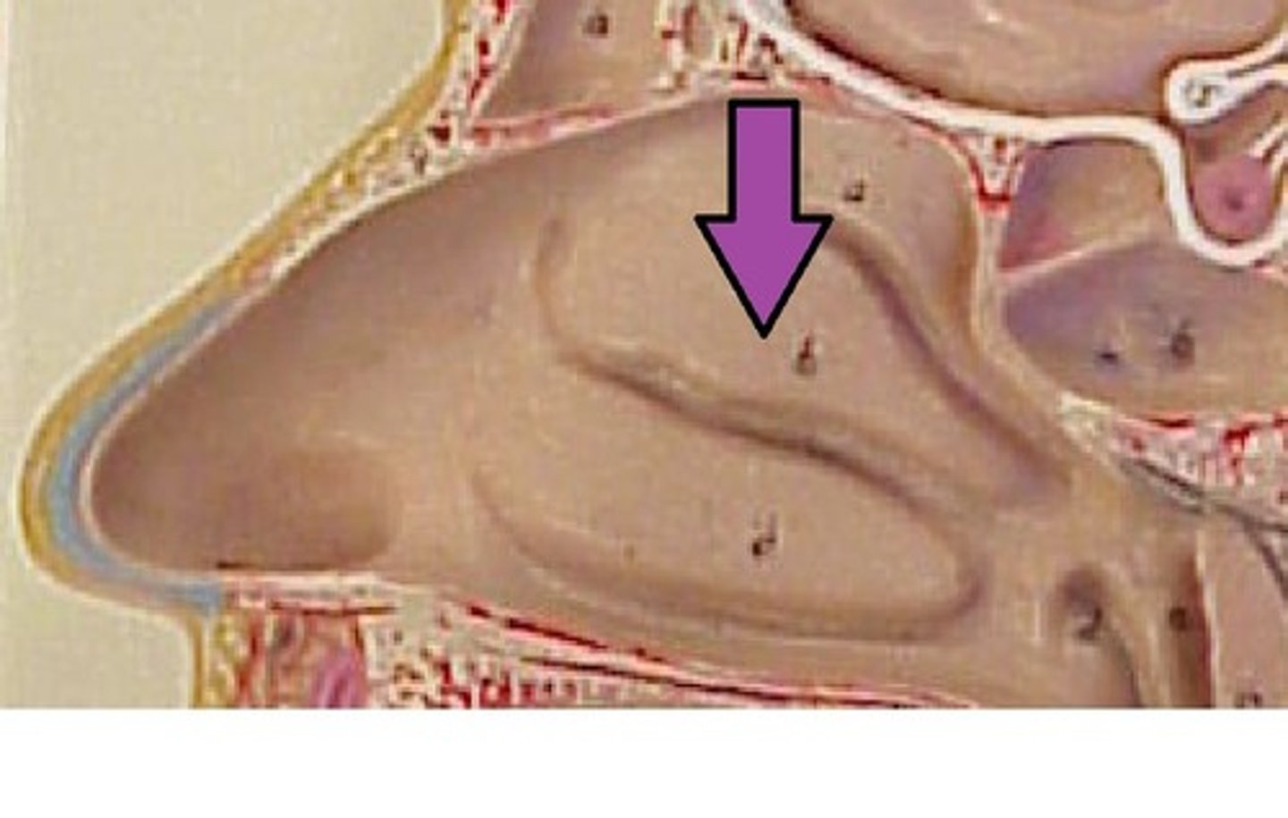
<p>part of the ethmoid bone.</p><p>-on lateral walls.</p><p>-increase surface area of mucus membrane (moistens air).</p>
6
New cards

superior conchae

small, curved bony plates located in the nasal cavity, forming part of the ethmoid bone and playing a crucial role in filtering, warming, and moistening inhaled air
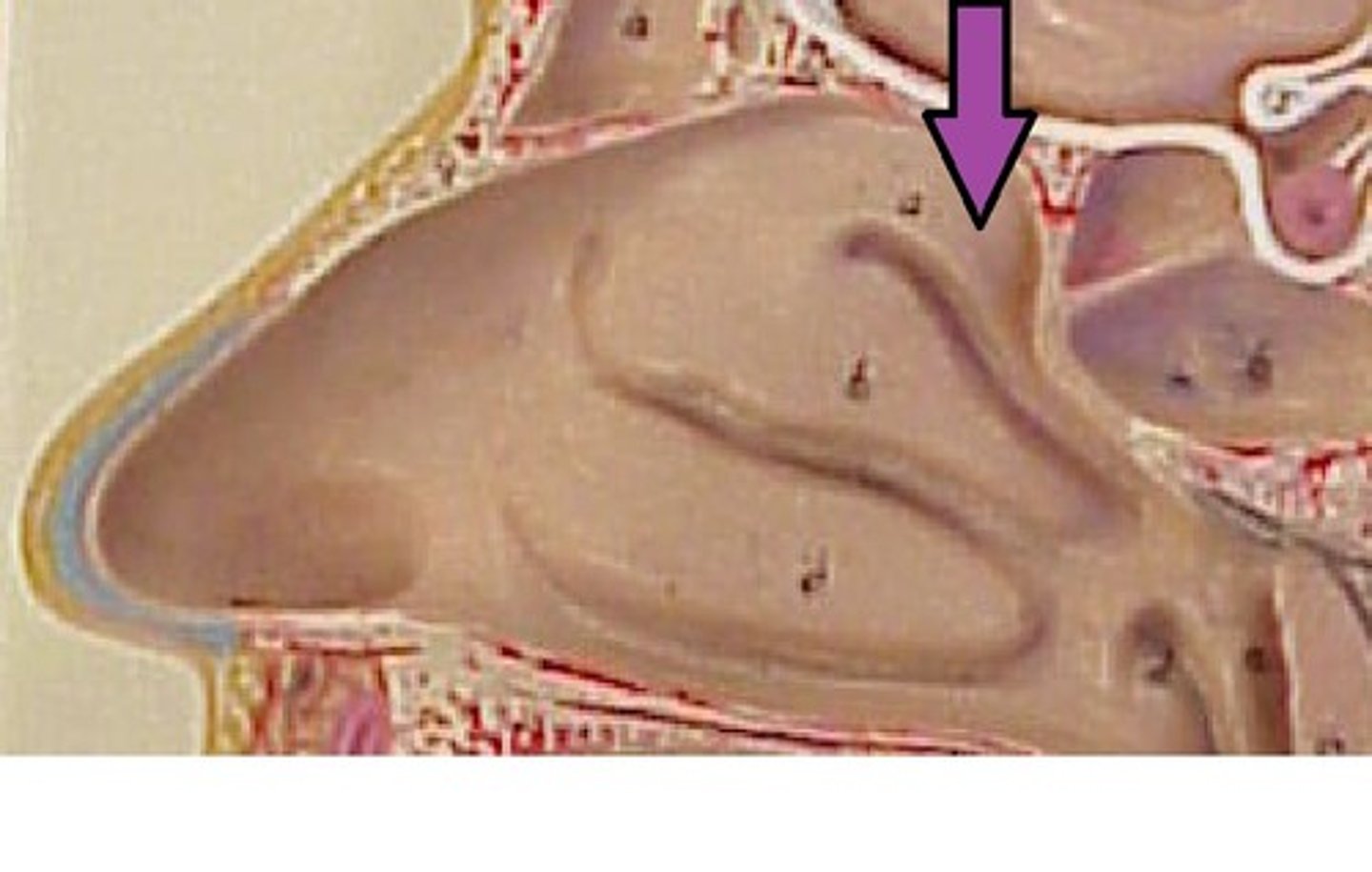
<p>small, curved bony plates located in the nasal cavity, forming part of the ethmoid bone and playing a crucial role in filtering, warming, and moistening inhaled air</p>
7
New cards

Pharynx

commonly referred to as the throat, a passage way in the head and neck that is part of both the digestive system and respiratory system
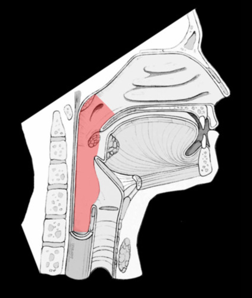
<p>commonly referred to as the throat, a passage way in the head and neck that is part of both the digestive system and respiratory system</p>
8
New cards

nasopharynx

region of the pharynx at the back of the nose and above the soft palate, starts from the top of the nasal cavity and ends towards the soft palate (uvula)
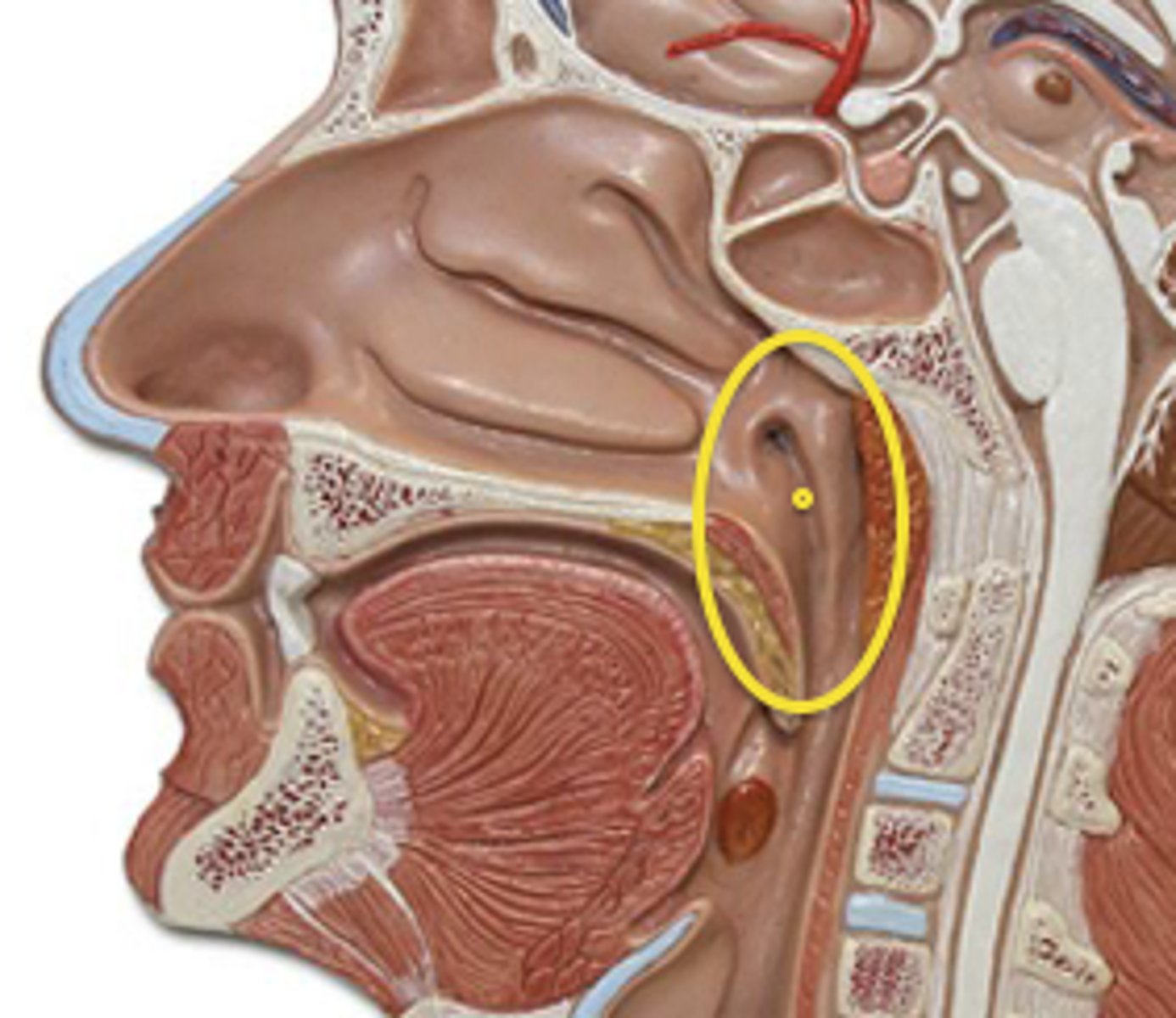
<p>region of the pharynx at the back of the nose and above the soft palate, starts from the top of the nasal cavity and ends towards the soft palate (uvula)</p>
9
New cards

oropharynx

central portion of the pharynx between the roof of the mouth and the upper edge of the epiglottis.

Starts from the uvula down to the epiglottis, located between the naso and laryngopharynx
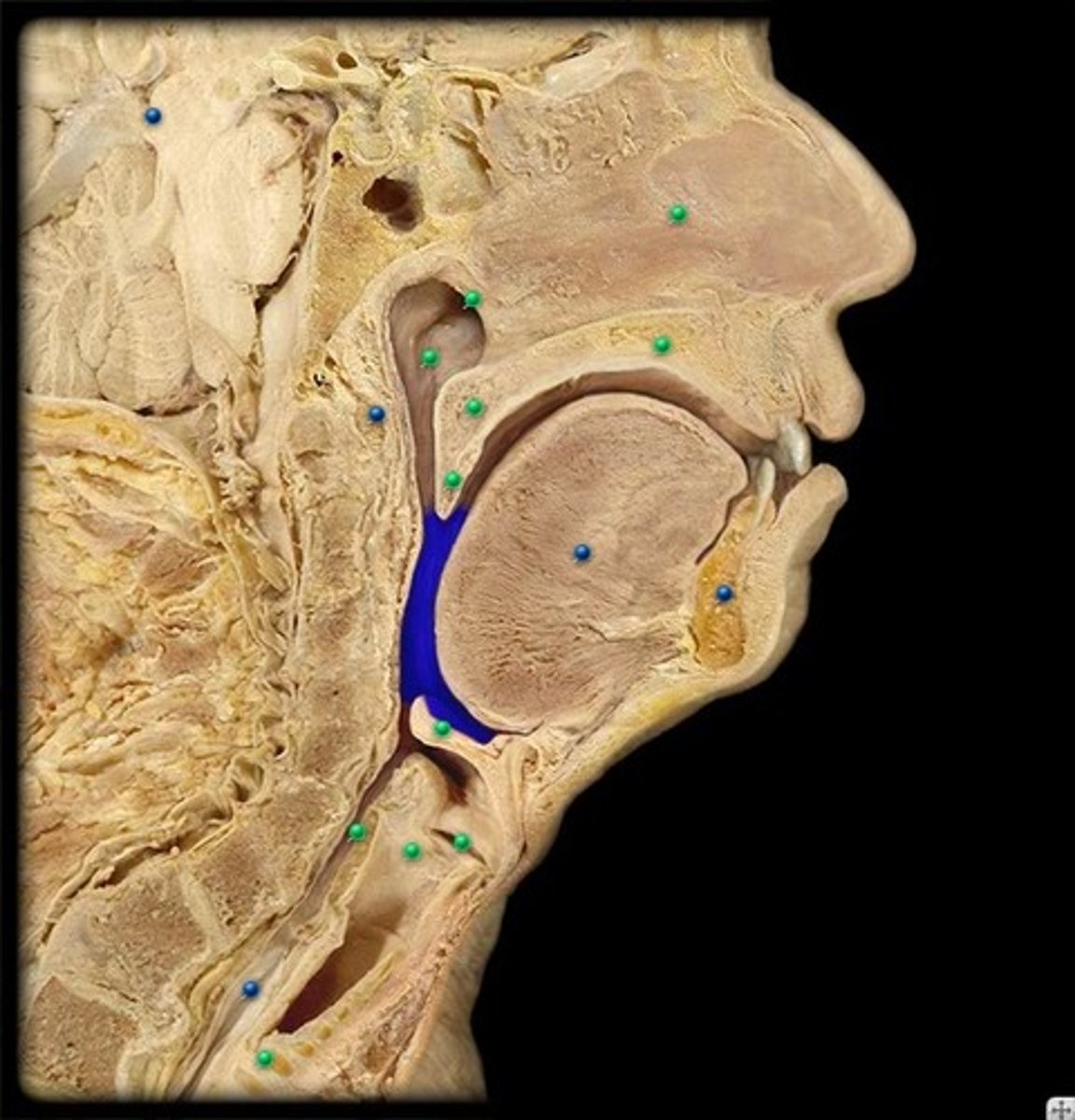
<p>central portion of the pharynx between the roof of the mouth and the upper edge of the epiglottis.</p><p>Starts from the uvula down to the epiglottis, located between the naso and laryngopharynx</p>
10
New cards

laryngopharynx

lower part of the pharynx, just below the oropharyngeal opening into the larynx and esophagus
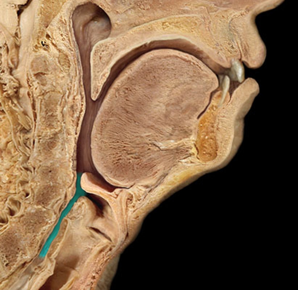
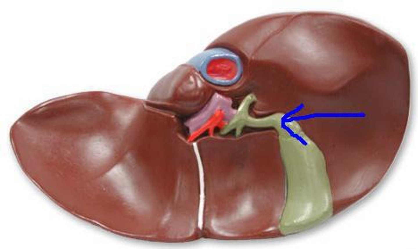
<p>lower part of the pharynx, just below the oropharyngeal opening into the larynx and esophagus</p>
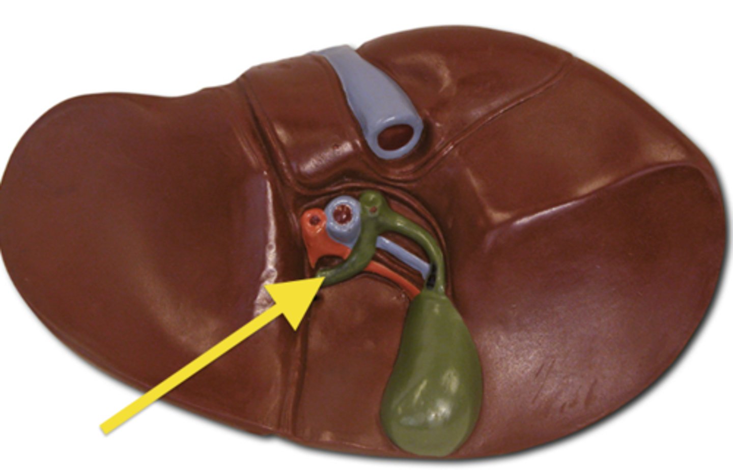
11
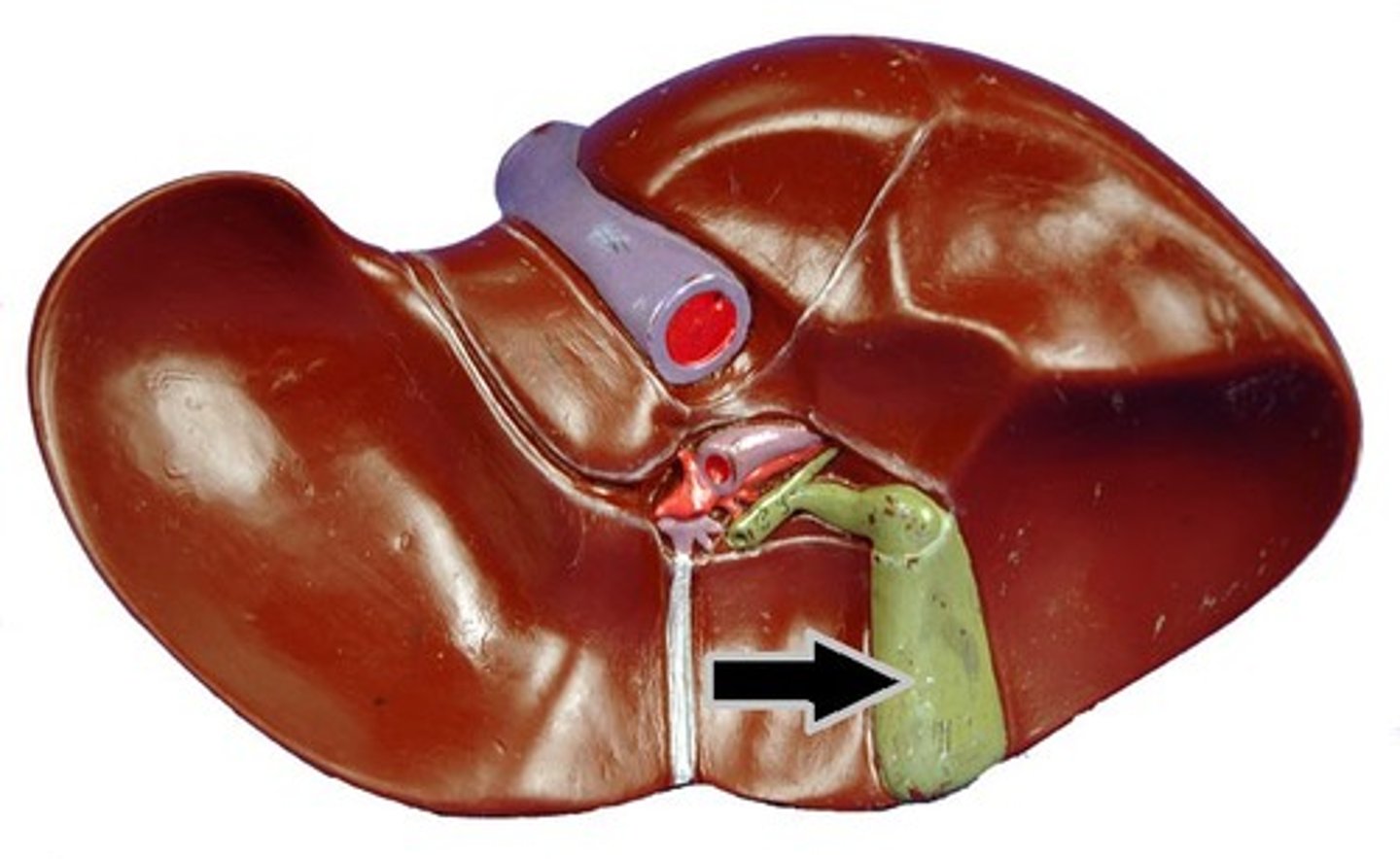
New cards

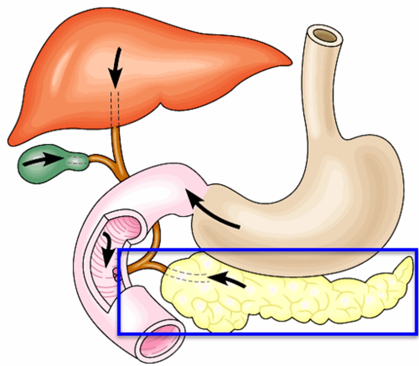
Larynx

voice box; passageway for air moving from pharynx to trachea; contains vocal cords
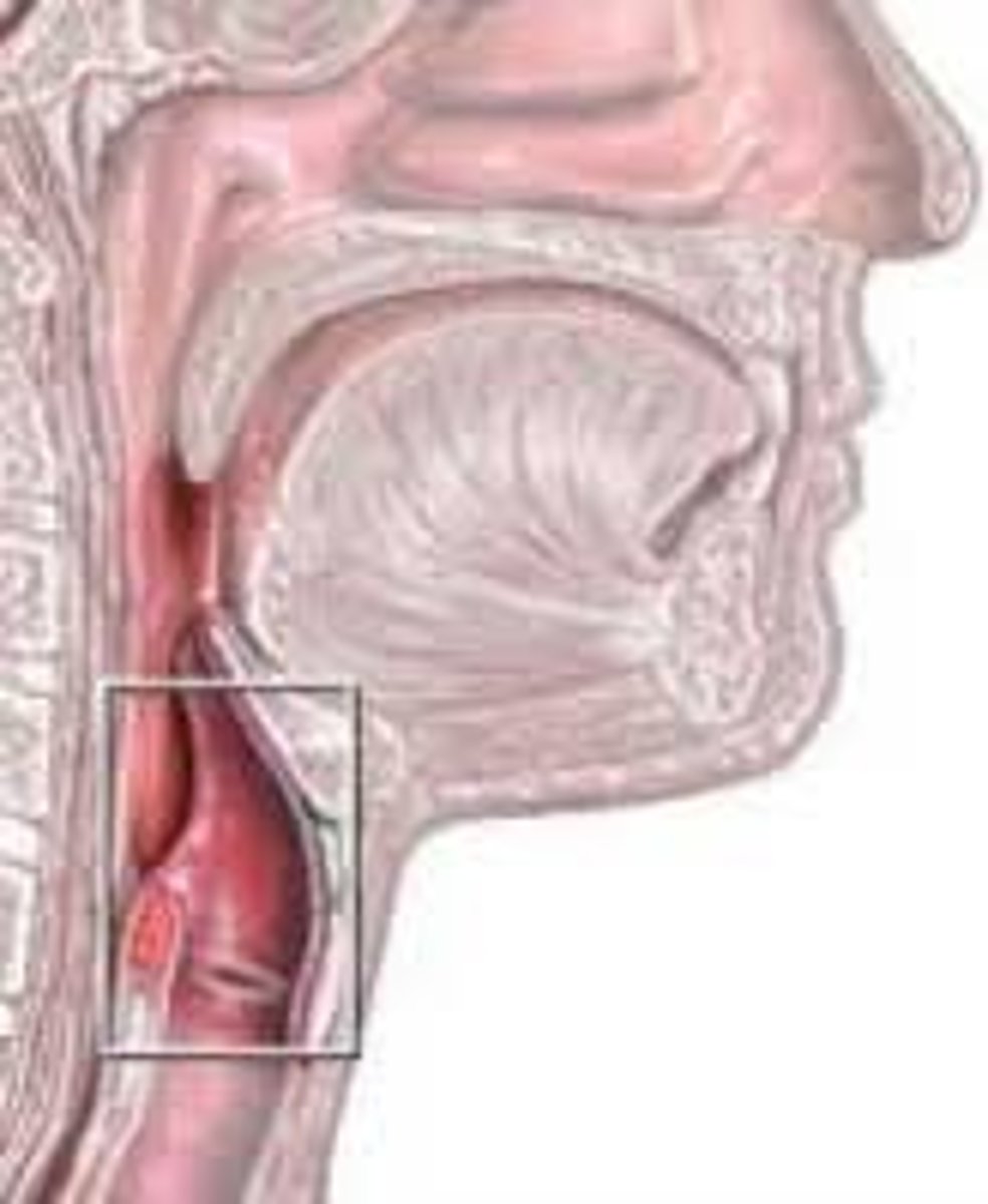
<p>voice box; passageway for air moving from pharynx to trachea; contains vocal cords</p>
12
New cards

Epiglottis

a flap of cartilage at the root of the tongue, which is depressed during swallowing to cover the opening of the windpipe.
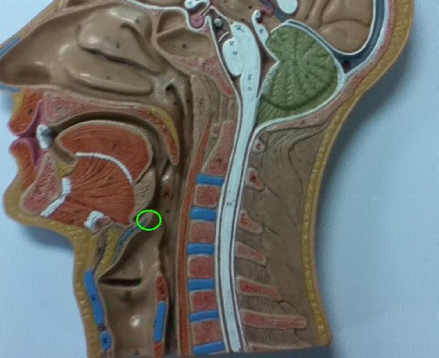
<p>a flap of cartilage at the root of the tongue, which is depressed during swallowing to cover the opening of the windpipe.</p>
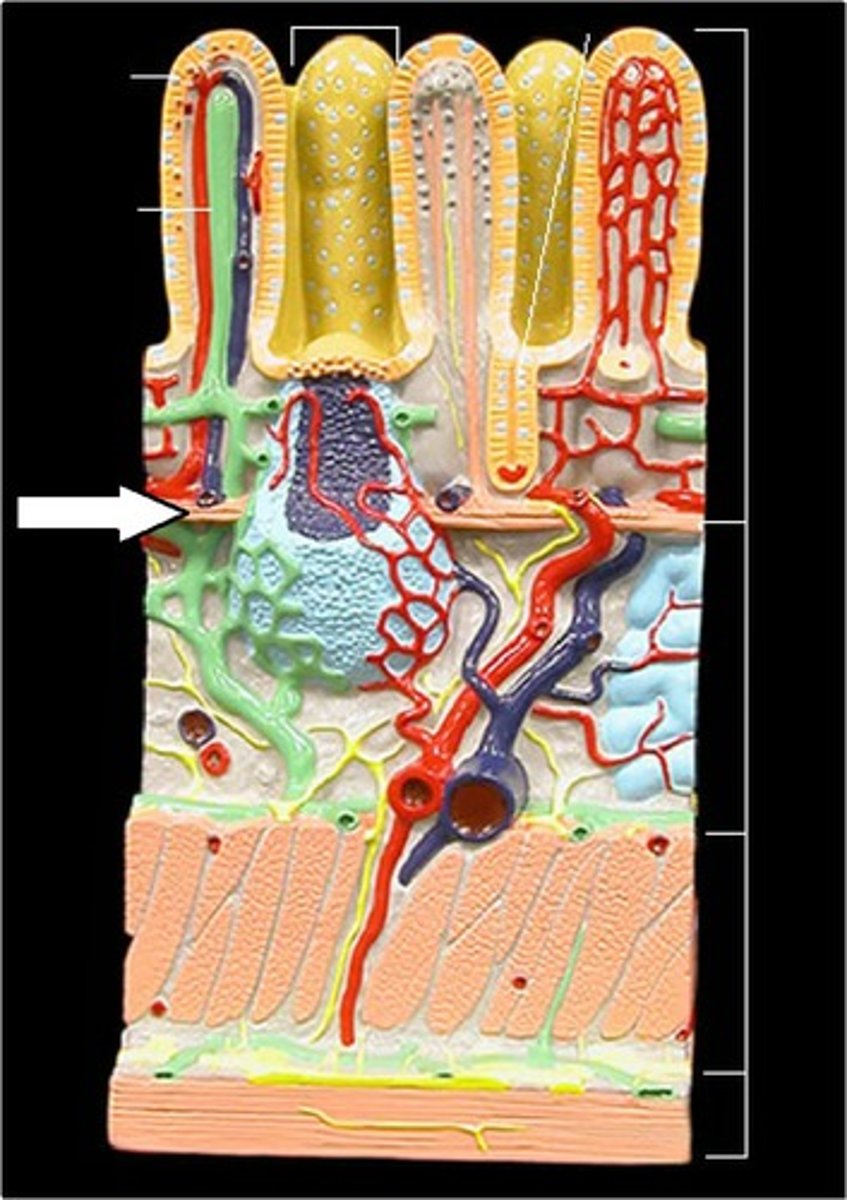
13
New cards

thyroid cartilage

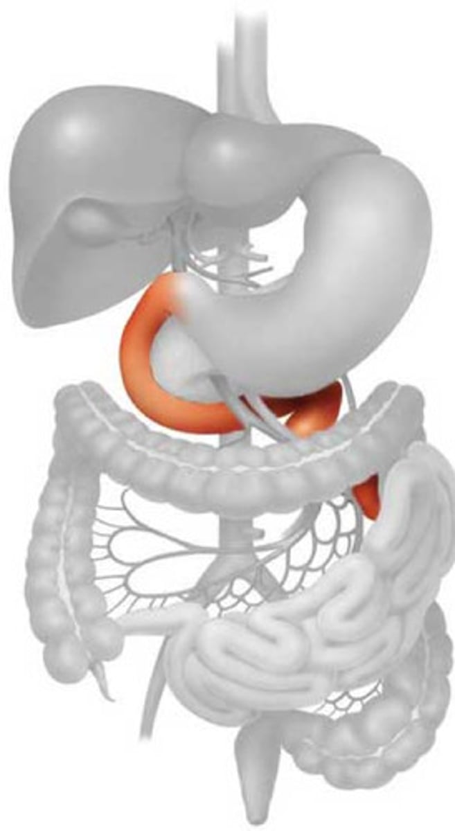
The shield-shaped plate of cartilage that sits anterior to the larynx and forms the Adam's apple.
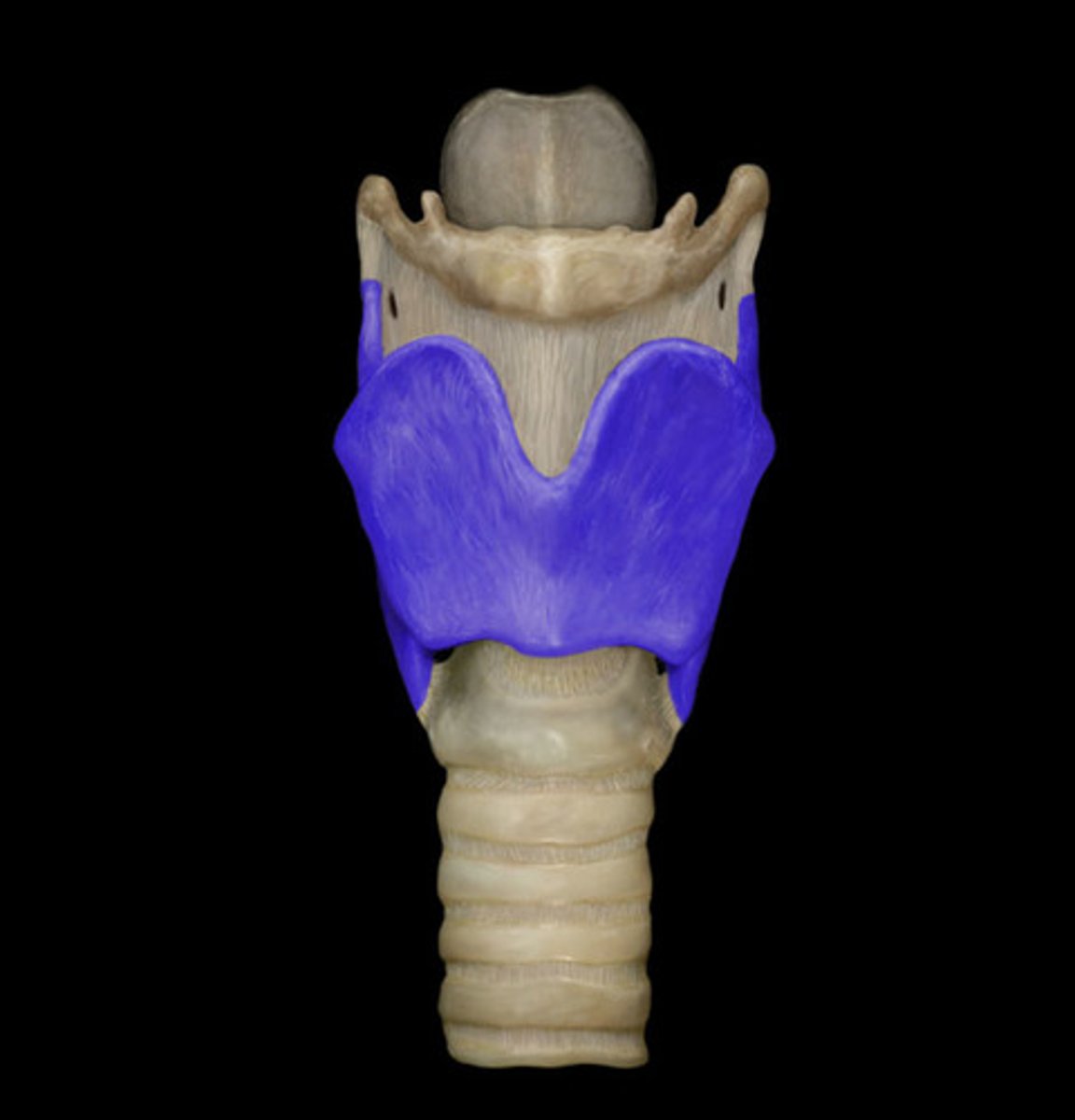
<p>The shield-shaped plate of cartilage that sits anterior to the larynx and forms the Adam's apple.</p>
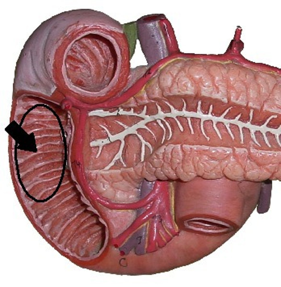
14
New cards

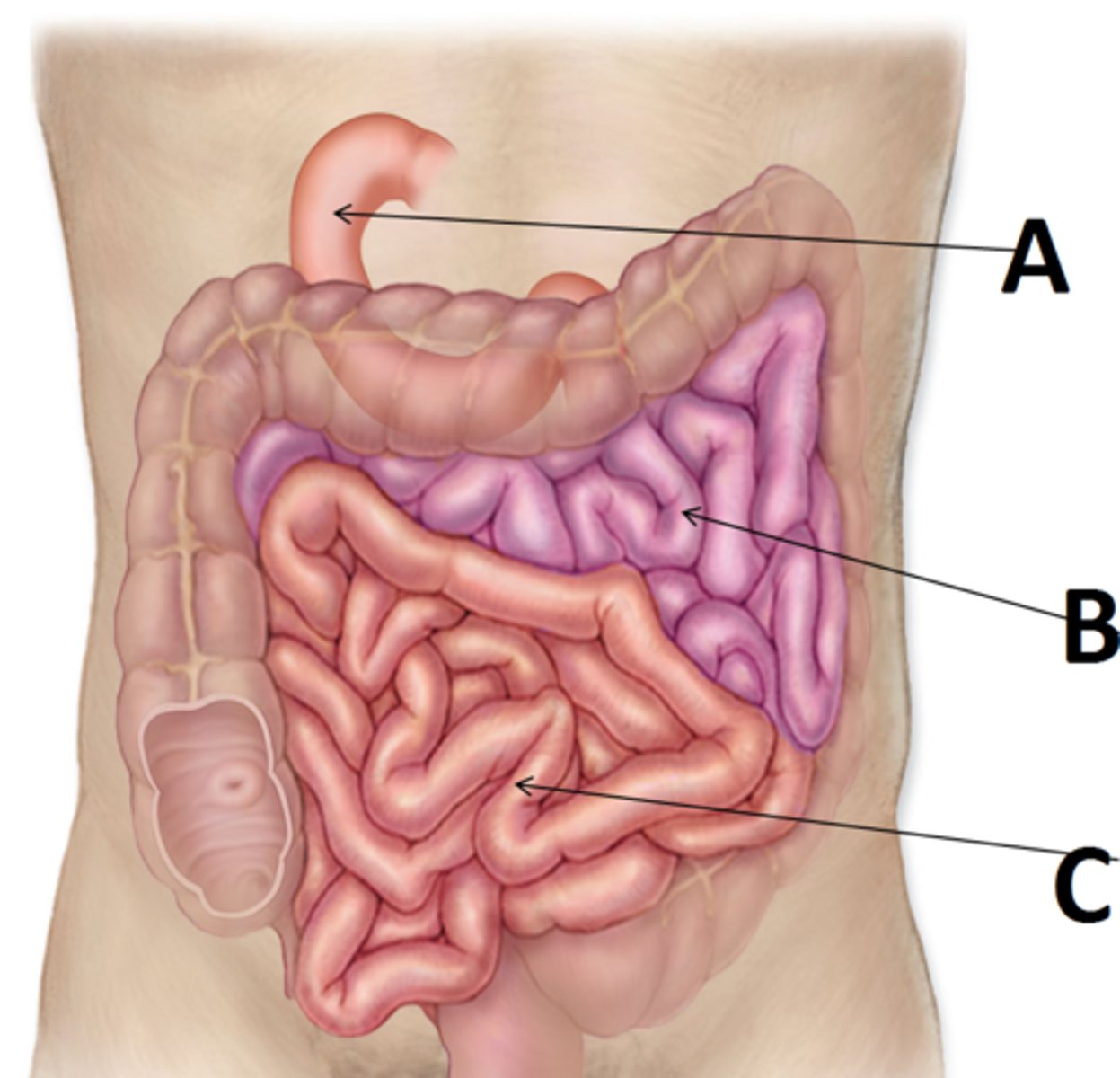
cricothyroid membrane

A thin sheet of fascia that connects the thyroid and cricoid cartilages that make up the larynx.
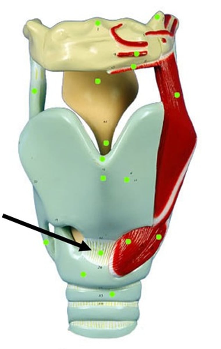
<p>A thin sheet of fascia that connects the thyroid and cricoid cartilages that make up the larynx.</p>
15
New cards

cricoid cartilage

A firm ring shaped ridge of cartilage that forms the lower part of the larynx.
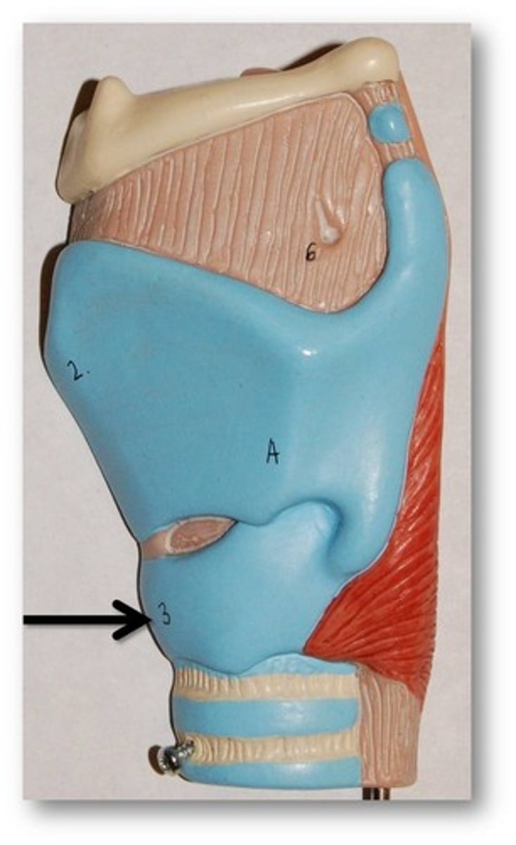
<p>A firm ring shaped ridge of cartilage that forms the lower part of the larynx.</p>
16
New cards

Arytenoid cartilage

Two small cartilages in the larynx, the movements of which abduct and adduct the vocal folds.
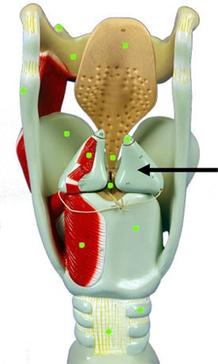
<p>Two small cartilages in the larynx, the movements of which abduct and adduct the vocal folds.</p>
17
New cards

Vestibular folds (false folds)

Located superior to the glottis and true folds, this structure functions to close off the larynx during swallowing.
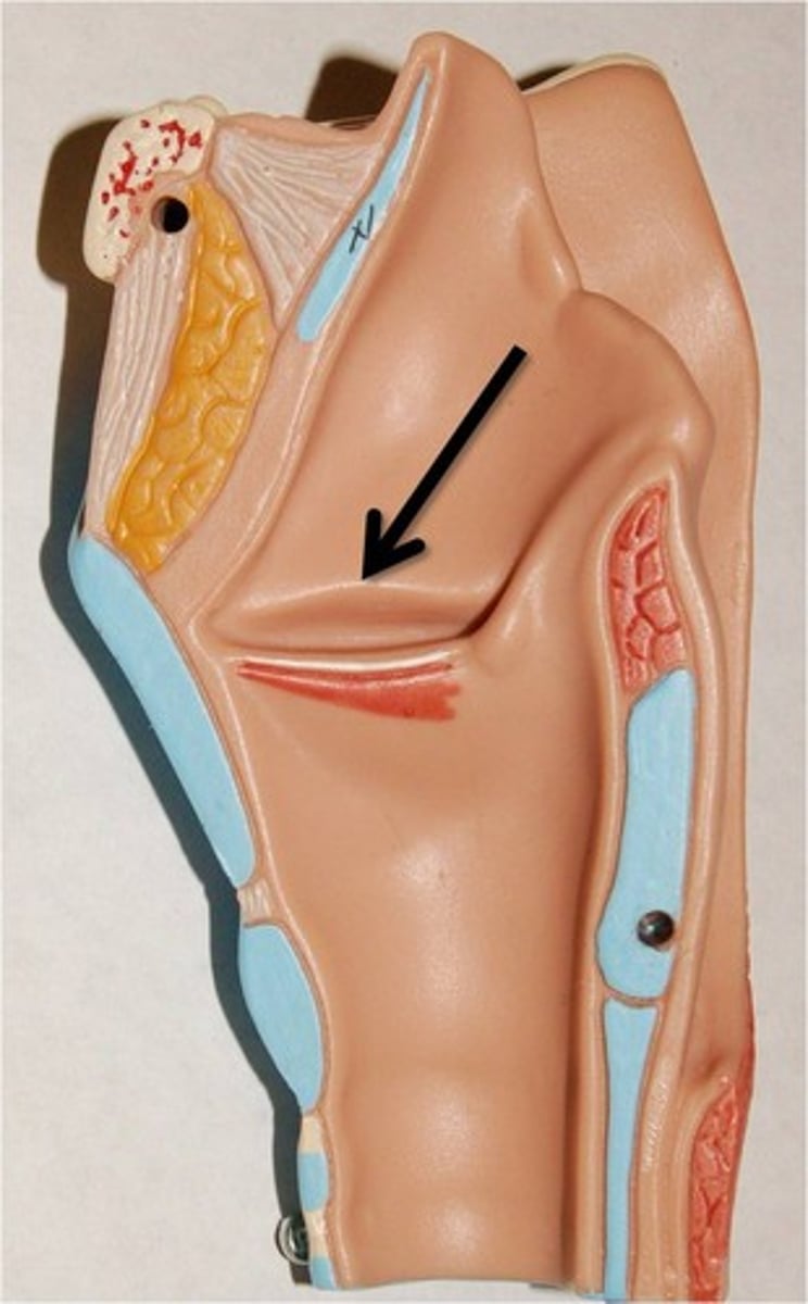
<p>Located superior to the glottis and true folds, this structure functions to close off the larynx during swallowing.</p>
18
New cards

Glottis

The opening between the vocal cords at the upper part of the larynx. functions and aids in respiration, voice production, protecting the airway, and modulating (controlling) airflow.
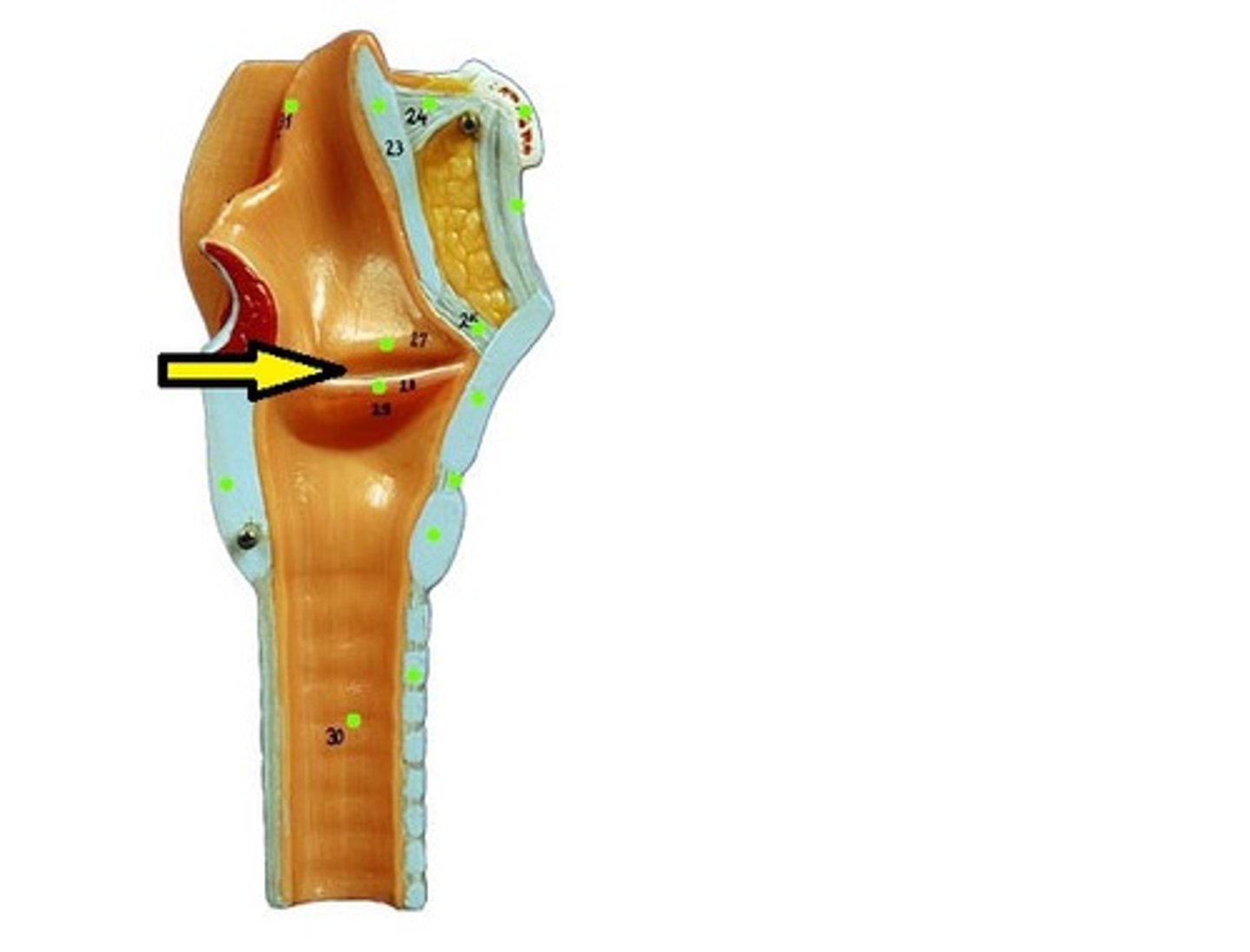
<p>The opening between the vocal cords at the upper part of the larynx. functions and aids in respiration, voice production, protecting the airway, and modulating (controlling) airflow.</p>
19
New cards

Vocal folds (true folds)

found inferior to the glottis, responsible for sound production through vibration when air passes through them, and they also act as a laryngeal sphincter to protect the airway
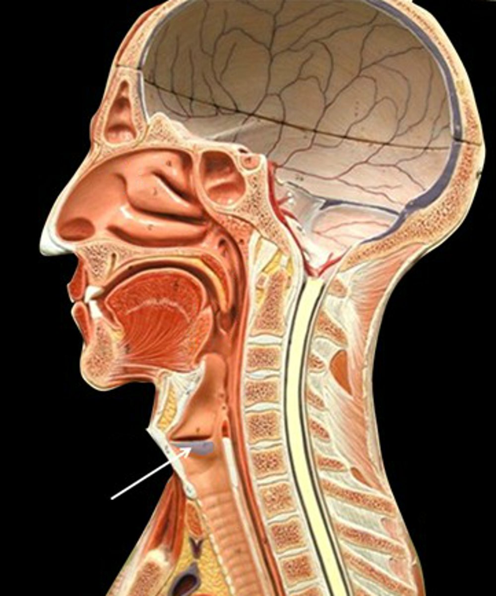
<p>found inferior to the glottis, responsible for sound production through vibration when air passes through them, and they also act as a laryngeal sphincter to protect the airway</p>
20
New cards

Trachea

commonly known as the windpipe, found inferior to the larynx, part of the lower respiratory tract and the conducting zone.

Lined by C shaped cartilage in the front and towards the back its is a concaved shape lined by the trachealis muscle.
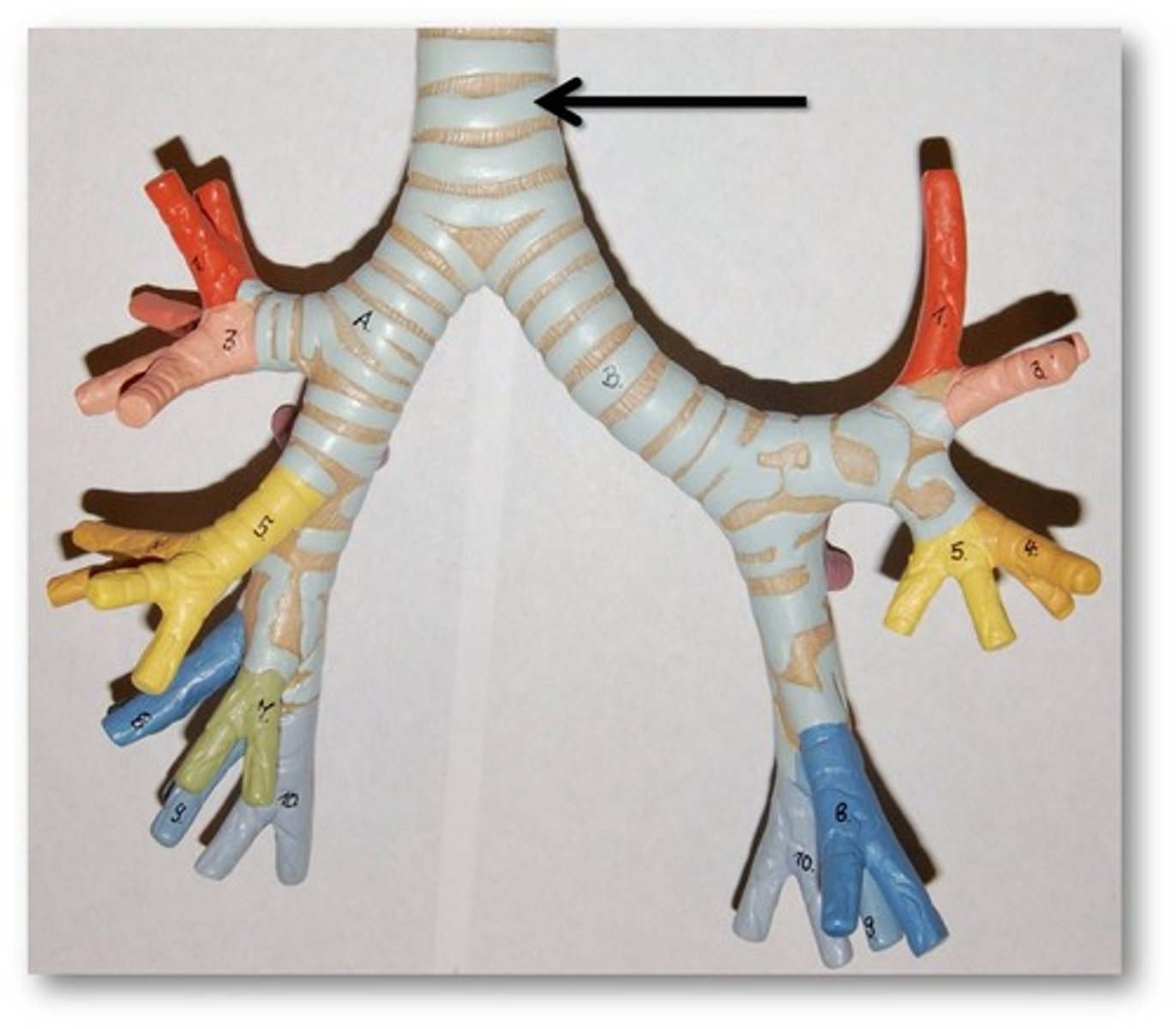
<p>commonly known as the windpipe, found inferior to the larynx, part of the lower respiratory tract and the conducting zone.</p><p>Lined by C shaped cartilage in the front and towards the back its is a concaved shape lined by the trachealis muscle.</p>
21
New cards

Primary Bronchus

a pair of branches of the trachea that lead to the right and left lung; consist of incomplete rings of cartilage and are lined by pseudostratified ciliated columnar epithelium
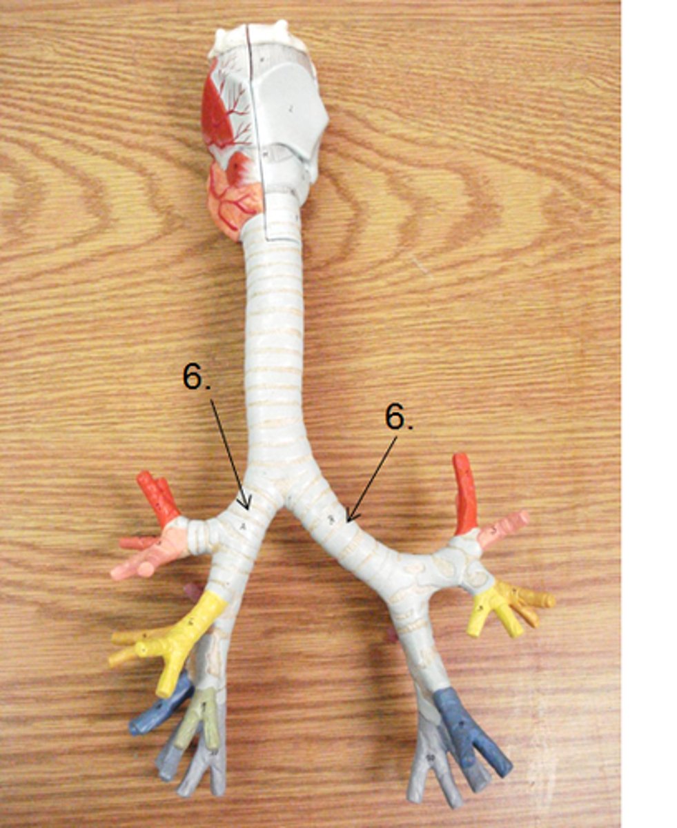
<p>a pair of branches of the trachea that lead to the right and left lung; consist of incomplete rings of cartilage and are lined by pseudostratified ciliated columnar epithelium</p>
22
New cards

Secondary bronchus

branches of the primary bronchi which supply each lobe of lung; there are 2 in the left lung and 3 in the right lung
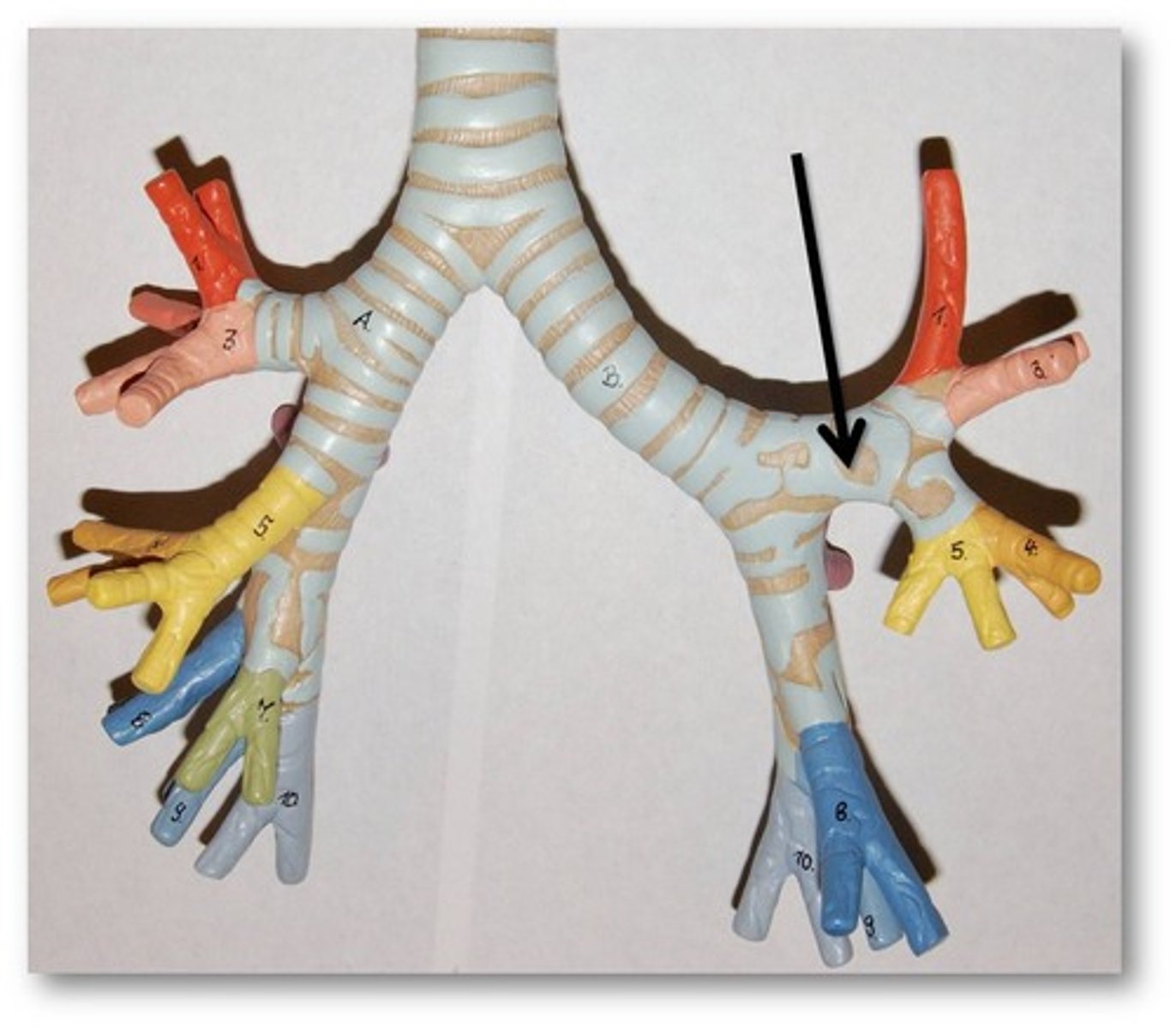
<p>branches of the primary bronchi which supply each lobe of lung; there are 2 in the left lung and 3 in the right lung</p>
23
New cards

Tertiary bronchus

Extends from the secondary bronchus and conducts air to each lobule of the lungs.
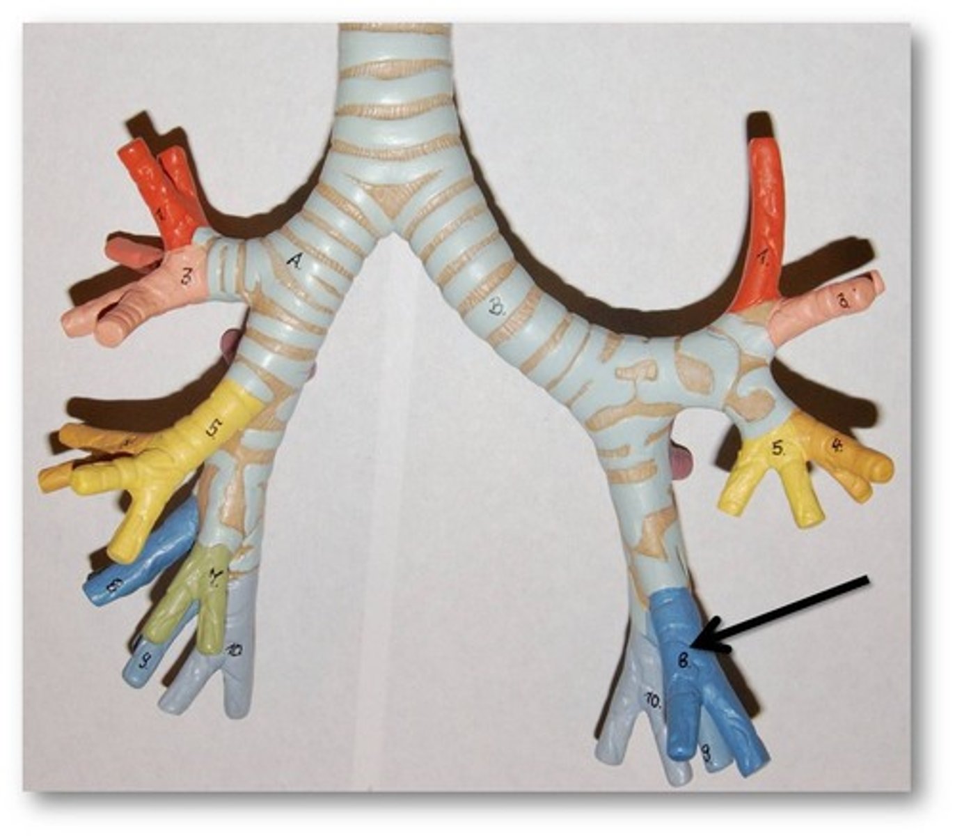
<p>Extends from the secondary bronchus and conducts air to each lobule of the lungs.</p>
24
New cards

Right lung

contains 3 lobes (superior, middle, and inferior), and known as the largest of the 2 lungs, also contains 2 fissures (horizontal and oblique)
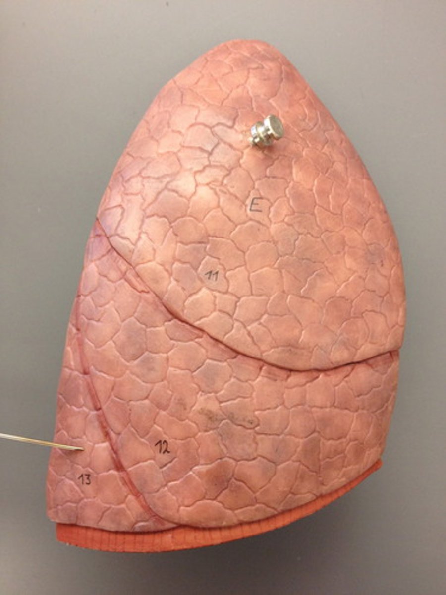
<p>contains 3 lobes (superior, middle, and inferior), and known as the largest of the 2 lungs, also contains 2 fissures (horizontal and oblique)</p>
25
New cards

superior lobe of right lung

Separated from middle lobe by horizontal fissure: receives secondary bronchus
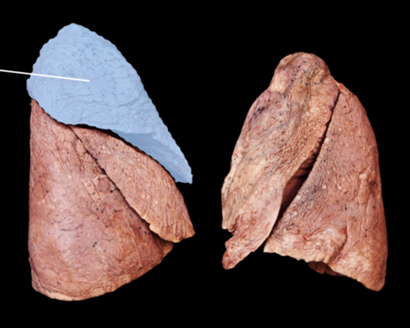
<p>Separated from middle lobe by horizontal fissure: receives secondary bronchus</p>
26
New cards

middle lobe of right lung

Separated from superior lobe by horizontal fissure, and lower lobe by oblique fissure: receives secondary bronchus
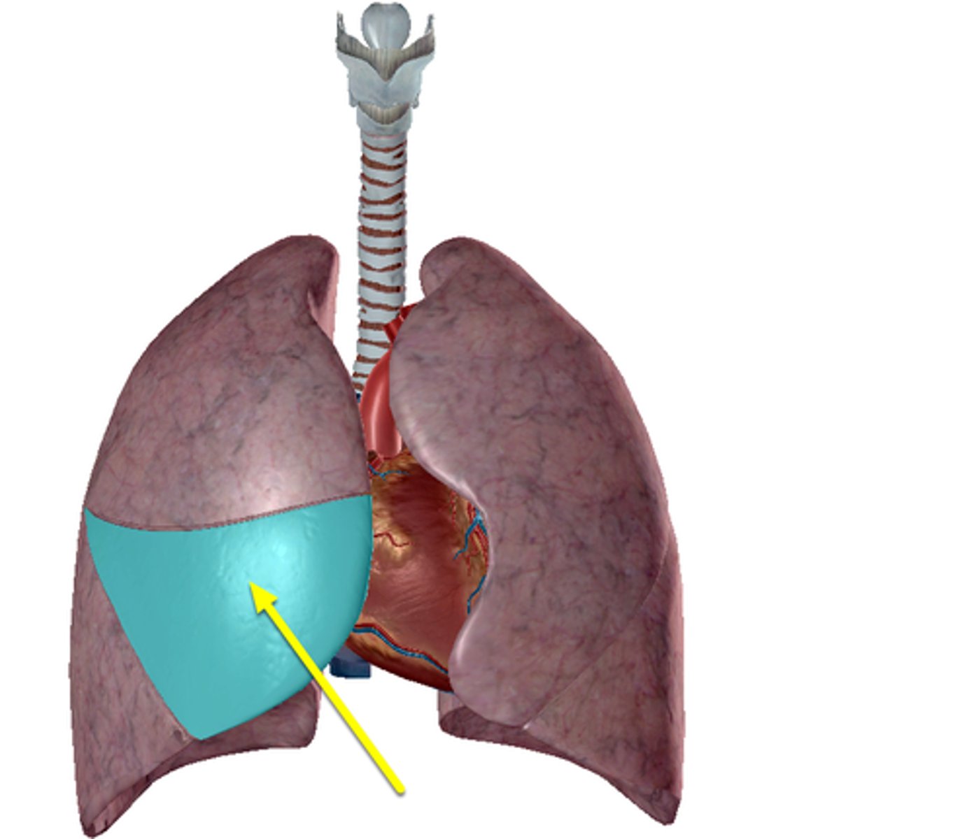
<p>Separated from superior lobe by horizontal fissure, and lower lobe by oblique fissure: receives secondary bronchus</p>
27
New cards

inferior lobe of right lung

Separated from middle lobe by oblique fissure: receives secondary bronchus
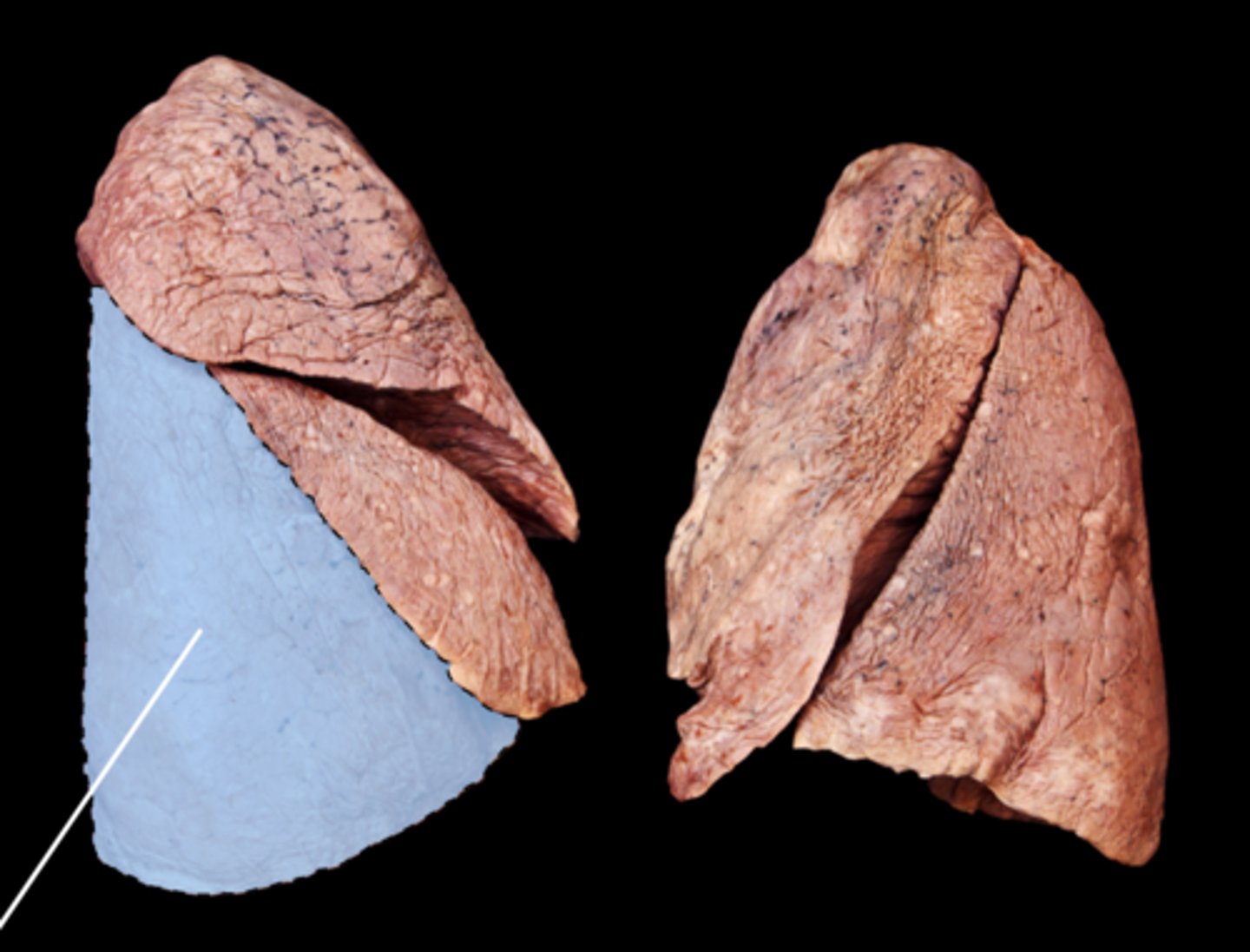
<p>Separated from middle lobe by oblique fissure: receives secondary bronchus</p>
28
New cards

horizontal fissure of right lung

Separates upper and middle lobes, joins oblique fissure laterally
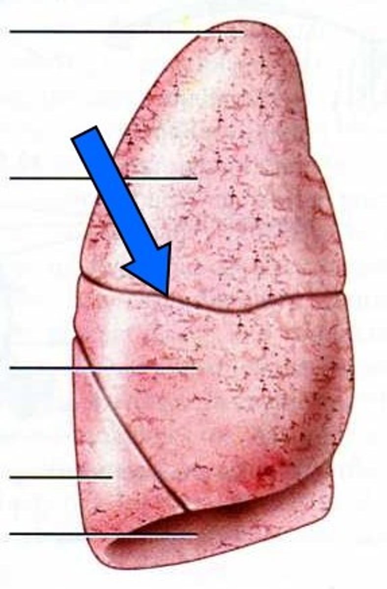
<p>Separates upper and middle lobes, joins oblique fissure laterally</p>
29
New cards

oblique fissure of right lung

separates the middle lobe from the inferior lobe
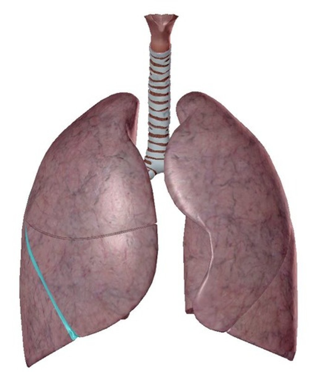
<p>separates the middle lobe from the inferior lobe</p>
30
New cards

superior lobe of left lung

the upper portion of the left lung, situated above the oblique fissure
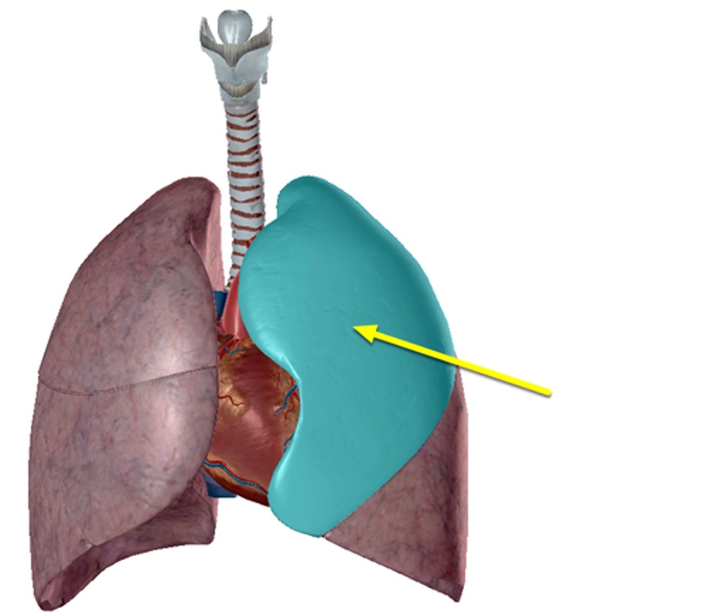
<p>the upper portion of the left lung, situated above the oblique fissure</p>
31
New cards

inferior lobe of left lung

Rests on diaphragm, separated from upper by oblique fissure: receives secondary bronchus
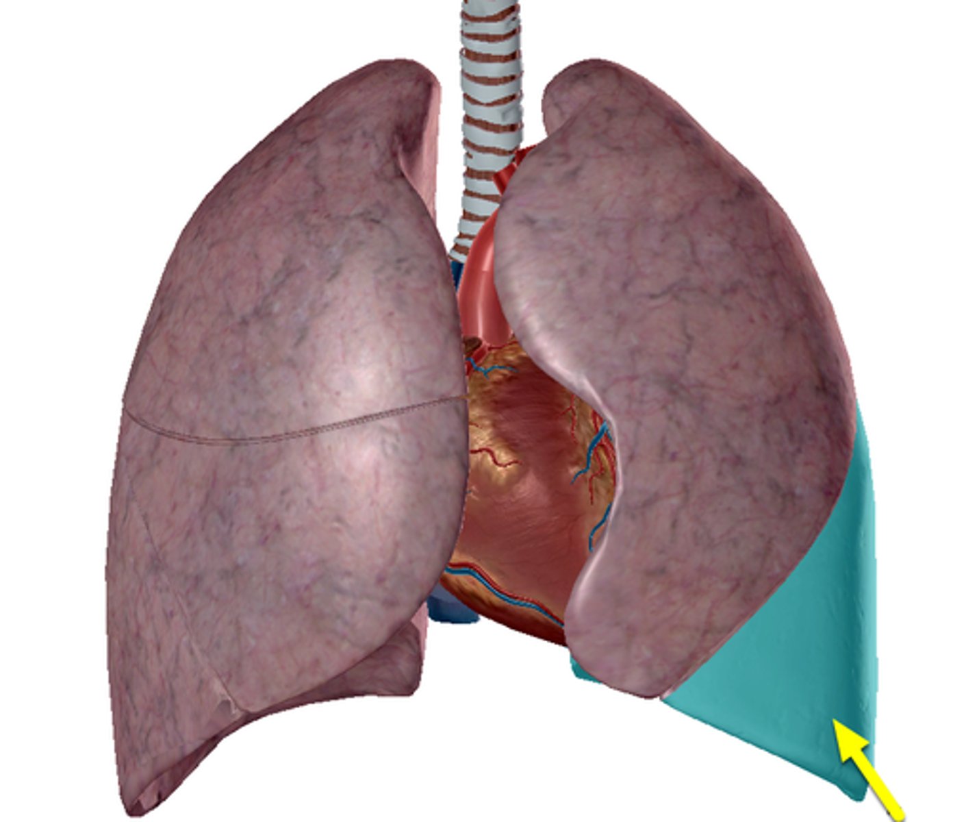
<p>Rests on diaphragm, separated from upper by oblique fissure: receives secondary bronchus</p>
32
New cards

oblique fissure of left lung

Located on the left lung, it separates the superior and inferior lobes
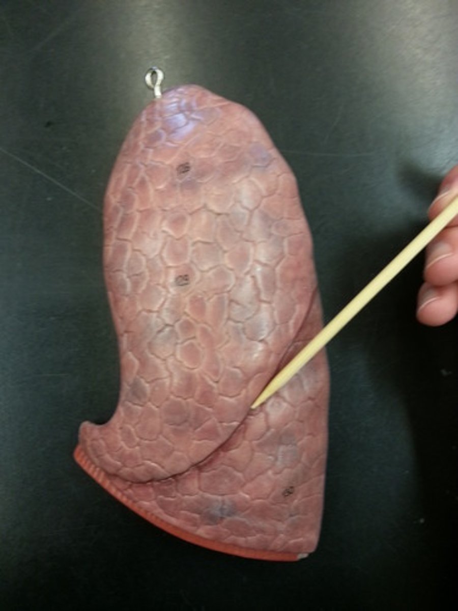
<p>Located on the left lung, it separates the superior and inferior lobes</p>
33
New cards

Diaphragm

a dome-shaped, muscular partition separating the thorax from the abdomen in mammals. It plays a major role in breathing, as its contraction increases the volume of the thorax and allows the lungs to inflate.
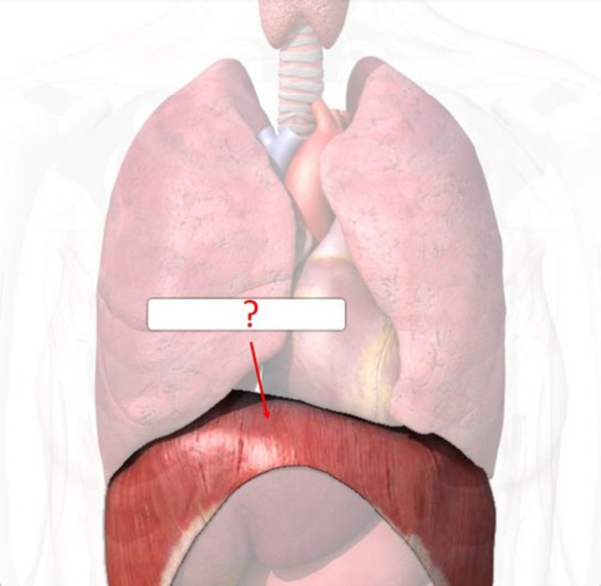
<p>a dome-shaped, muscular partition separating the thorax from the abdomen in mammals. It plays a major role in breathing, as its contraction increases the volume of the thorax and allows the lungs to inflate.</p>
34
New cards

internal intercostal muscles

muscles responsible in lowering the rib cage during forced expiration
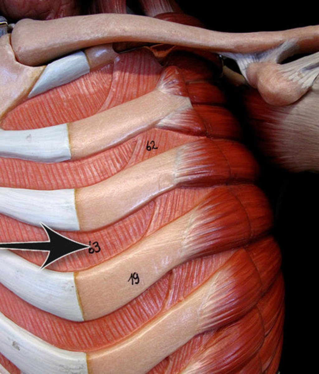
<p>muscles responsible in lowering the rib cage during forced expiration</p>
35
New cards

external intercoastal muscles

Muscles assist with breathing by moving the ribs up and outward to increase the total volume in the thoracic cavity.
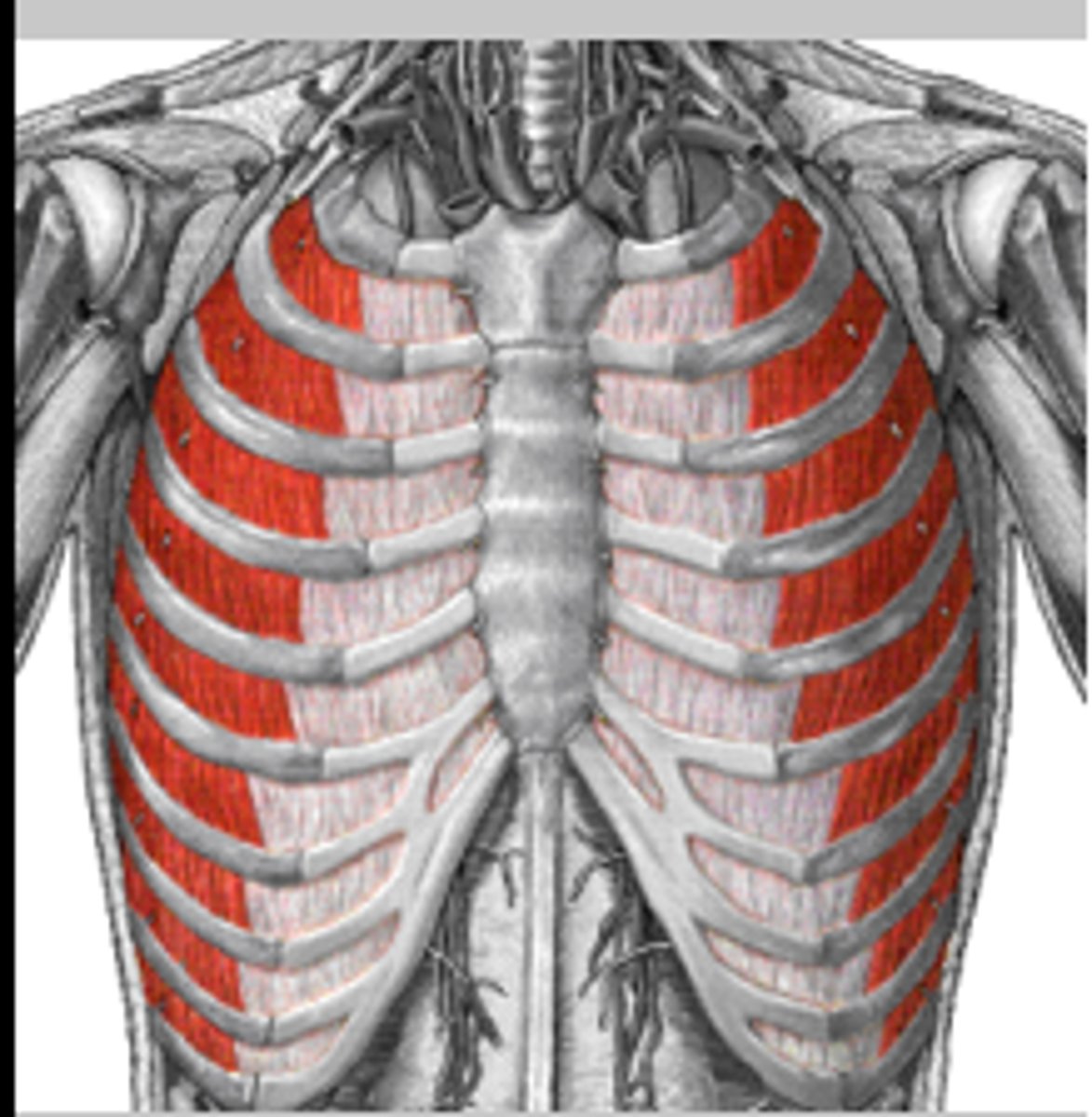
<p>Muscles assist with breathing by moving the ribs up and outward to increase the total volume in the thoracic cavity.</p>
36
New cards

Phrenic Nerve

located on both right and left sides this nerve directly innervates the diaphragm. Helps to provide motor function to the diaphragm. Tells the diaphragm to contract and relax.
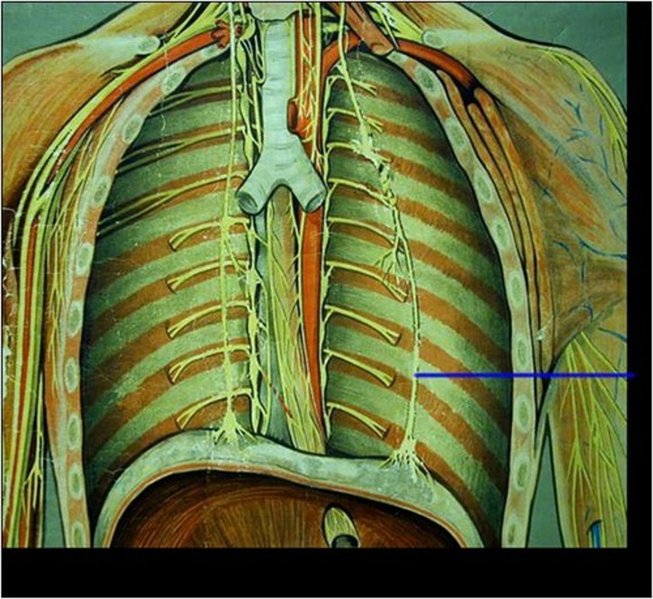
<p>located on both right and left sides this nerve directly innervates the diaphragm. Helps to provide motor function to the diaphragm. Tells the diaphragm to contract and relax.</p>
37
New cards

Orbicularis Oris

round shaped muscle that surrounds the lips,

closes the lips, purses, and protrudes lips,

Innervated by Facial nerve
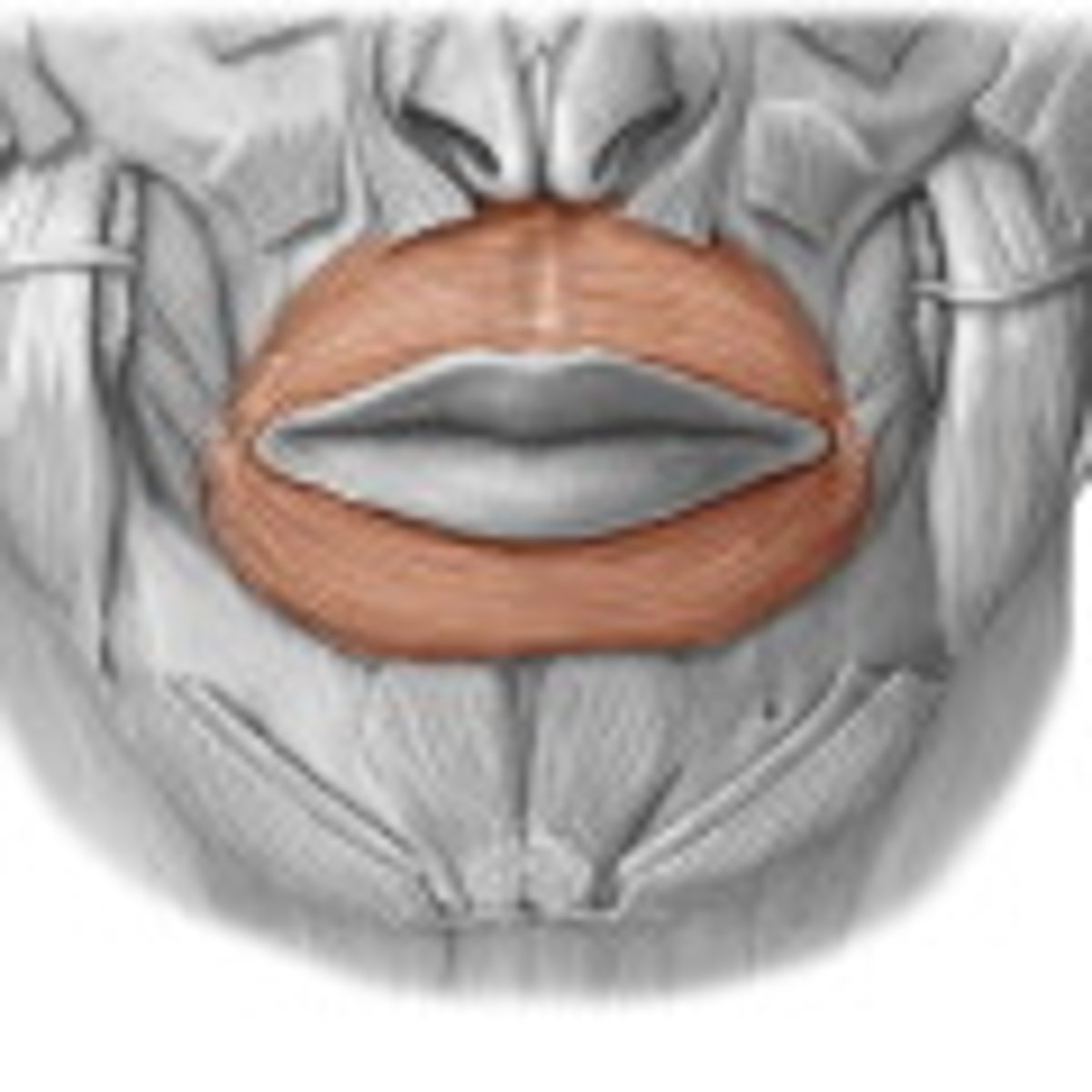
<p>round shaped muscle that surrounds the lips,</p><p>closes the lips, purses, and protrudes lips,</p><p>Innervated by Facial nerve</p>
38
New cards

Buccinator

Muscle responsible for compressing the cheeks,

innervated by Facial nerve
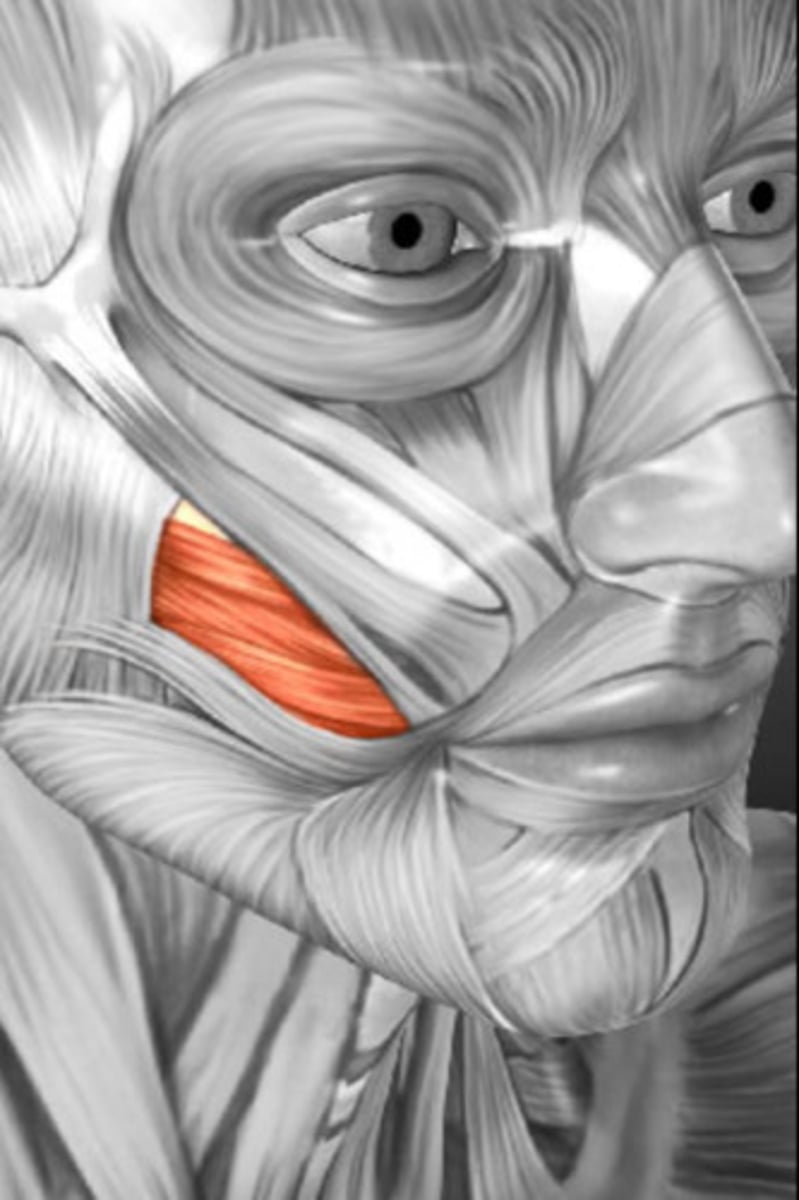
<p>Muscle responsible for compressing the cheeks,</p><p>innervated by Facial nerve</p>
39
New cards

Temporalis

large muscle found along the temporal bone,

responsible for closing the jaw

innervated by the Trigeminal nerve
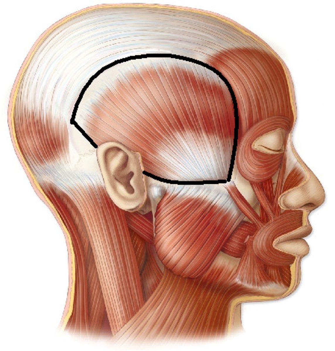
<p>large muscle found along the temporal bone,</p><p>responsible for closing the jaw</p><p>innervated by the Trigeminal nerve</p>
40
New cards

Masseter

One of the strongest muscles in the body,

Prime mover of jaw closure, elevates mandible,

Innervated by the Trigeminal nerve
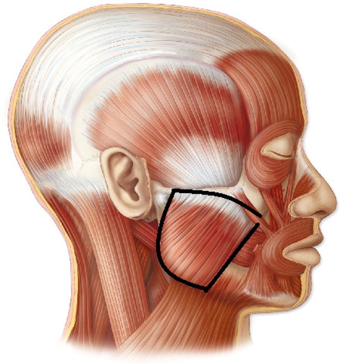
<p>One of the strongest muscles in the body,</p><p>Prime mover of jaw closure, elevates mandible,</p><p>Innervated by the Trigeminal nerve</p>
41
New cards

Central incisor

the first tooth starting from the midline; used to cut or bite the food that is ingested
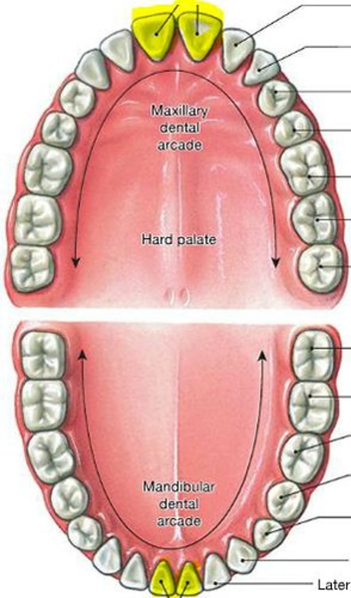
<p>the first tooth starting from the midline; used to cut or bite the food that is ingested</p>
42
New cards

Lateral inscisors

the two teeth located to the right and left sides of the central incisors, Function to shear or cut food during mastication
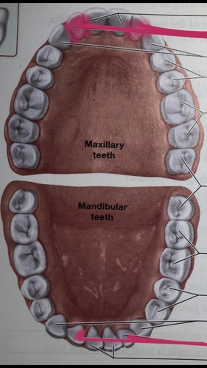
<p>the two teeth located to the right and left sides of the central incisors, Function to shear or cut food during mastication</p>
43
New cards

Canines

Fanglike teeth in front of the premolars that rip and tear food.
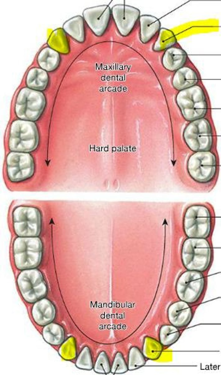
<p>Fanglike teeth in front of the premolars that rip and tear food.</p>
44
New cards

First premolars

one of two transitional teeth (also called bicuspids) located between the canine and molar teeth
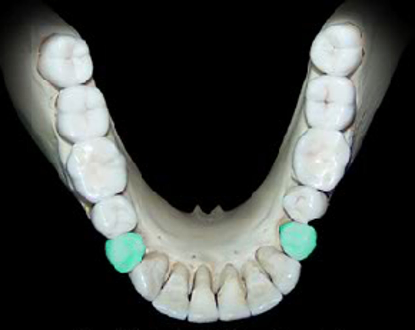
<p>one of two transitional teeth (also called bicuspids) located between the canine and molar teeth</p>
45
New cards

Second premolars

known as a bicuspid, is a tooth located between the first premolar and the first molar in the upper and lower jaws
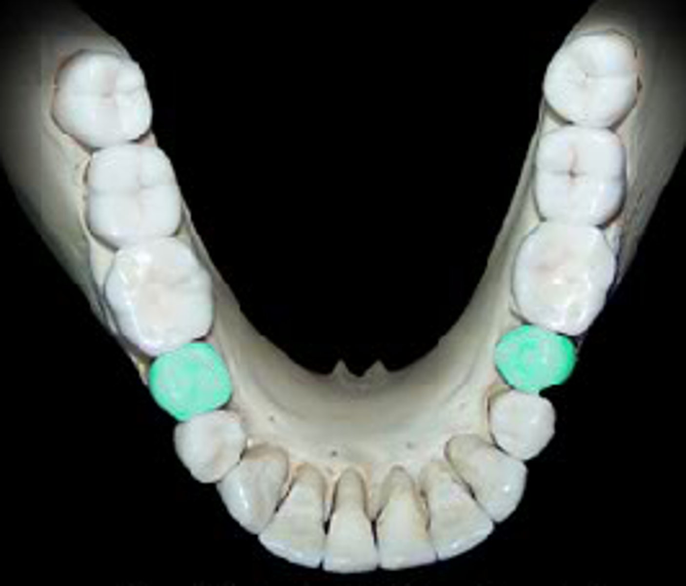
<p>known as a bicuspid, is a tooth located between the first premolar and the first molar in the upper and lower jaws</p>
46
New cards

First molars

the first permanent teeth to erupt in the mouth, function to grind down food into small pieces
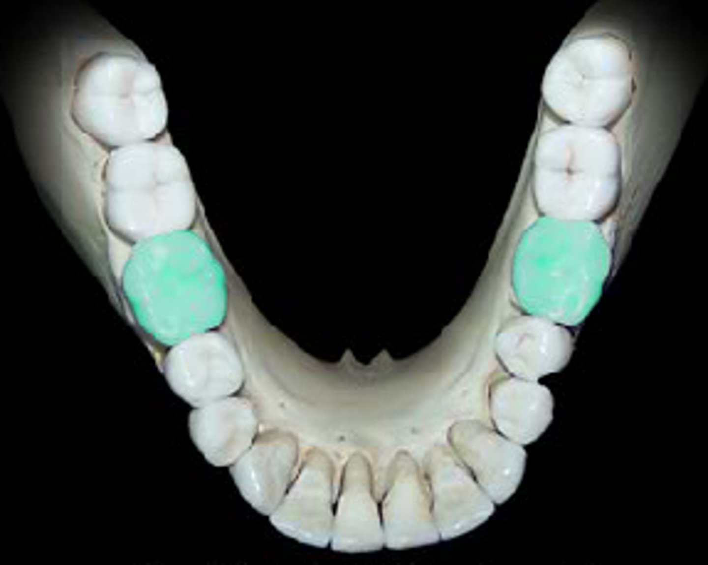
<p>the first permanent teeth to erupt in the mouth, function to grind down food into small pieces</p>
47
New cards

Second molars

sandwhiched between the 1st molar and the wisdom teeth
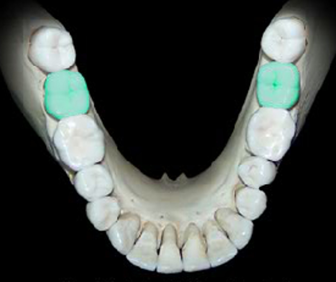
<p>sandwhiched between the 1st molar and the wisdom teeth</p>
48
New cards

Third molars

Known as the wisdom teeth, the last row of your molars
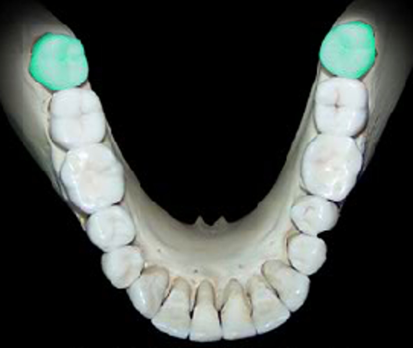
<p>Known as the wisdom teeth, the last row of your molars</p>
49
New cards

Hard palate

Bony part of the mouth also known as the roof of the mouth.

Separates the oral cavity from the nasal cavity.

Comprised of the palatine bone and the maxilla.
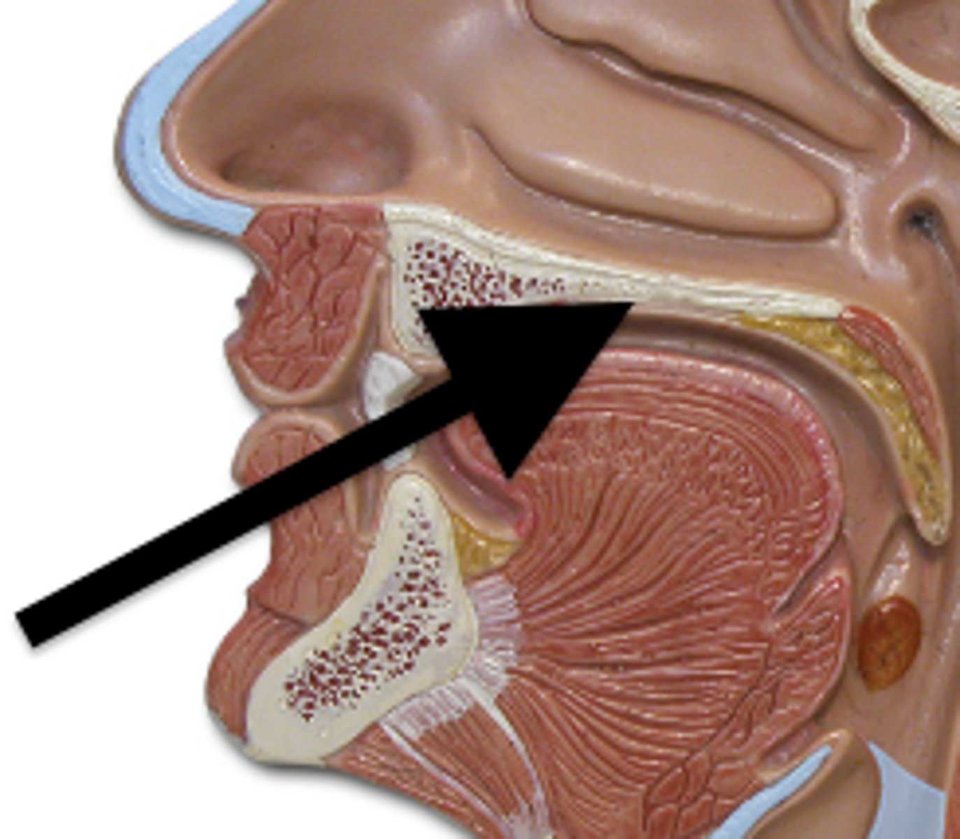
<p>Bony part of the mouth also known as the roof of the mouth.</p><p>Separates the oral cavity from the nasal cavity.</p><p>Comprised of the palatine bone and the maxilla.</p>
50
New cards

Soft palate

the fleshy, flexible part toward the back of the roof of the mouth. functions to separate the oral and nasal cavities, playing a crucial role in feeding, breathing, swallowing, and speech.
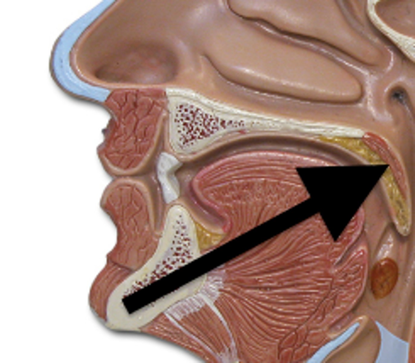
<p>the fleshy, flexible part toward the back of the roof of the mouth. functions to separate the oral and nasal cavities, playing a crucial role in feeding, breathing, swallowing, and speech.</p>
51
New cards

Uvula

small projection hanging from the back middle edge of the soft palate. Functions to prevent food or drink from entering the nasal cavity
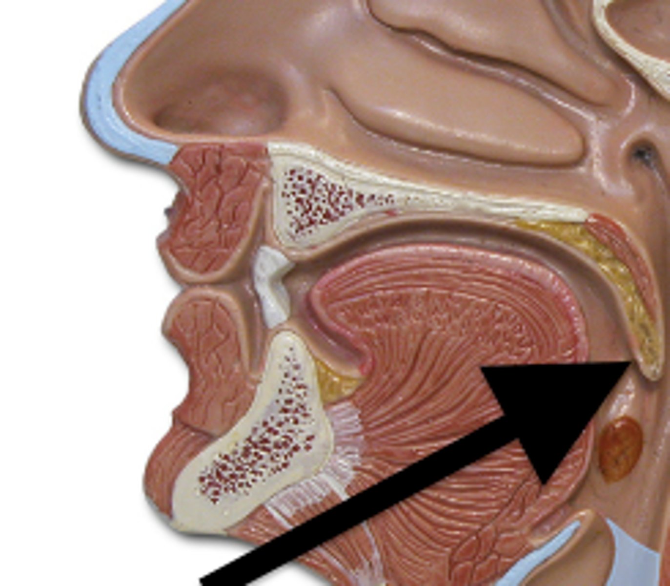
<p>small projection hanging from the back middle edge of the soft palate. Functions to prevent food or drink from entering the nasal cavity</p>
52
New cards

Parotid gland

paired salivary glands that sit just in front of the ears. Functions to produce/ secrete saliva, also helps to lubricate the mouth. Secretes amylase to help breakdown starches and carbohydrates.
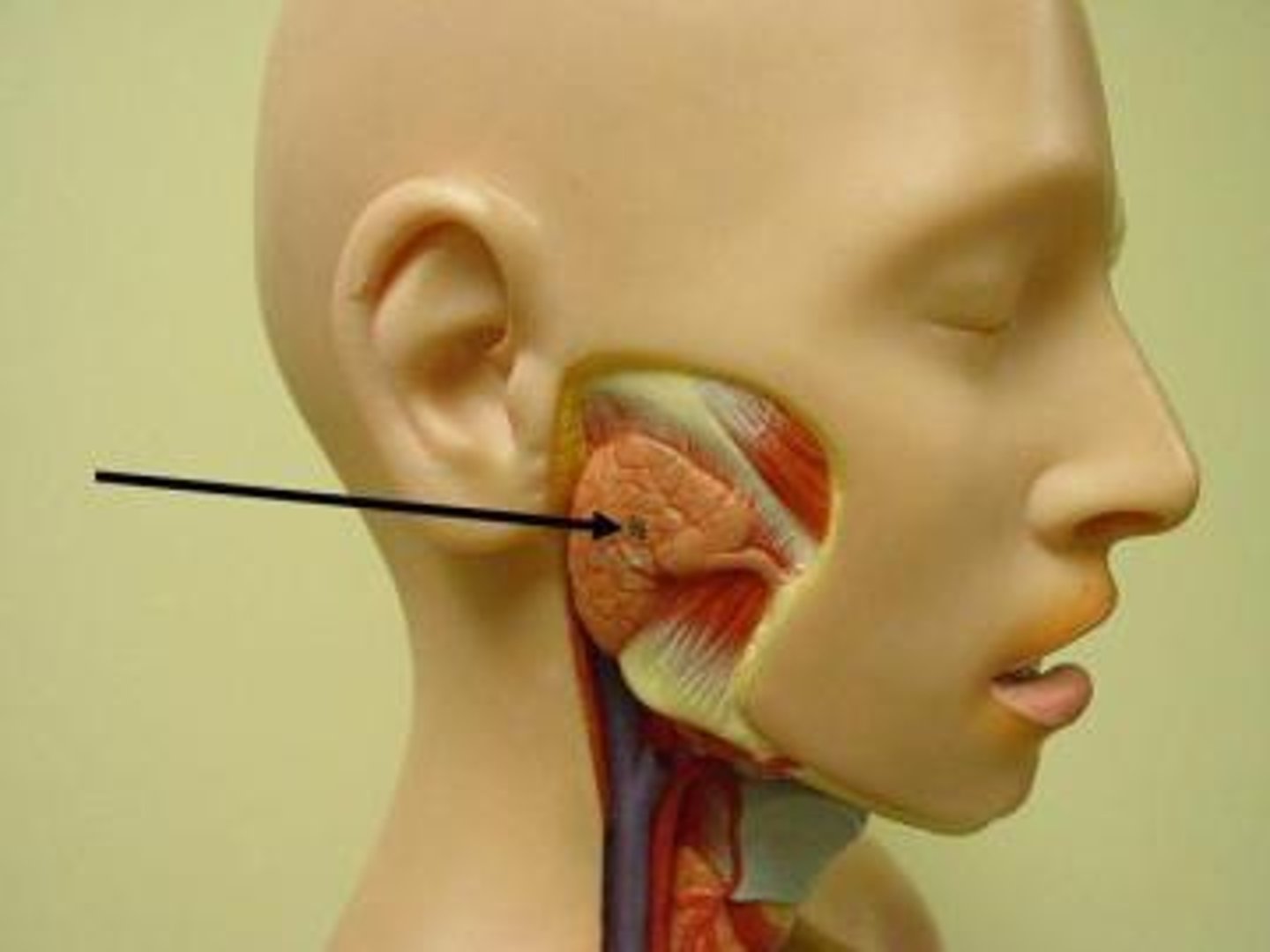
<p>paired salivary glands that sit just in front of the ears. Functions to produce/ secrete saliva, also helps to lubricate the mouth. Secretes amylase to help breakdown starches and carbohydrates.</p>
53
New cards

Submandibular gland

a salivary gland found below the lower jaw on either side that produces most of the nocturnal saliva, secretes amylase to help breakdown starches and carbohydrates.
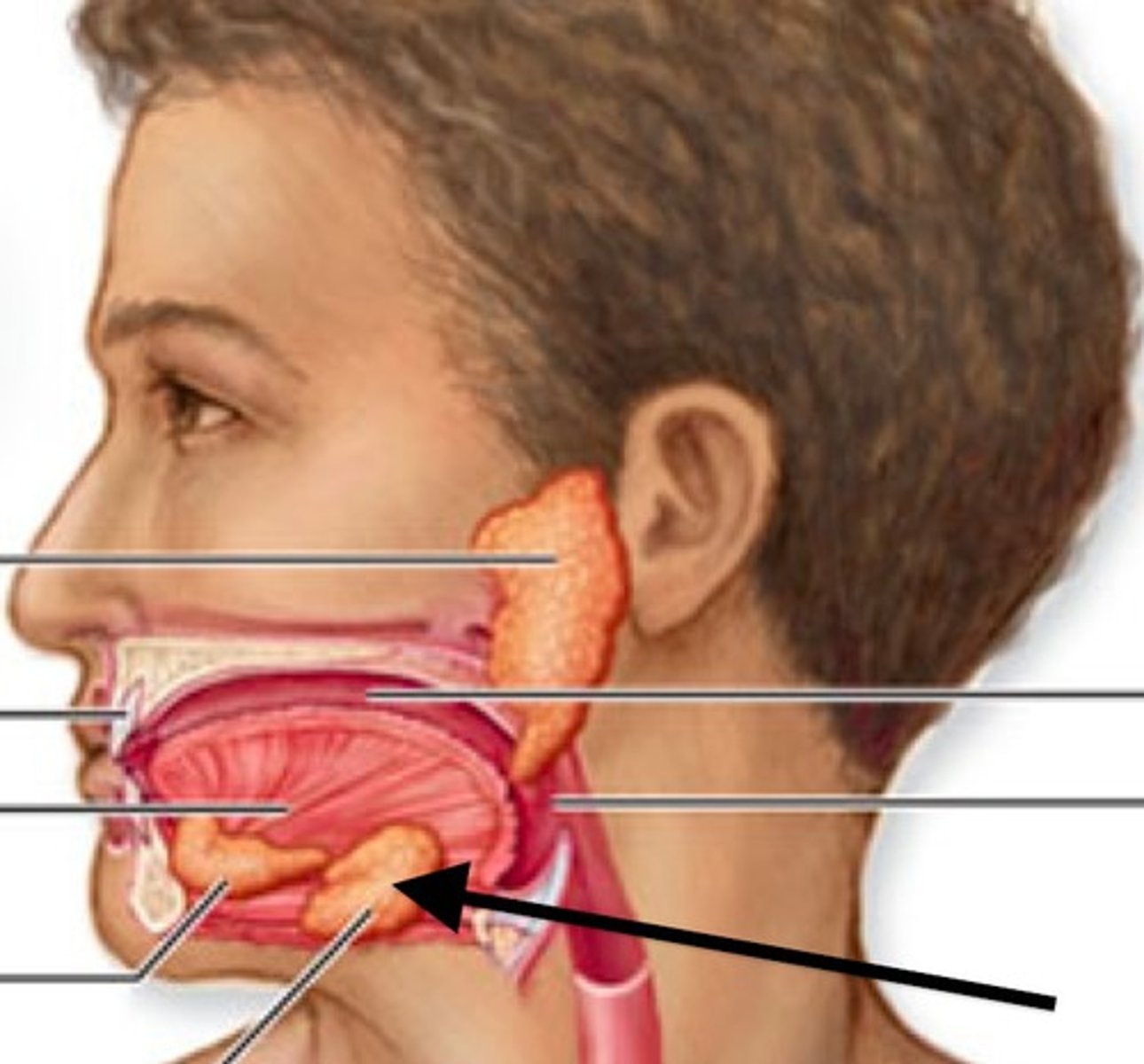
<p>a salivary gland found below the lower jaw on either side that produces most of the nocturnal saliva, secretes amylase to help breakdown starches and carbohydrates.</p>
54
New cards

Sublingual gland

smallest salivary gland found under the tongue, primarily secretes mucins, and a minimal amount of saliva and amylase
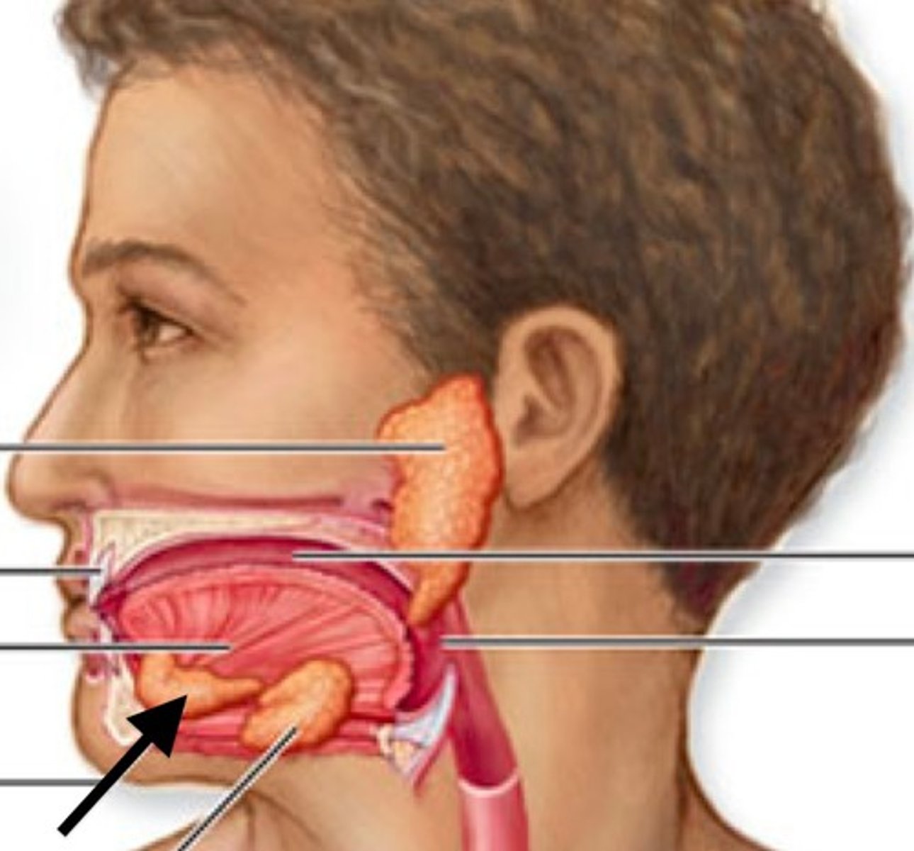
<p>smallest salivary gland found under the tongue, primarily secretes mucins, and a minimal amount of saliva and amylase</p>
55
New cards

Nasopharynx

region of the pharynx at the back of the nose and above the soft palate
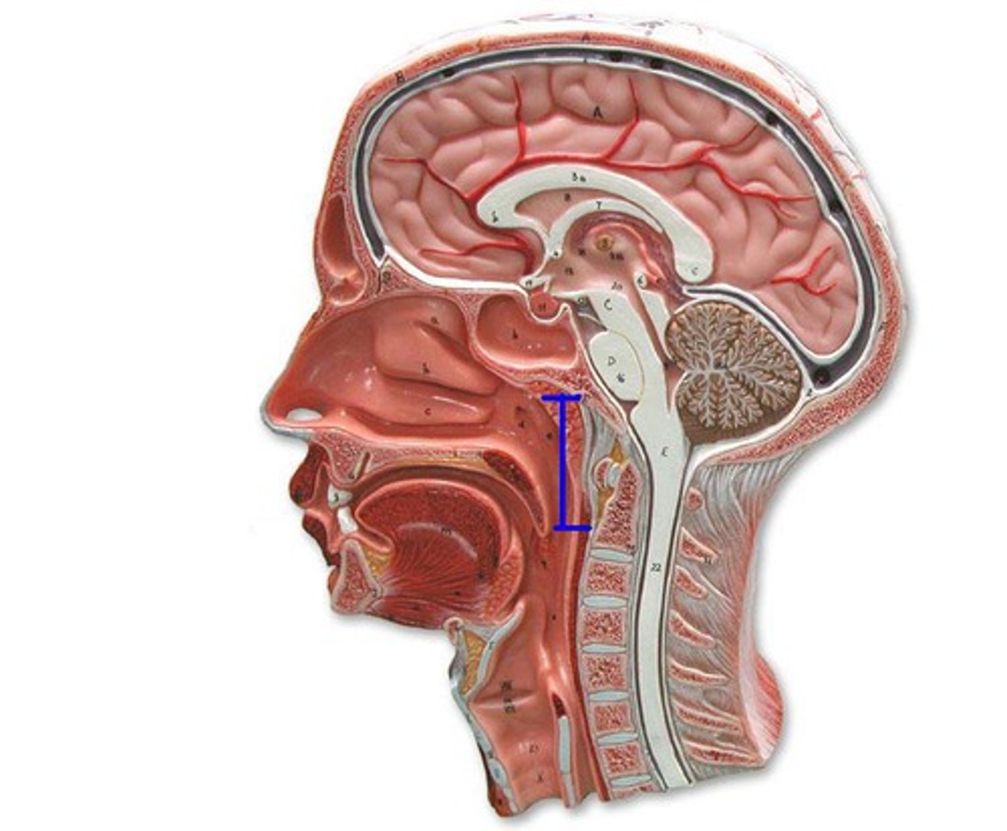
<p>region of the pharynx at the back of the nose and above the soft palate</p>
56
New cards

Oropharynx

central portion of the pharynx between the roof of the mouth and the upper edge of the epiglottis
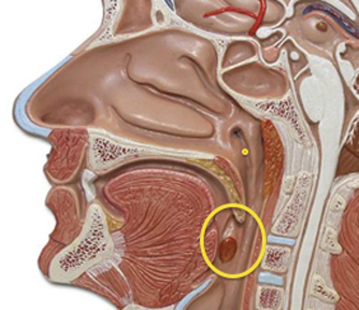
<p>central portion of the pharynx between the roof of the mouth and the upper edge of the epiglottis</p>
57
New cards

Laryngopharynx

the third division of the pharynx, is shared by both the respiratory and digestive systems
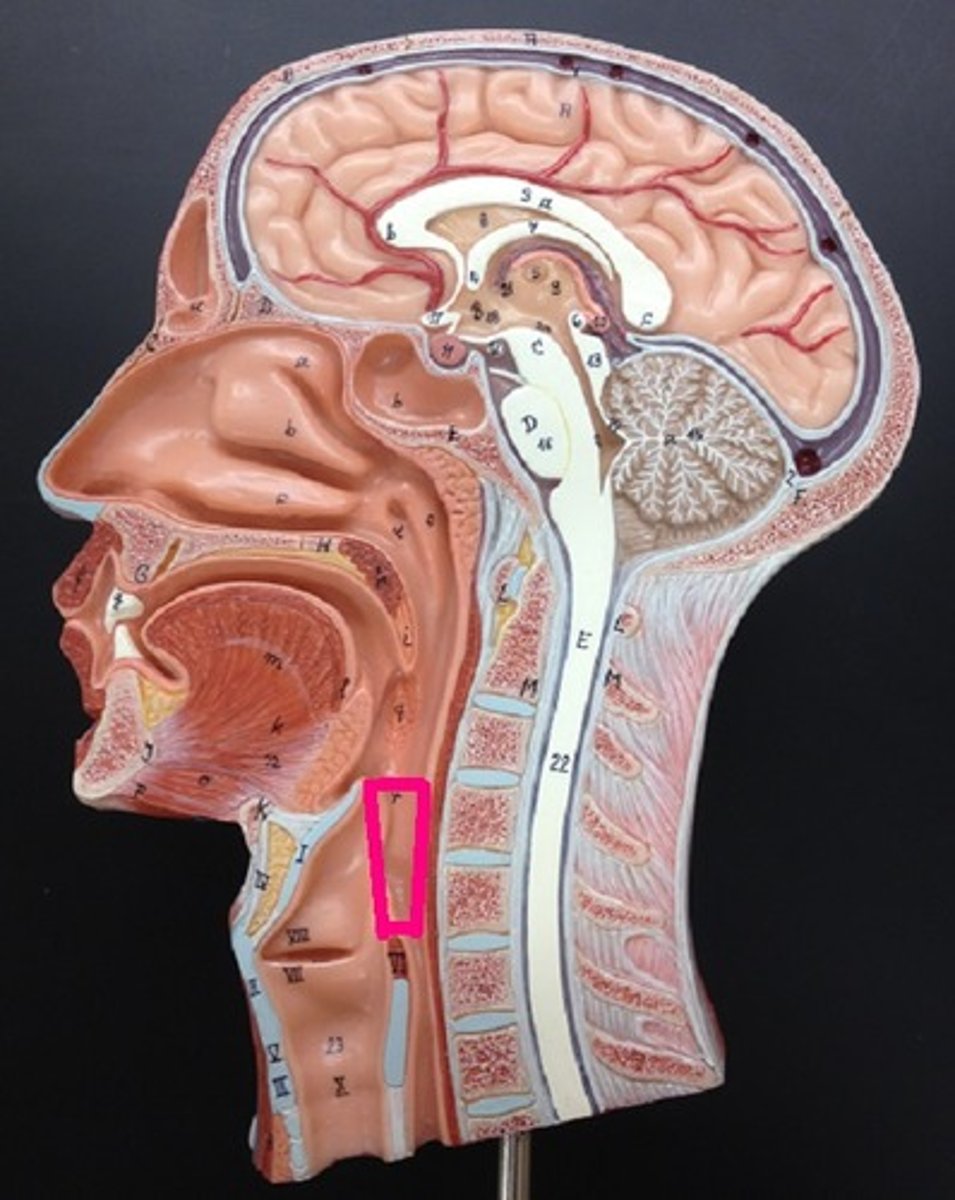
<p>the third division of the pharynx, is shared by both the respiratory and digestive systems</p>
58
New cards

Esophagus

A muscular tube that connects the mouth to the stomach. consists of 4 layers: mucosa, submucosa, muscularis propria, and adventitia
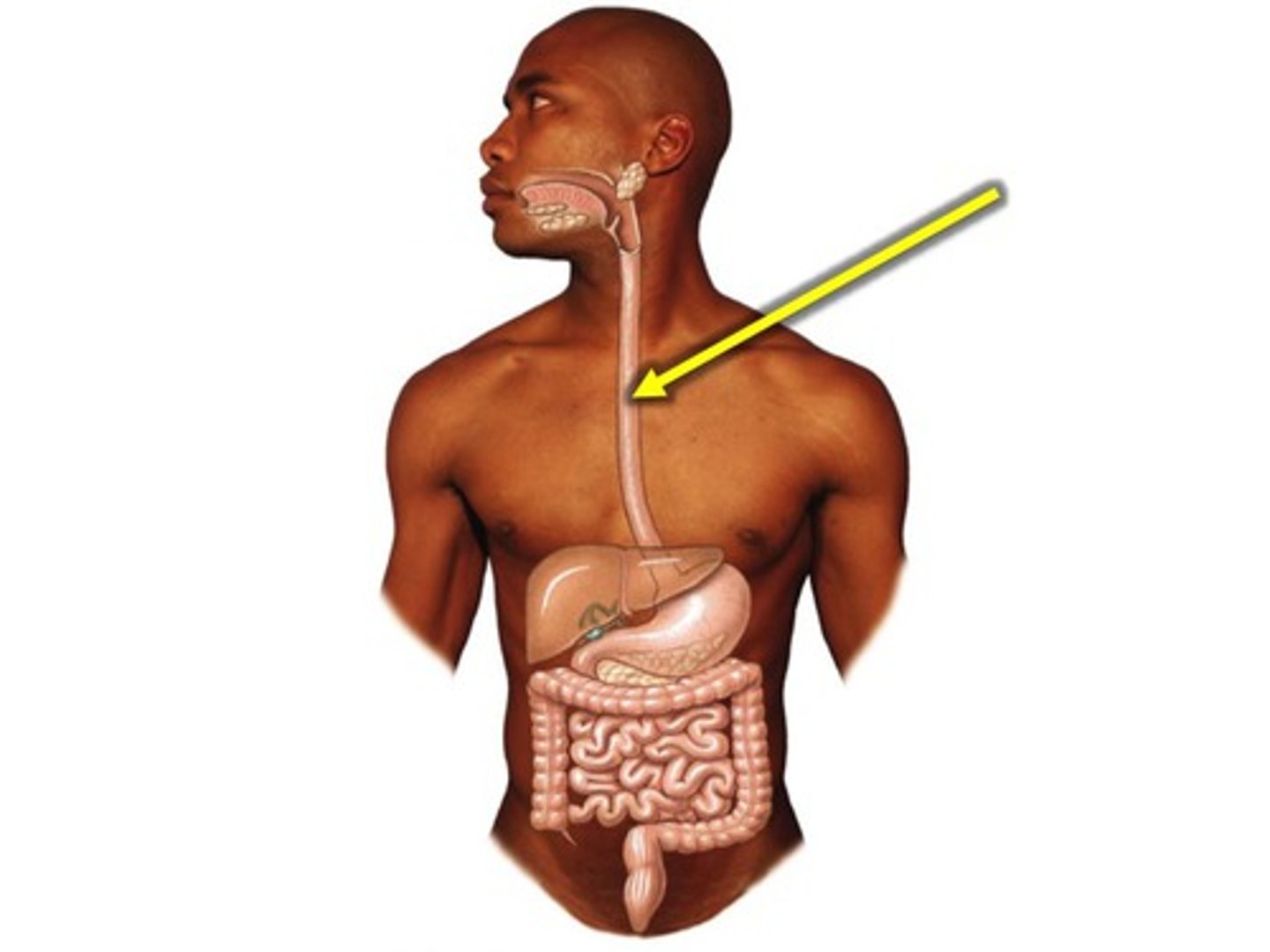
<p>A muscular tube that connects the mouth to the stomach. consists of 4 layers: mucosa, submucosa, muscularis propria, and adventitia</p>
59
New cards

Stomach

A muscular and elastic sac that serves mainly to store food, break it up mechanically, and begin chemical digestion of proteins and fat. Consists of mucosa, submucosa, muscularis externa, and serosa
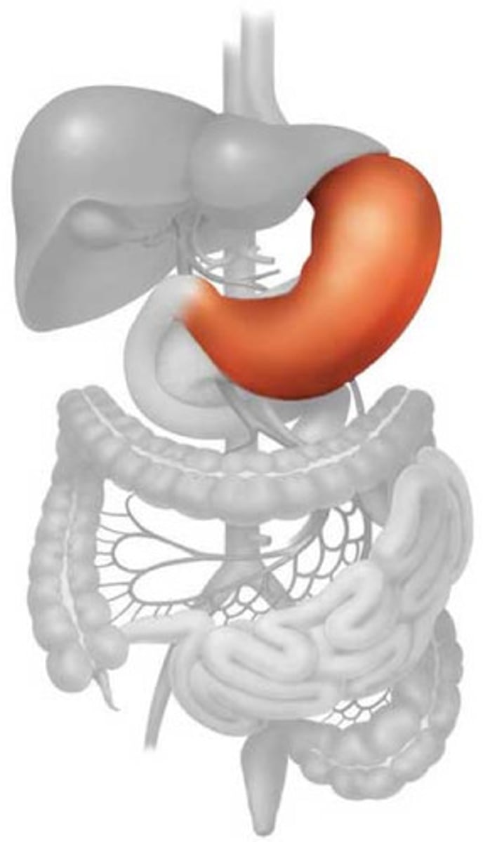
<p>A muscular and elastic sac that serves mainly to store food, break it up mechanically, and begin chemical digestion of proteins and fat. Consists of mucosa, submucosa, muscularis externa, and serosa</p>
60
New cards

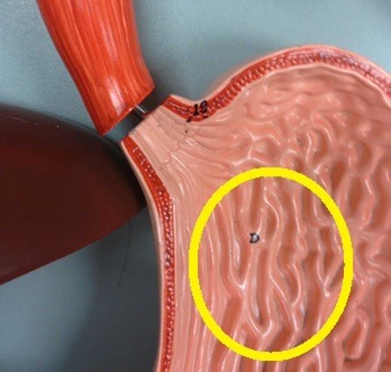
Rugae

the folds in the mucosa lining the stomach
<p>the folds in the mucosa lining the stomach</p>
61
New cards

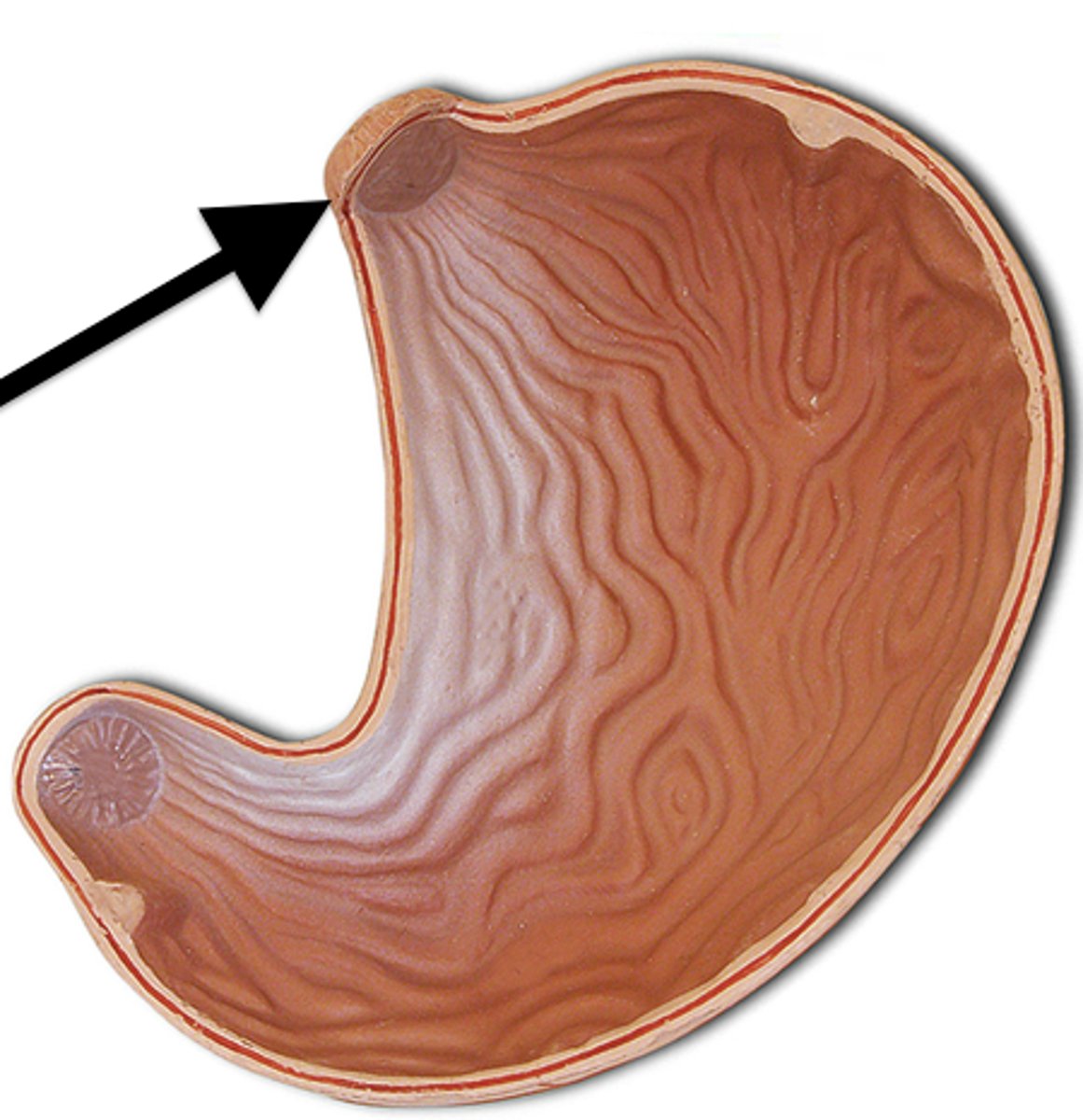
Cardiac sphincter

circular muscle located at the base of the esophagus and functions to prevent food materials from entering or leaving the esophagus from the stomach
<p>circular muscle located at the base of the esophagus and functions to prevent food materials from entering or leaving the esophagus from the stomach</p>
62
New cards

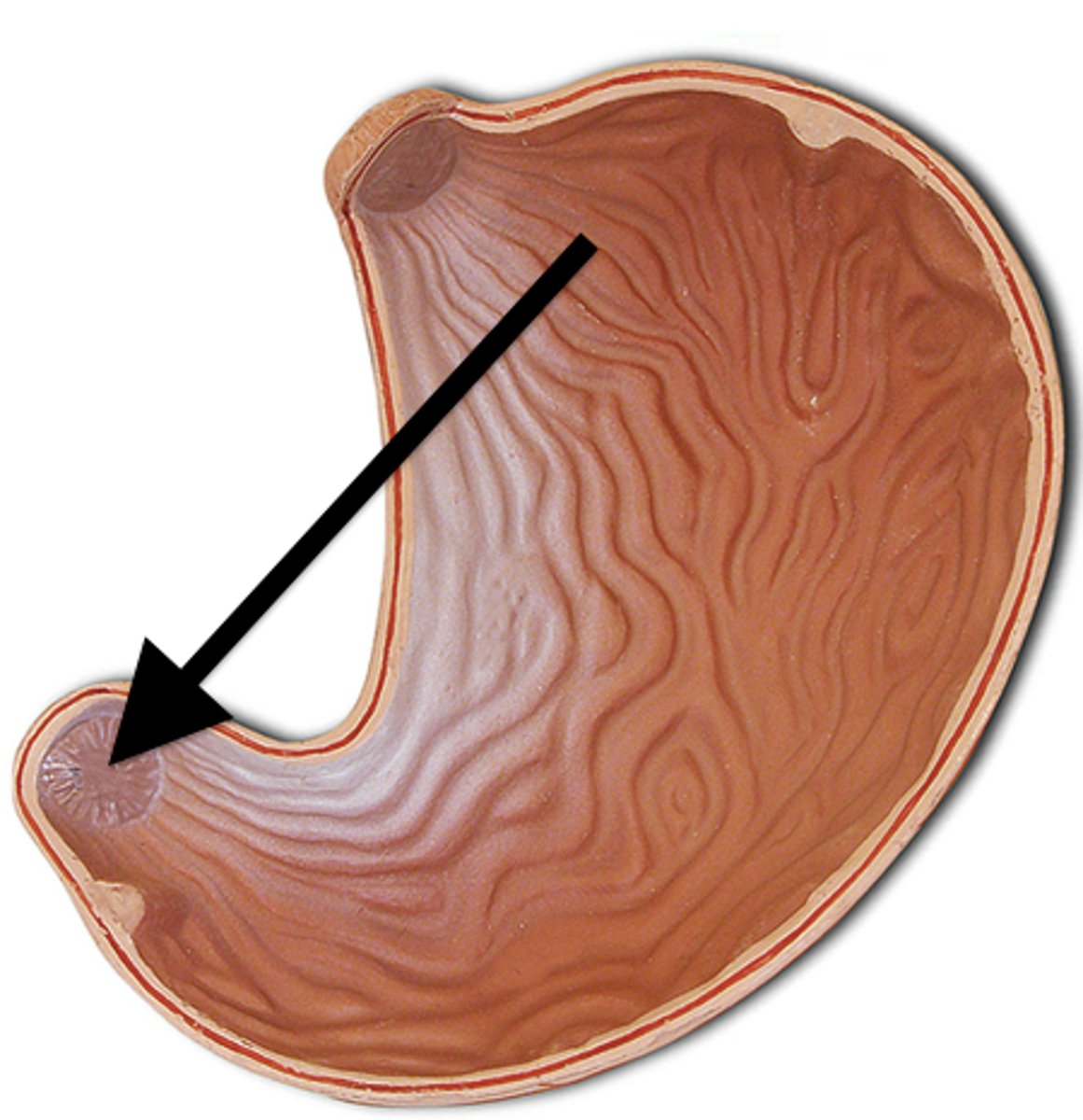
Pyloric sphincter

ring of muscle that guards the opening between the stomach and the duodenum
<p>ring of muscle that guards the opening between the stomach and the duodenum</p>
63
New cards

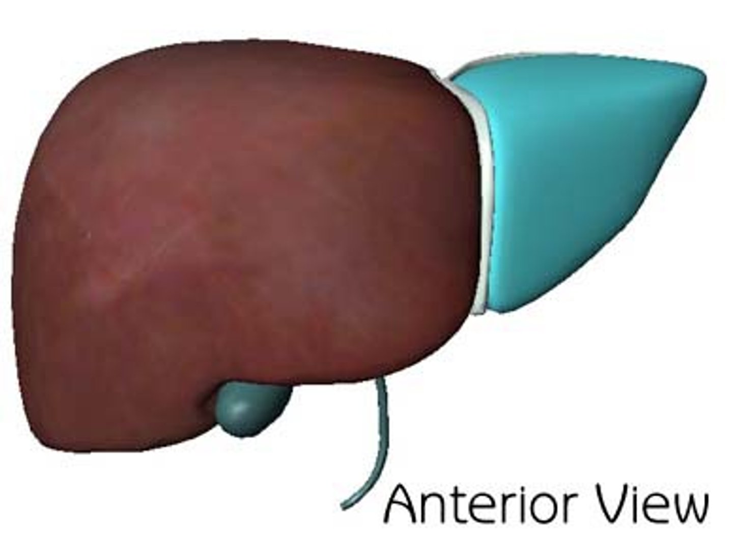
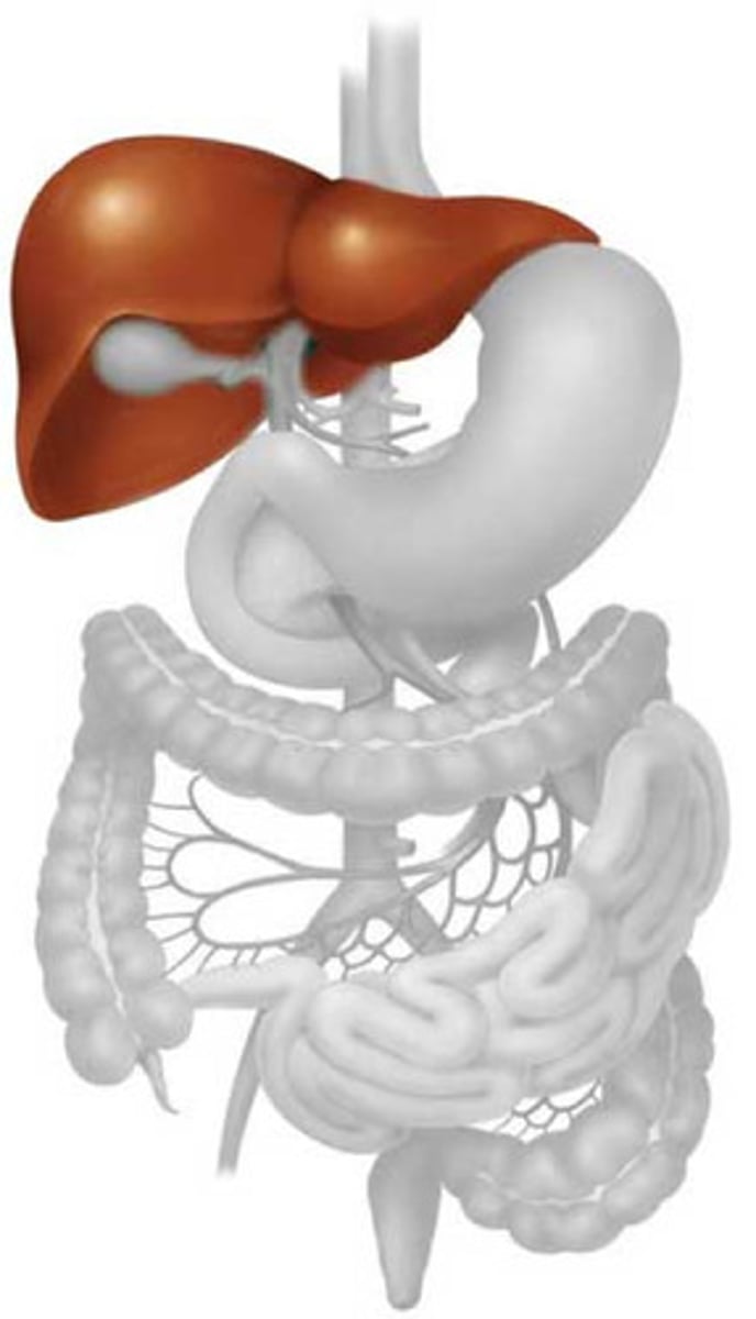
Liver

A large organ located in the upper abdomen (RUQ), this organ has 4 major lobes, and functions to filter blood, metabolize drugs, stores nutrients such as vitamins and minerals and glucose in the form of glycogen, produces Bile, and synthesizes proteins, process hemoglobin, regulates blood sugar levels, immune functions.
<p>A large organ located in the upper abdomen (RUQ), this organ has 4 major lobes, and functions to filter blood, metabolize drugs, stores nutrients such as vitamins and minerals and glucose in the form of glycogen, produces Bile, and synthesizes proteins, process hemoglobin, regulates blood sugar levels, immune functions.</p>
64
New cards

Right lobe

The largest lobe of the liver, separated from the left lobe above and in front by the falciform ligament, and separated from the caudate and quadrate lobes by the sulcus for the vena cava and by the fossa for the gallbladder.
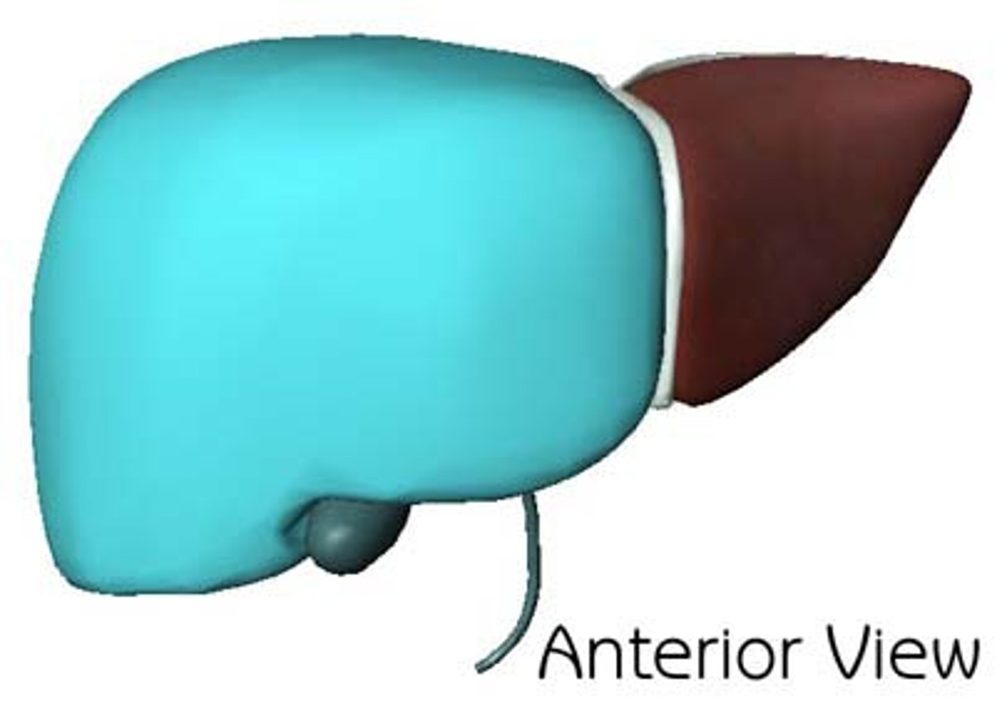
<p>The largest lobe of the liver, separated from the left lobe above and in front by the falciform ligament, and separated from the caudate and quadrate lobes by the sulcus for the vena cava and by the fossa for the gallbladder.</p>
65
New cards

Left lobe

smaller lobe of liver

<p>smaller lobe of liver</p>
66
New cards

Caudate lobe

smallest lobe of the liver situated on the posterosuperior surface of the left lobe; the ligamentum venosum is the anterior border (located posteriorly to the quadrate lobe)
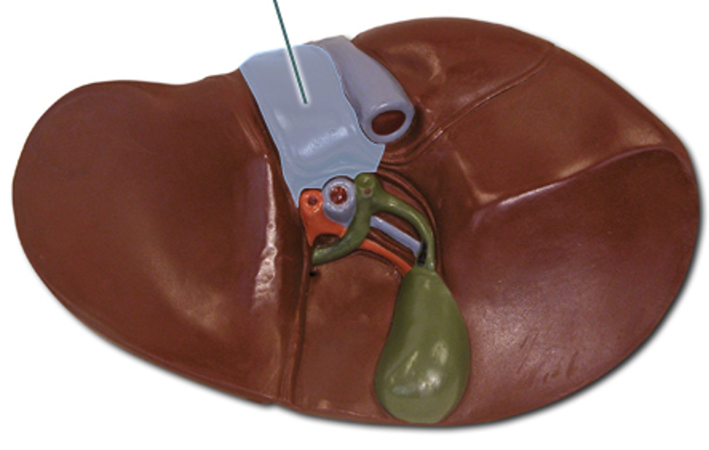
<p>smallest lobe of the liver situated on the posterosuperior surface of the left lobe; the ligamentum venosum is the anterior border (located posteriorly to the quadrate lobe)</p>
67
New cards

Quadrate lobe

square like lobe located in between the gallbladder and left lobe.
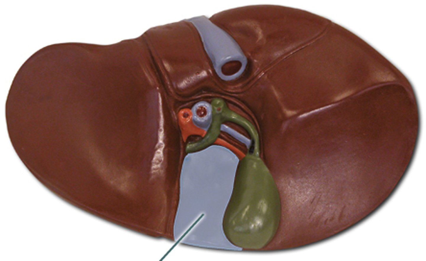
<p>square like lobe located in between the gallbladder and left lobe.</p>
68
New cards

Round ligament/ligamentum teres

A ligament that forms part of the free edge of the falciform ligament of the liver. It connects the liver to the umbilicus. It is the remnant of the left umbilical vein. located on the inferior side of the liver in-between the left and quadrate lobe
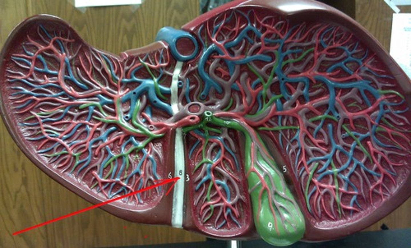
<p>A ligament that forms part of the free edge of the falciform ligament of the liver. It connects the liver to the umbilicus. It is the remnant of the left umbilical vein. located on the inferior side of the liver in-between the left and quadrate lobe</p>
69
New cards

Portal Triad

Comprised of the hepatic portal vein, proper hepatic artery, and common bile duct
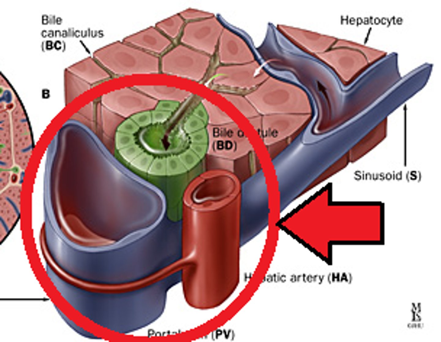
<p>Comprised of the hepatic portal vein, proper hepatic artery, and common bile duct</p>
70
New cards

Hepatic portal vein

the vein that collects blood from the GI tract and conducts it to the liver
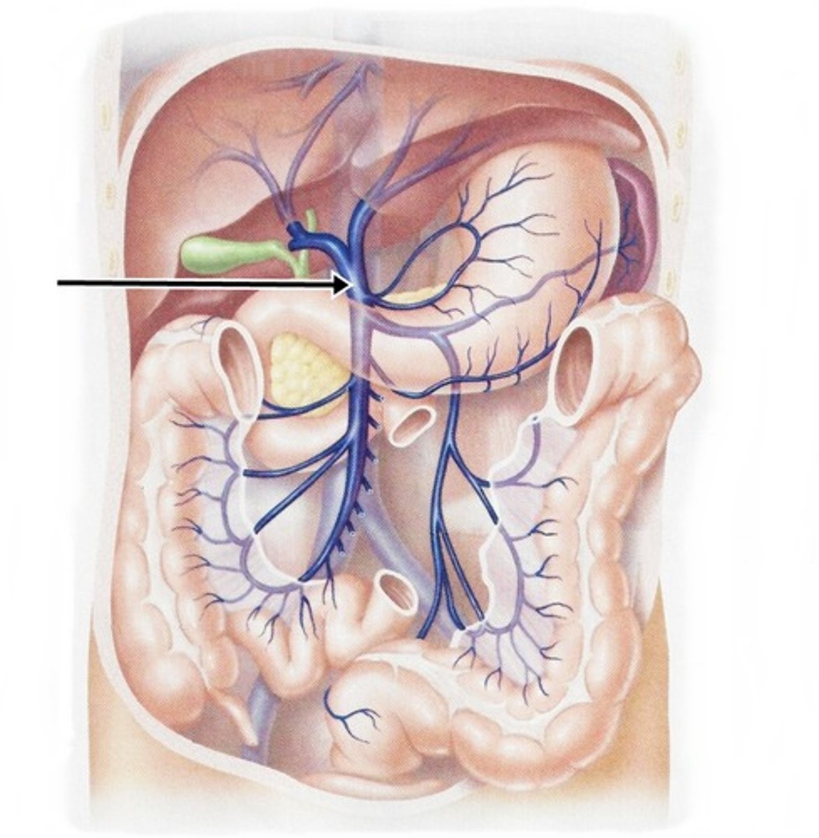
<p>the vein that collects blood from the GI tract and conducts it to the liver</p>
71
New cards

Proper Hepatic Artery (PHA)

Artery that supplies nutrients to the liver and gallbladder, branches off of the the common hepatic artery
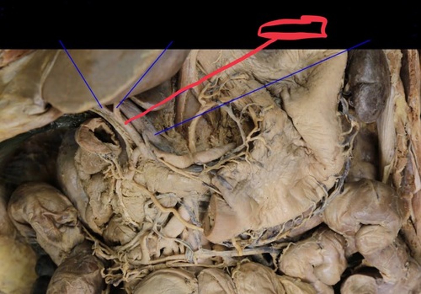
<p>Artery that supplies nutrients to the liver and gallbladder, branches off of the the common hepatic artery</p>
72
New cards

Common bile duct

Carries bile from the liver and gallbladder to the duodenum. Also called the choledochus.
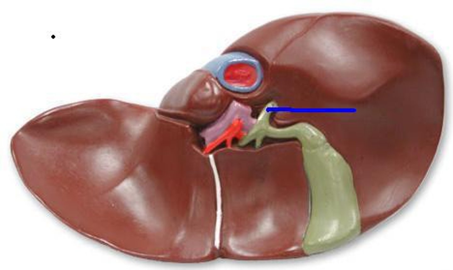
<p>Carries bile from the liver and gallbladder to the duodenum. Also called the choledochus.</p>
73
New cards

Cystic duct

a duct draining bile from the gallbladder; merges with the common hepatic duct to form the common bile duct
<p>a duct draining bile from the gallbladder; merges with the common hepatic duct to form the common bile duct</p>
74
New cards

Common hepatic duct

large bile duct leading from liver; joins with the cystic duct to form the common bile duct
<p>large bile duct leading from liver; joins with the cystic duct to form the common bile duct</p>
75
New cards

Gallbladder

A muscular sac attached to the liver that secretes bile and stores it until needed for digestion
<p>A muscular sac attached to the liver that secretes bile and stores it until needed for digestion</p>
76
New cards

Pancreas

An organs in the abdominal cavity with two roles. The first is an exocrine role: to produce digestive enzymes and bicarbonate, which are delivered to the small intestine via the pancreatic duct. The second is an endocrine role: to secrete insulin and glucagon into the bloodstream to help regulate blood glucose levels.
<p>An organs in the abdominal cavity with two roles. The first is an exocrine role: to produce digestive enzymes and bicarbonate, which are delivered to the small intestine via the pancreatic duct. The second is an endocrine role: to secrete insulin and glucagon into the bloodstream to help regulate blood glucose levels.</p>
77
New cards

Pancreatic duct

The main duct of the pancreas. The pancreatic duct carries the exocrine secretions of the pancreas (enzymes and bicarbonate) to the small intestine (dueodenum).
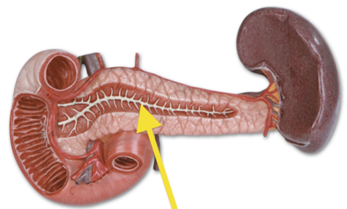
<p>The main duct of the pancreas. The pancreatic duct carries the exocrine secretions of the pancreas (enzymes and bicarbonate) to the small intestine (dueodenum).</p>
78
New cards

Greater/Major duodenal papilla

a rounded projection in the duodenum into which the common bile duct and pancreatic duct drain
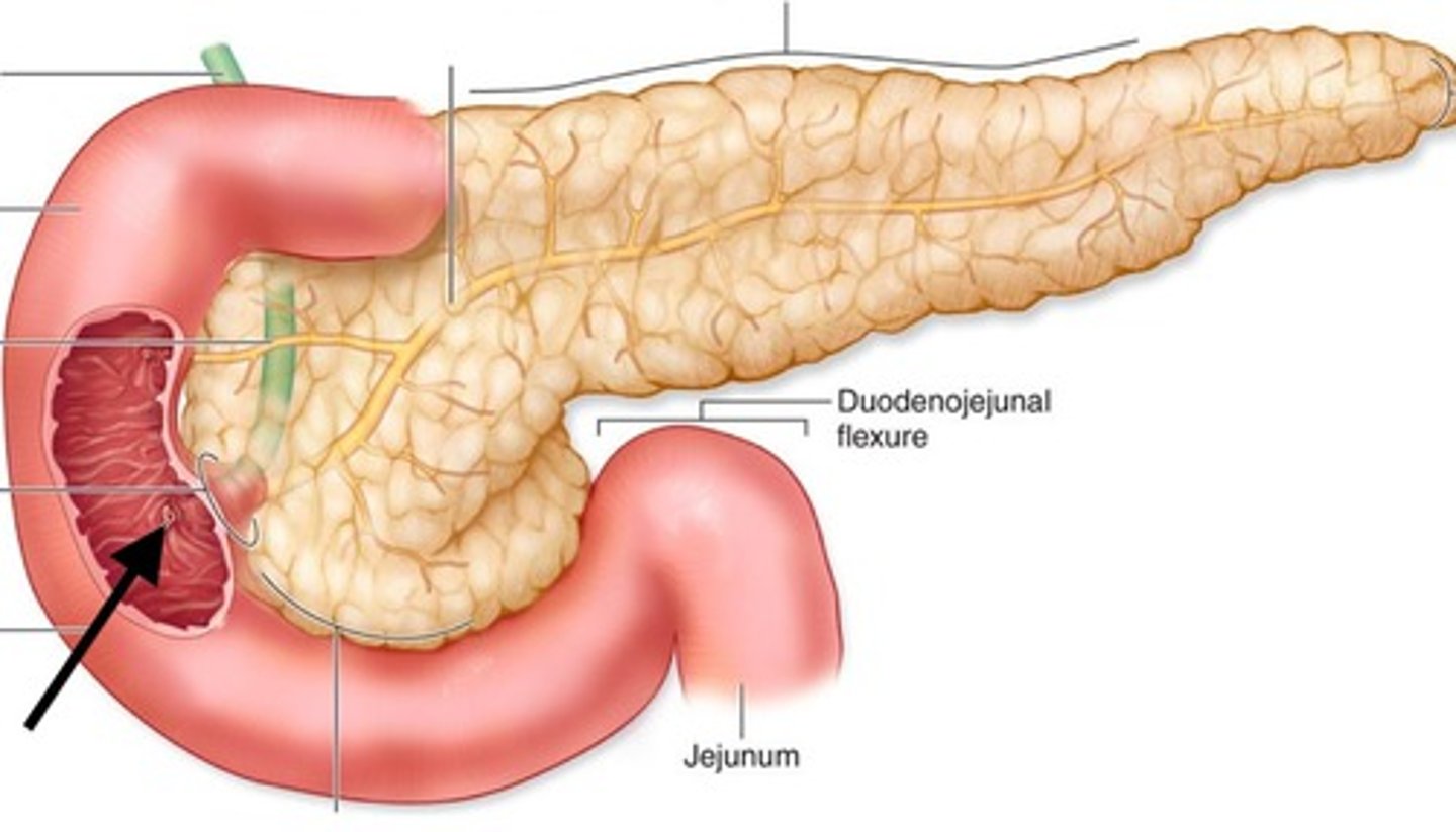
<p>a rounded projection in the duodenum into which the common bile duct and pancreatic duct drain</p>
79
New cards

Small intestine

Digestive organ where most chemical digestion and absorption of food takes place, has 3 distinct regions called the duodenum, jejunum, and ileum.

Known as the longest intestine.
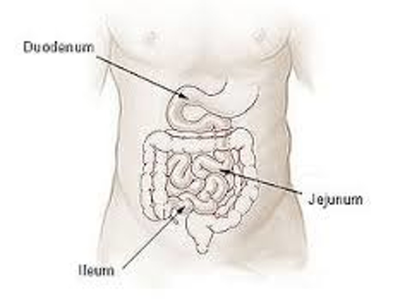
<p>Digestive organ where most chemical digestion and absorption of food takes place, has 3 distinct regions called the duodenum, jejunum, and ileum.</p><p>Known as the longest intestine.</p>
80
New cards

Villi

Fingerlike extensions of the intestinal mucosa that increase the surface area for absorption
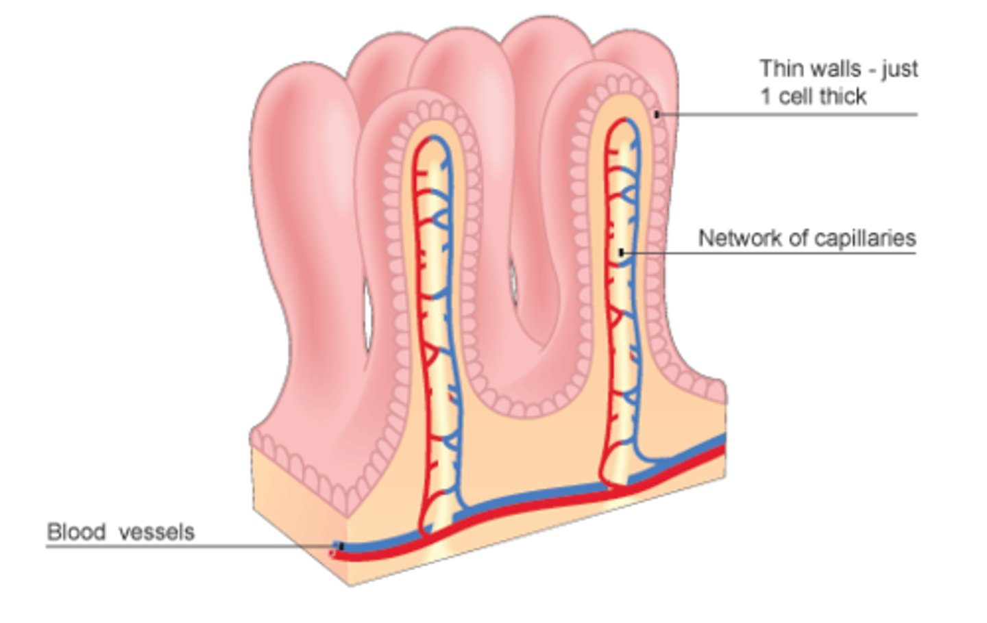
<p>Fingerlike extensions of the intestinal mucosa that increase the surface area for absorption</p>
81
New cards

Lymphatic duct (small intestine)

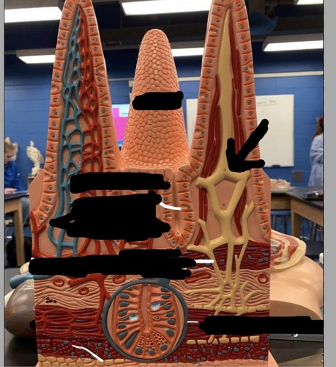
lacteals in the small intestine's villi, play a crucial role in absorbing dietary fats and fat-soluble vitamins
<p>lacteals in the small intestine's villi, play a crucial role in absorbing dietary fats and fat-soluble vitamins</p>
82
New cards

Intestinal Crypt/Crypts of Lieberkühn

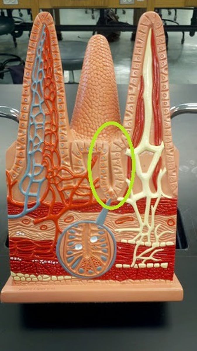
invaginations in the intestinal lining between villi, containing stem cells and specialized cells that regenerate and maintain the intestinal epithelium
<p>invaginations in the intestinal lining between villi, containing stem cells and specialized cells that regenerate and maintain the intestinal epithelium</p>
83
New cards

Longitudinal layer muscle

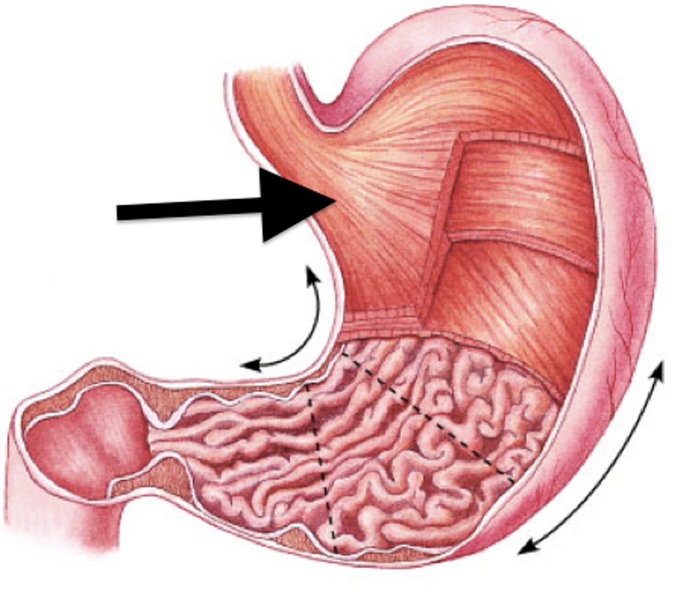
outer layer of smooth muscle that helps propel food through the gut
<p>outer layer of smooth muscle that helps propel food through the gut</p>
84
New cards

Circular layer muscle

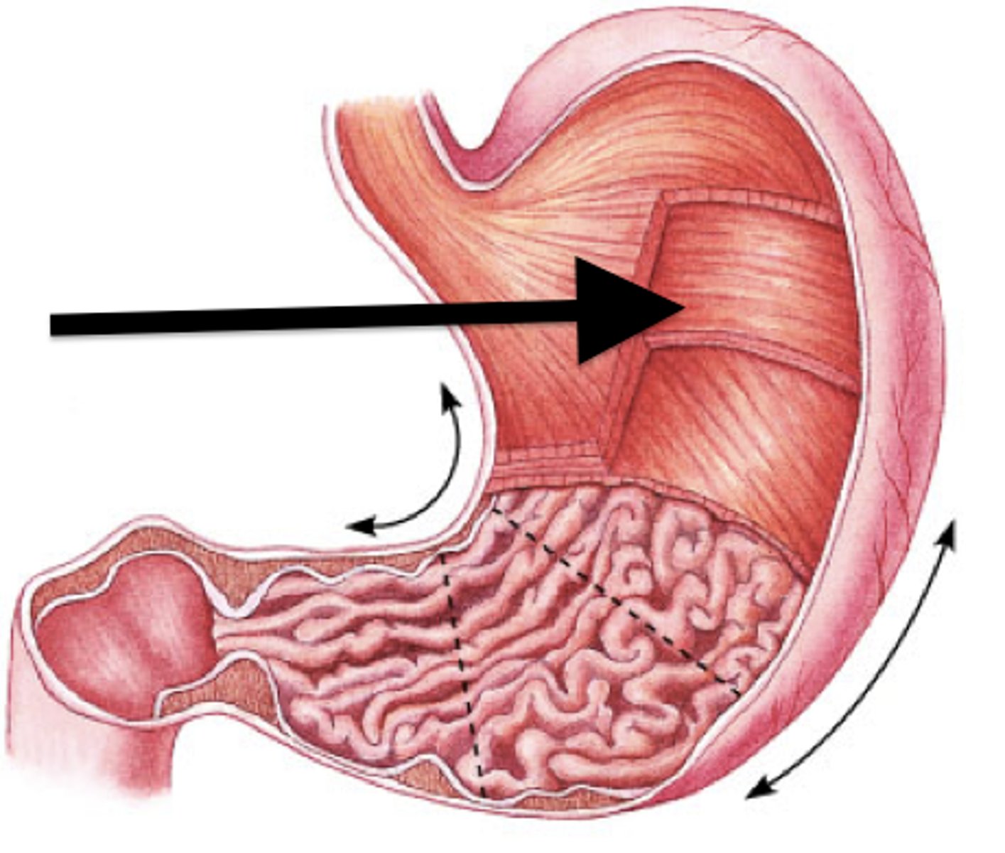
inner layer which orients around the circumference of the canal
<p>inner layer which orients around the circumference of the canal</p>
85
New cards

Muscularis mucosa

thin layer of smooth muscle,

tenses mucosa creating grooves and ridges that enhance surface area and contact with food,

improves efficiency of digestion and nutrient absorption
<p>thin layer of smooth muscle,</p><p>tenses mucosa creating grooves and ridges that enhance surface area and contact with food,</p><p>improves efficiency of digestion and nutrient absorption</p>
86
New cards

Duodenum

first portion of the small intestine
<p>first portion of the small intestine</p>
87
New cards

Plicae circulares

circular folds in small intestine
<p>circular folds in small intestine</p>
88
New cards

Jejunum

second part/ region of the small intestine (B)
<p>second part/ region of the small intestine (B)</p>
89
New cards

Ileum

the last and longest portion of the small intestine (C)
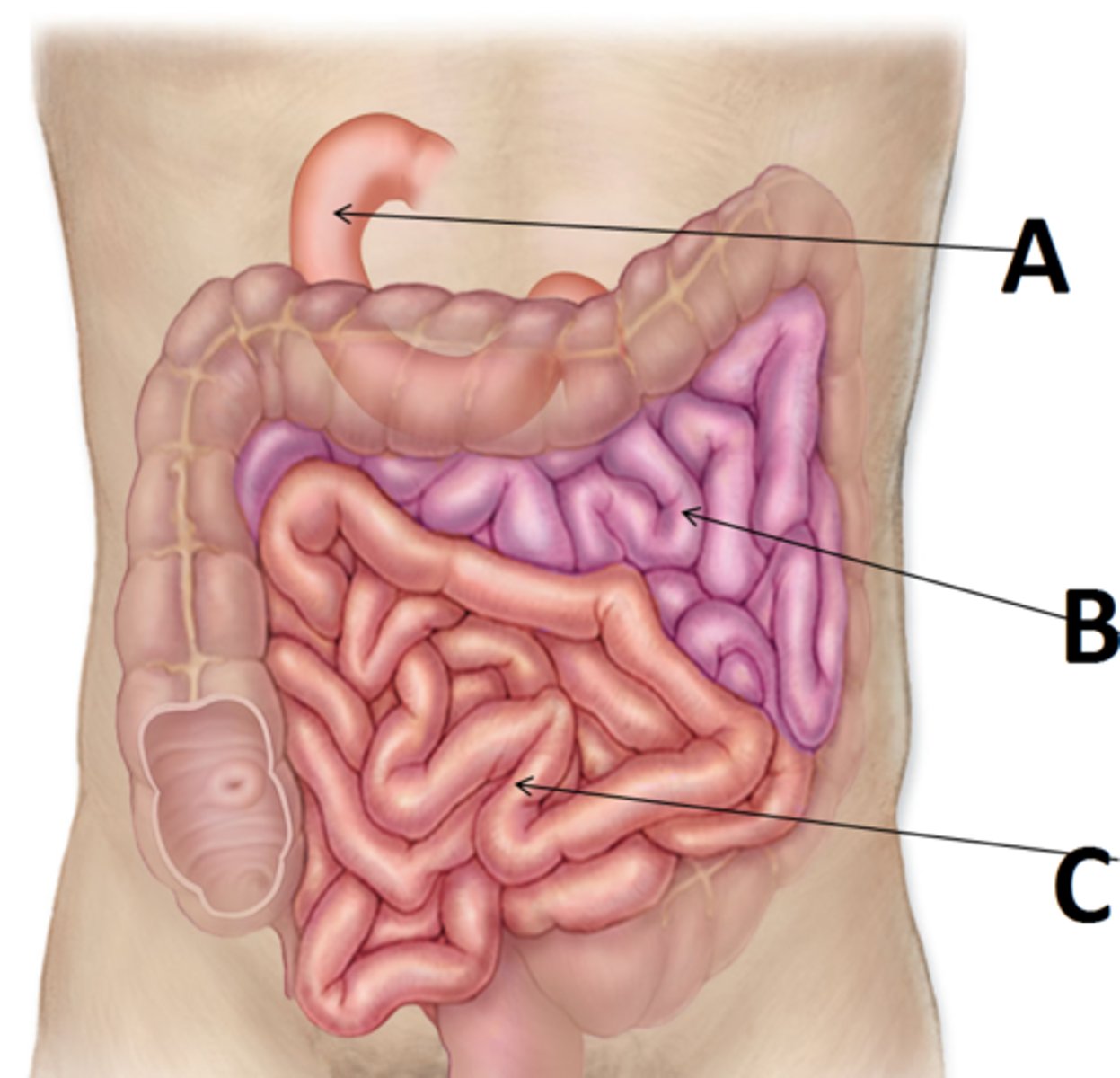
<p>the last and longest portion of the small intestine (C)</p>
90
New cards

Ileocecal junction

the end of the small intestine where the ileum joins the cecum of the large intestine
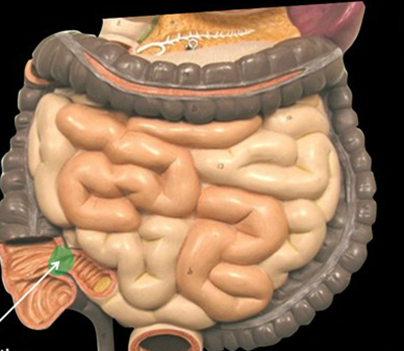
<p>the end of the small intestine where the ileum joins the cecum of the large intestine</p>
91
New cards

Large intestine/Colon

The long, tube-like organ that is connected to the small intestine at one end and the anus at the other. absorbing water and electrolytes, producing and absorbing vitamins, and forming and propelling feces toward the rectum for elimination.
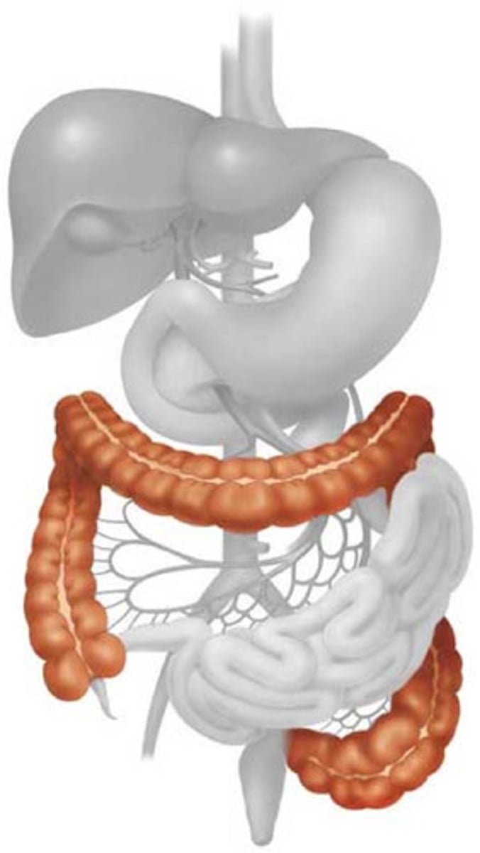
<p>The long, tube-like organ that is connected to the small intestine at one end and the anus at the other. absorbing water and electrolytes, producing and absorbing vitamins, and forming and propelling feces toward the rectum for elimination.</p>
92
New cards

Cecum

a pouch connected to the junction of the small and large intestines. receives undigested food from the small intestine, absorbs water and salts, and lubricates the waste material with mucus, ultimately contributing to the formation of feces
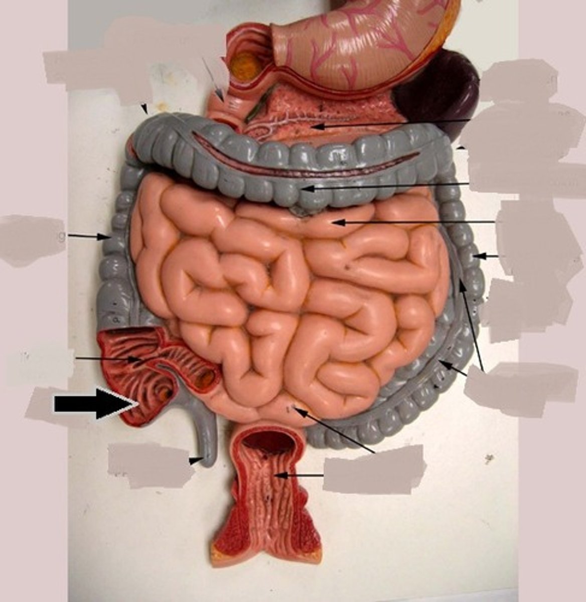
<p>a pouch connected to the junction of the small and large intestines. receives undigested food from the small intestine, absorbs water and salts, and lubricates the waste material with mucus, ultimately contributing to the formation of feces</p>
93
New cards

Vermiform appendix

a vestigial process that extends from the lower end of the cecum and that resembles a small pouch, houses bacteria.
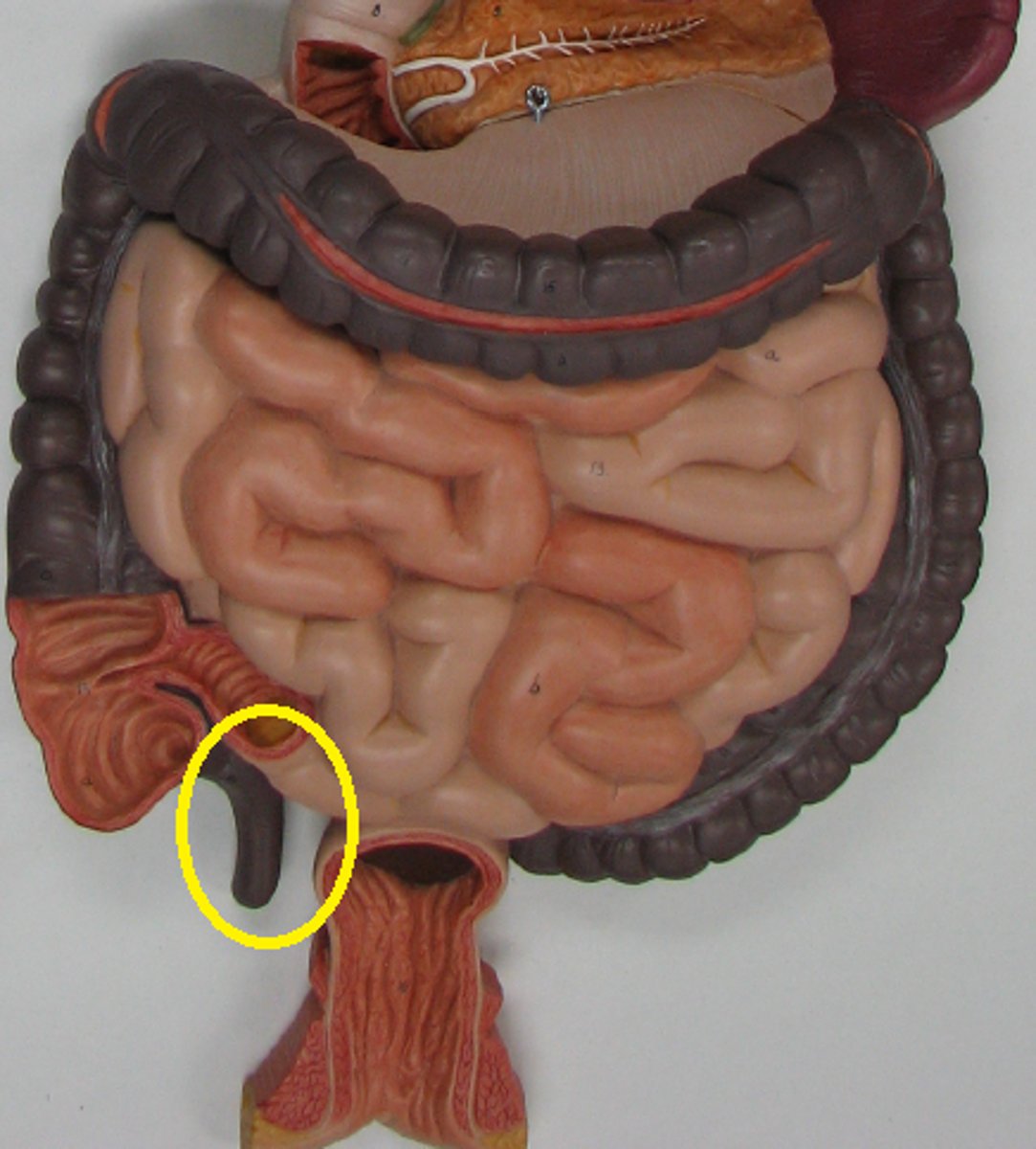
<p>a vestigial process that extends from the lower end of the cecum and that resembles a small pouch, houses bacteria.</p>
94
New cards

Ascending colon

Part of the large intestine that travels upward from the cecum to the undersurface of the liver
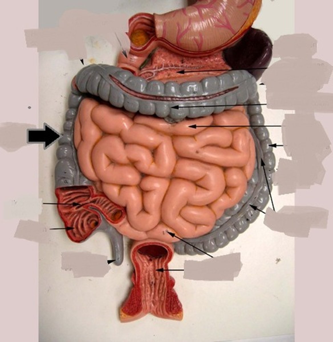
<p>Part of the large intestine that travels upward from the cecum to the undersurface of the liver</p>
95
New cards

Transverse colon

the middle part of the large intestine, passing across the abdomen from right to left below the stomach.
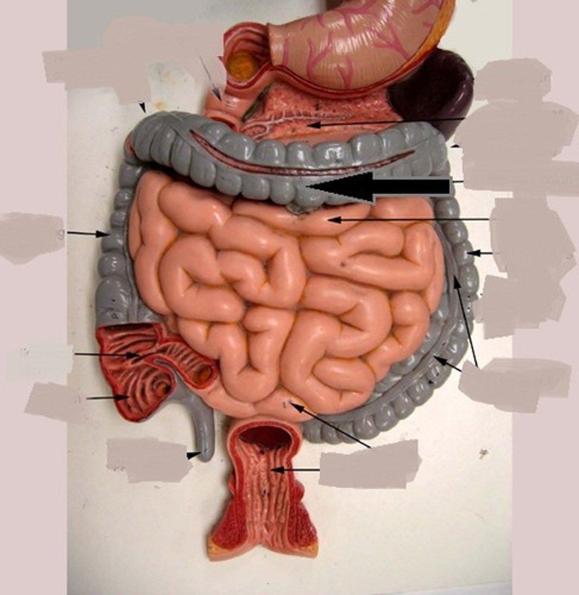
<p>the middle part of the large intestine, passing across the abdomen from right to left below the stomach.</p>
96
New cards

Descending colon

the part of the large intestine that passes downward on the left side of the abdomen toward the rectum.
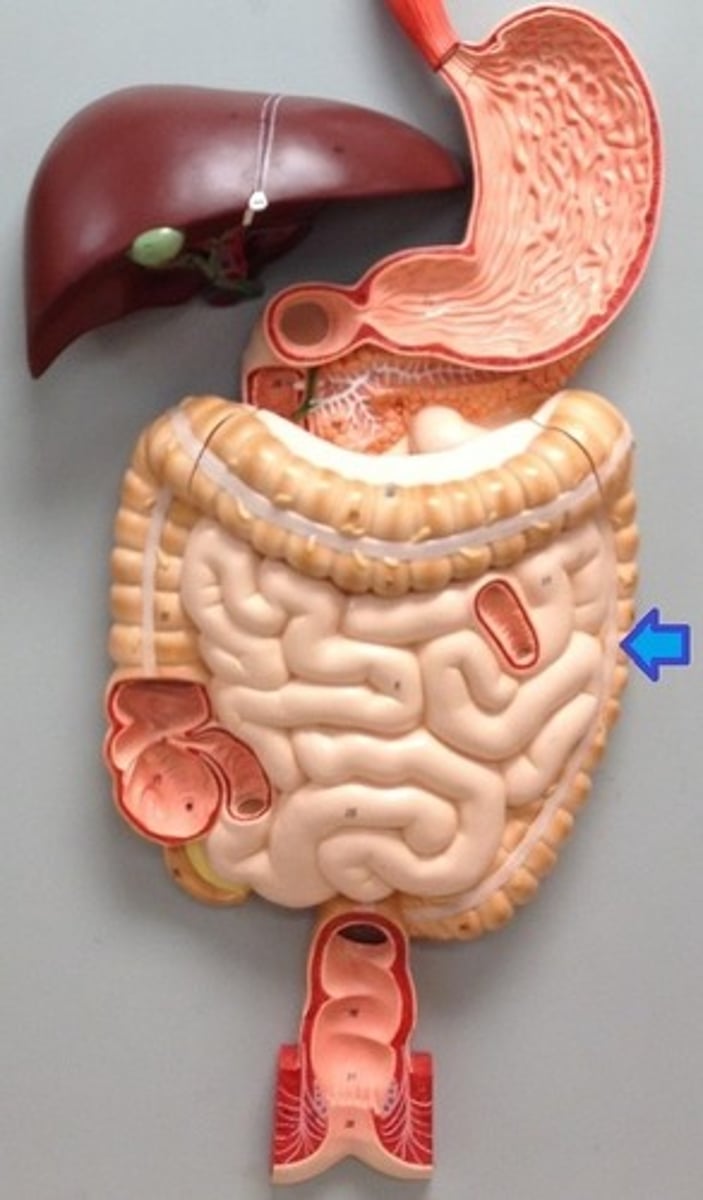
<p>the part of the large intestine that passes downward on the left side of the abdomen toward the rectum.</p>
97
New cards

Sigmoid colon

fourth and last, S-shaped segment of the colon, just before the rectum; empties into the rectum
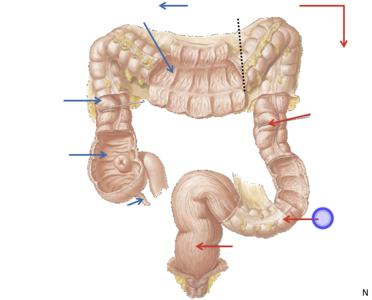
<p>fourth and last, S-shaped segment of the colon, just before the rectum; empties into the rectum</p>
98
New cards

Rectum

A short tube at the end of the large intestine where waste material is compressed into a solid form before being eliminated
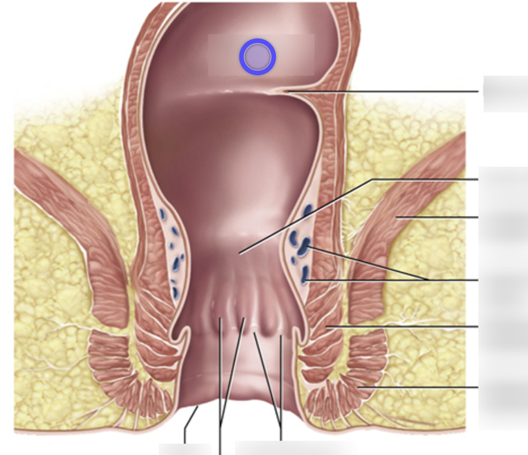
<p>A short tube at the end of the large intestine where waste material is compressed into a solid form before being eliminated</p>
99
New cards

Anal canal

the terminal region of the digestive tract, containing two sphincters, through which feces are expelled from the body
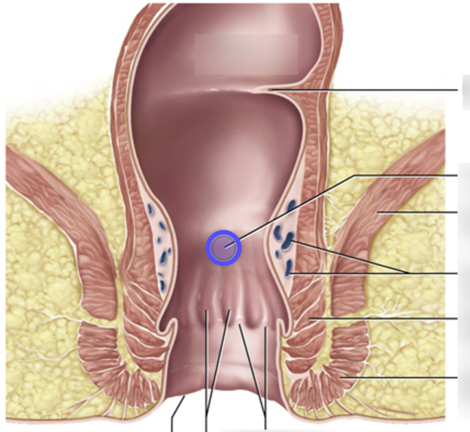
<p>the terminal region of the digestive tract, containing two sphincters, through which feces are expelled from the body</p>
100
New cards

Teniae coli

3 tiny shiny bands that spans the length of the colon (large intestine), comprised of smooth muscle functions to contract and shorten the colon, allowing for the formation of haustra and facilitating the movement of fecal matter through the large intestine.
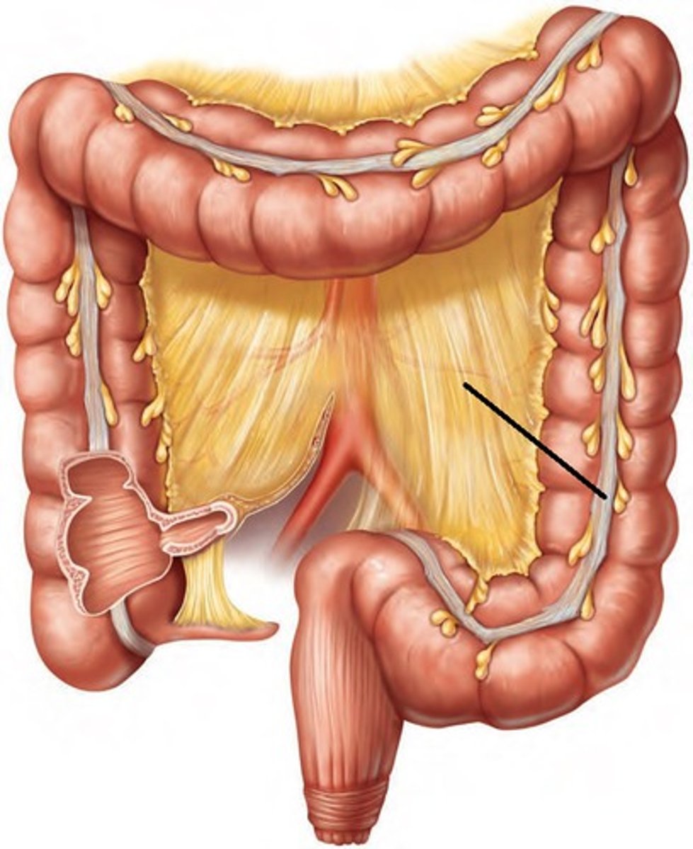
<p>3 tiny shiny bands that spans the length of the colon (large intestine), comprised of smooth muscle functions to contract and shorten the colon, allowing for the formation of haustra and facilitating the movement of fecal matter through the large intestine.</p>