Weeks 1 & 2 - Anatomy
 Call Kai
 Learn
Learn Practice Test
 Spaced Repetition
 Match
Match Flashcards
 Knowt Play
1/25

Earn XP
Description and Tags
Not an extensive list of all the topics in Anatomy, one's that was just taught in Human Neuroscience
Name | Mastery | Learn | Test | Matching | Spaced | Call with Kai | Chat |
|---|
No analytics yet
Send a link to your students to track their progress
26 Terms
What are the two types of cerebral cortex and what does it make up?
Neocortex which has 6 cell layers and makes up most of the cerebral cortex.
Allocortex which has less than 6 cell layers and makes up the olfactory cortex and hippocampus.
What are five important structures that make up the cerebrum?
Corpus callosum (the largest white matter structure which connects the left and right cerebral hemisphere).
Longitudinal fasciculi (connects frontal lobe to other lobes in the same hemispheres).
Anterior commissure (connects right and left cerebral hemispheres).
Internal capsule (links cerebral cortex to other parts of the CNS).
Basal nuclei (comprises of masses of grey matter deep in the brain).

Limbic System
Structures located along the border of the cerebrum and diencephalon which are involved in emotional states, linking conscious and unconscious brain functions, and memory.
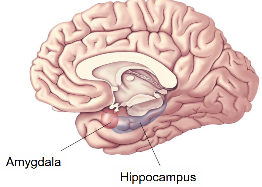
What are two key structures in the limbic system?
Hippocampus = A cerebral cortex structure that consists of allocortex and plays an important role in memory.
Amygdala (aka Amygdaloid body) = Acts as an interface between limbic system, cerebrum, and sensory organs which plays an important role in processing emotions, especially fear.
Ventricular System
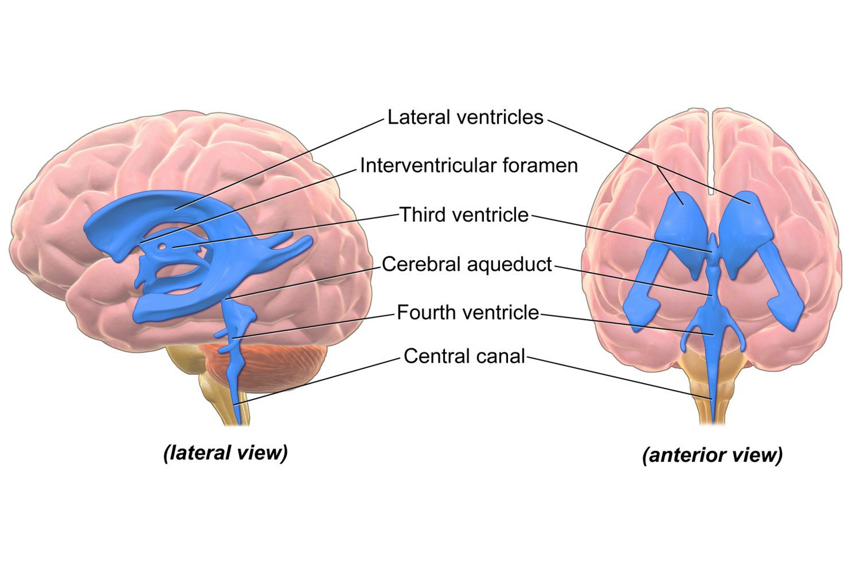
Four ventricles (lateral ventricles, third, and fourth ventricle) in the brain which all produce and circulate cerebrospinal fluid (CSF) that nourish brain tissue and provide nutrients for the brain to complete its metabolic activity.
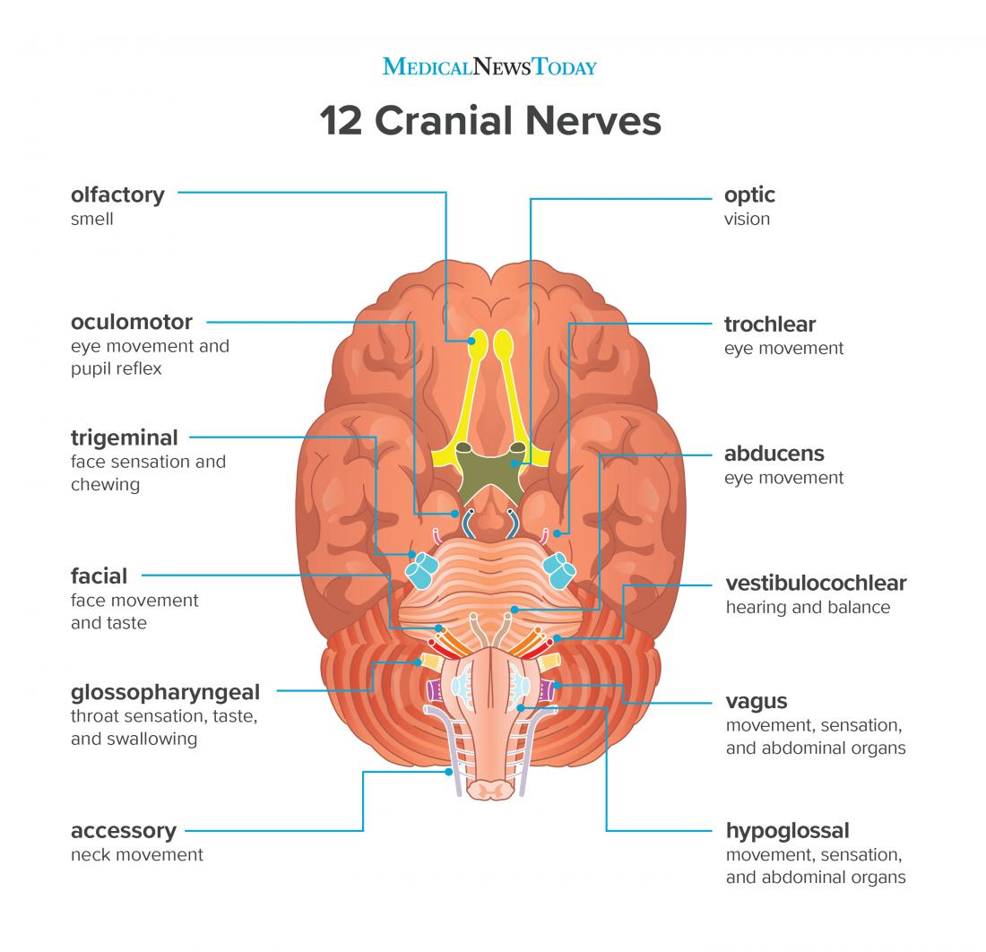
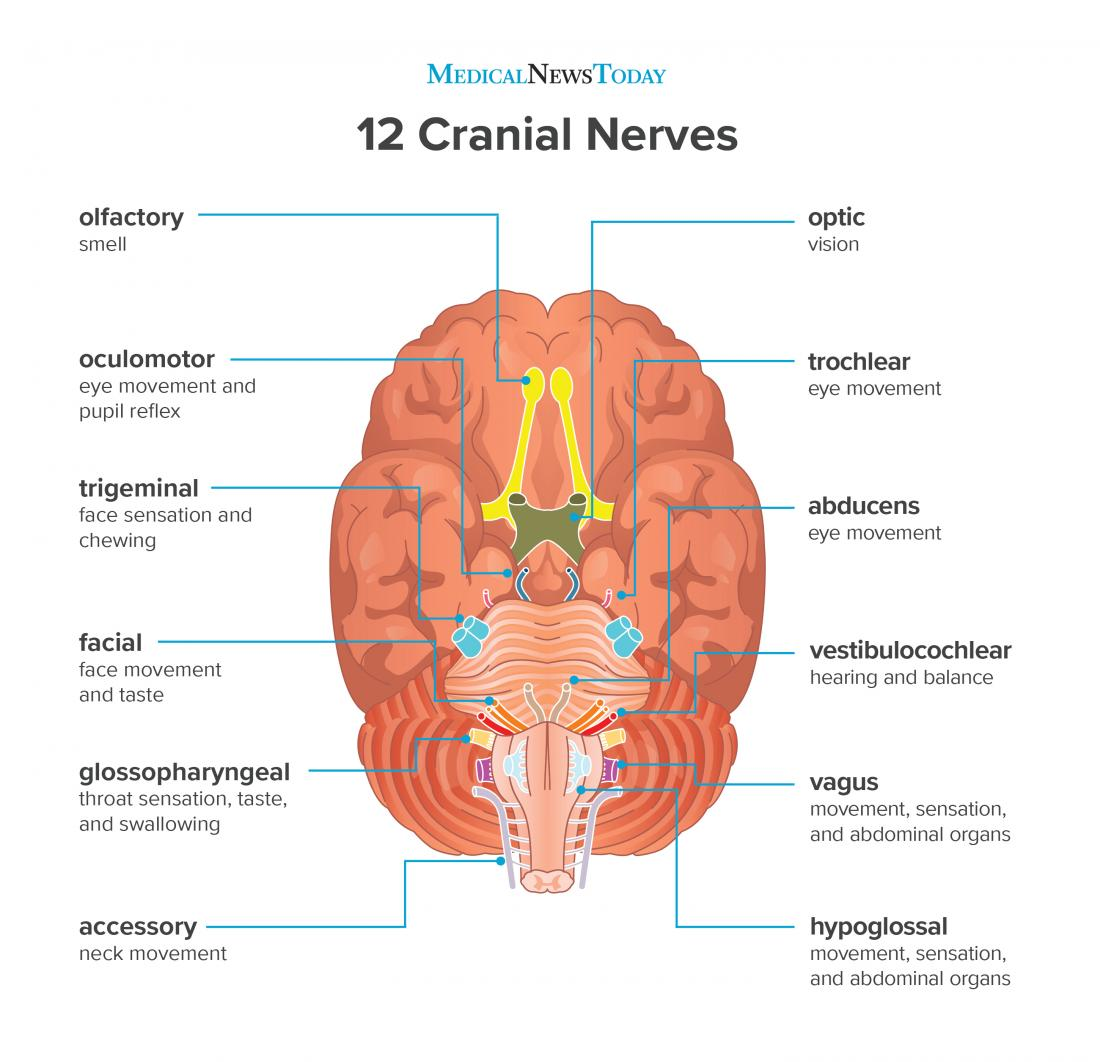
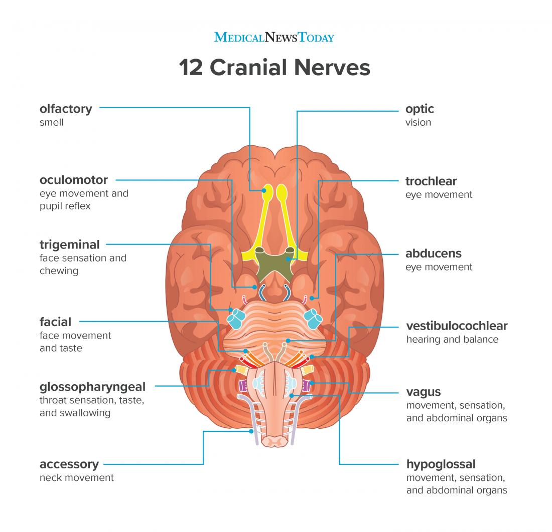
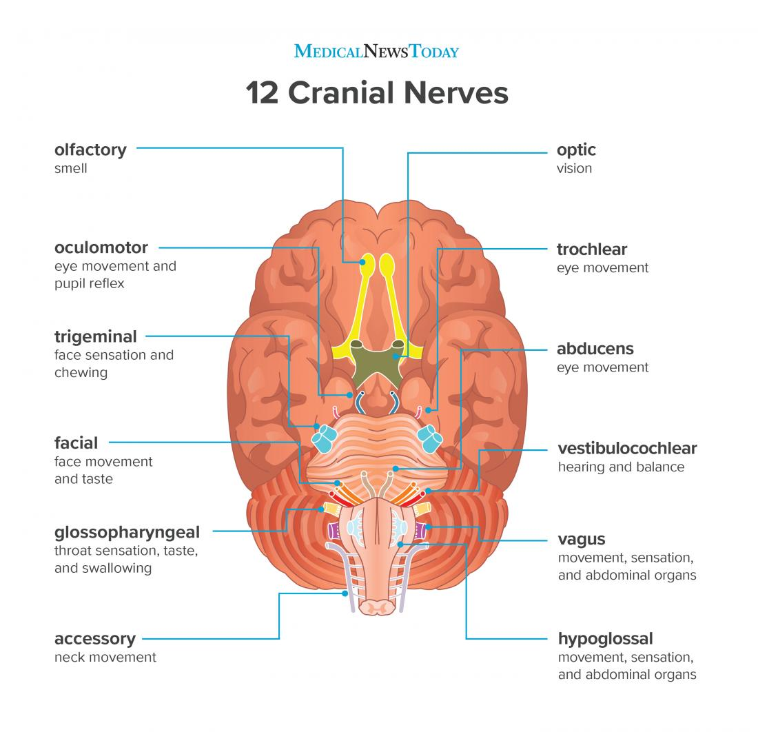
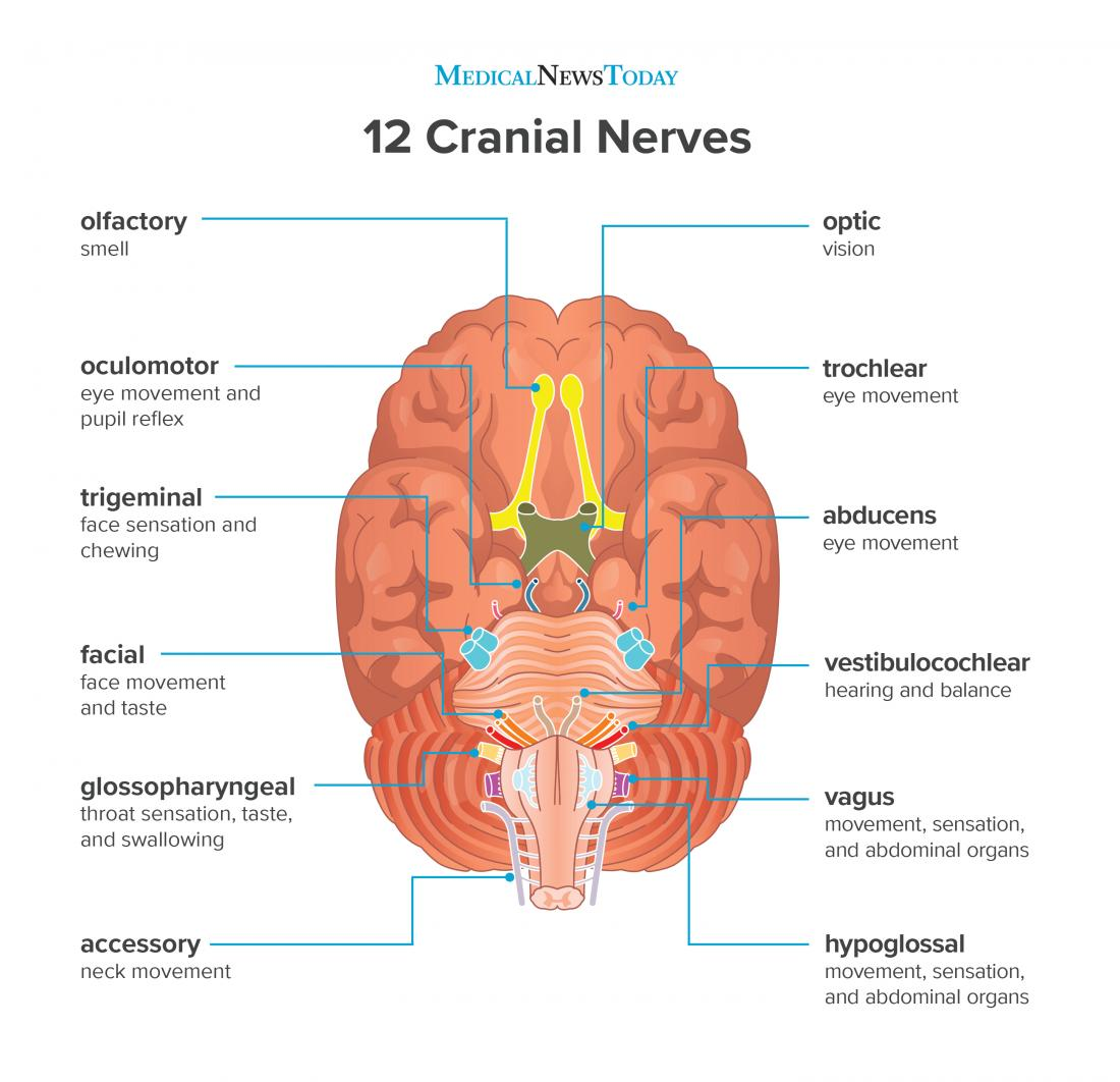
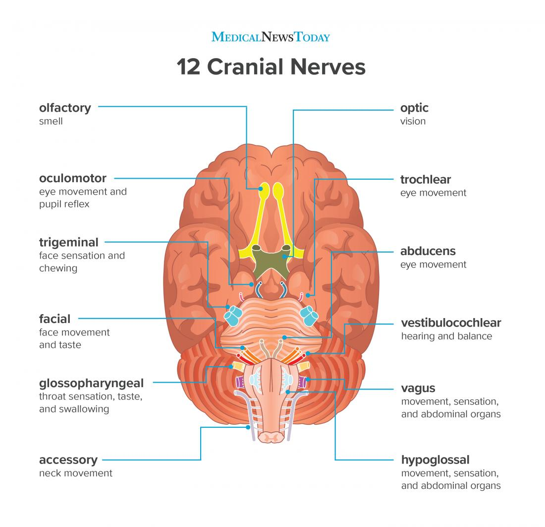
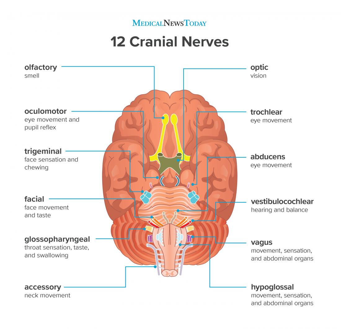
What are the twelve cranial nerves?
Olfactory nerve
Optic nerve
Oculomotor nerve
Trochlear nerve
Trigeminal nerve
Abducens nerve
Facial nerve
Vestibulocochlear (or auditory-vestibular) nerve
Glossopharyngeal nerve
Vagus nerve
Spinal accessory nerve
Hypoglossal nerve
Olfactory nerve
A cranial nerve located in the upper naval cavity which is responsible for smell.
Optic nerve
A cranial nerve apart of the CNS which is responsible for vision.
Oculomotor nerve
A cranial nerve located on midbrain (apart of brainstem) which is responsible for eye movement.
Trochlear nerve
A cranial nerve located on midbrain (apart of brainstem) which is responsible for eye movement of the superior oblique.
Trigeminal nerve
A cranial nerve located on pons (apart of brainstem) which is responsible for jaw movement and face sensation.

Abducens nerve
A cranial nerve located on pons (apart of brainstem) which is responsible for eye movement of the lateral rectus.
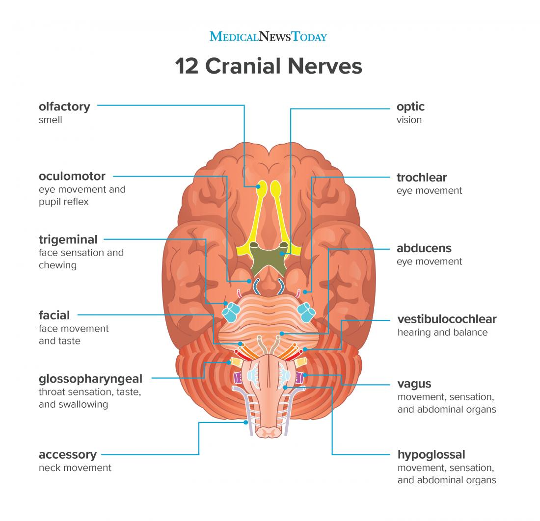
Facial nerve
A cranial nerve located on pons (apart of brainstem) which is responsible muscles of facial expression and taste from anterior 2/3 of tongue.
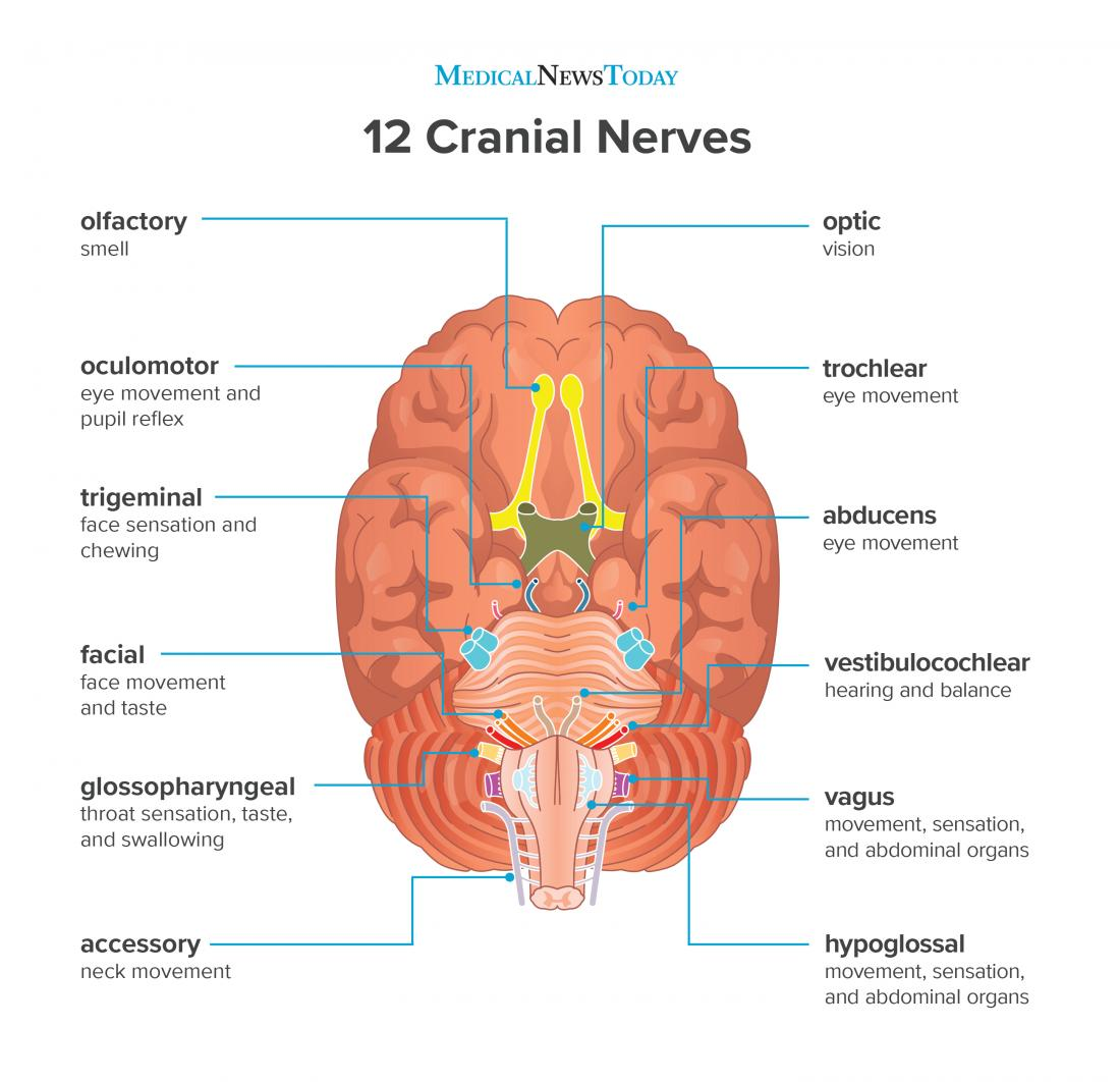
Vestibulocochlear nerve
A cranial nerve located on pons (apart of brainstem) which is responsible for balance, equilibrium, and hearing.

Glossopharyngeal nerve
A cranial nerve located on medulla oblongata (apart of brainstem) which is responsible for controlling throat muscles involved in swallowing and salivary glands, taste from posterior 1/3 of tongue, and monitoring blood pressure.
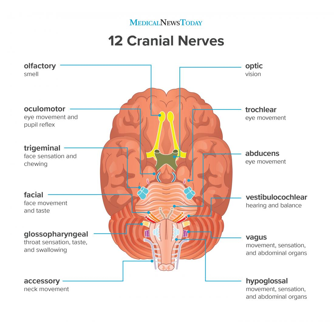
Vagus nerve
A cranial nerve located on medulla oblongata (apart of brainstem) which is responsible for controlling organ function, respiratory muscles, throat muscles; transmits sensory information from visceral organs, and rest-and-digest functions.
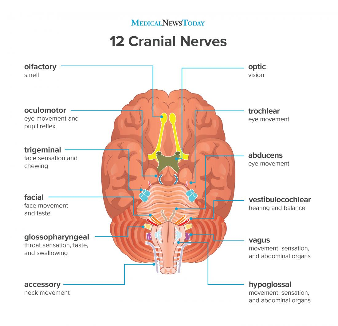
Spinal accessory nerve
A cranial nerve located on medulla oblongata (apart of brainstem) which is responsible for neck and upper back muscle movement.
Hypoglossal nerve
A cranial nerve located on medulla oblongata (apart of brainstem) which is responsible for tongue movement.
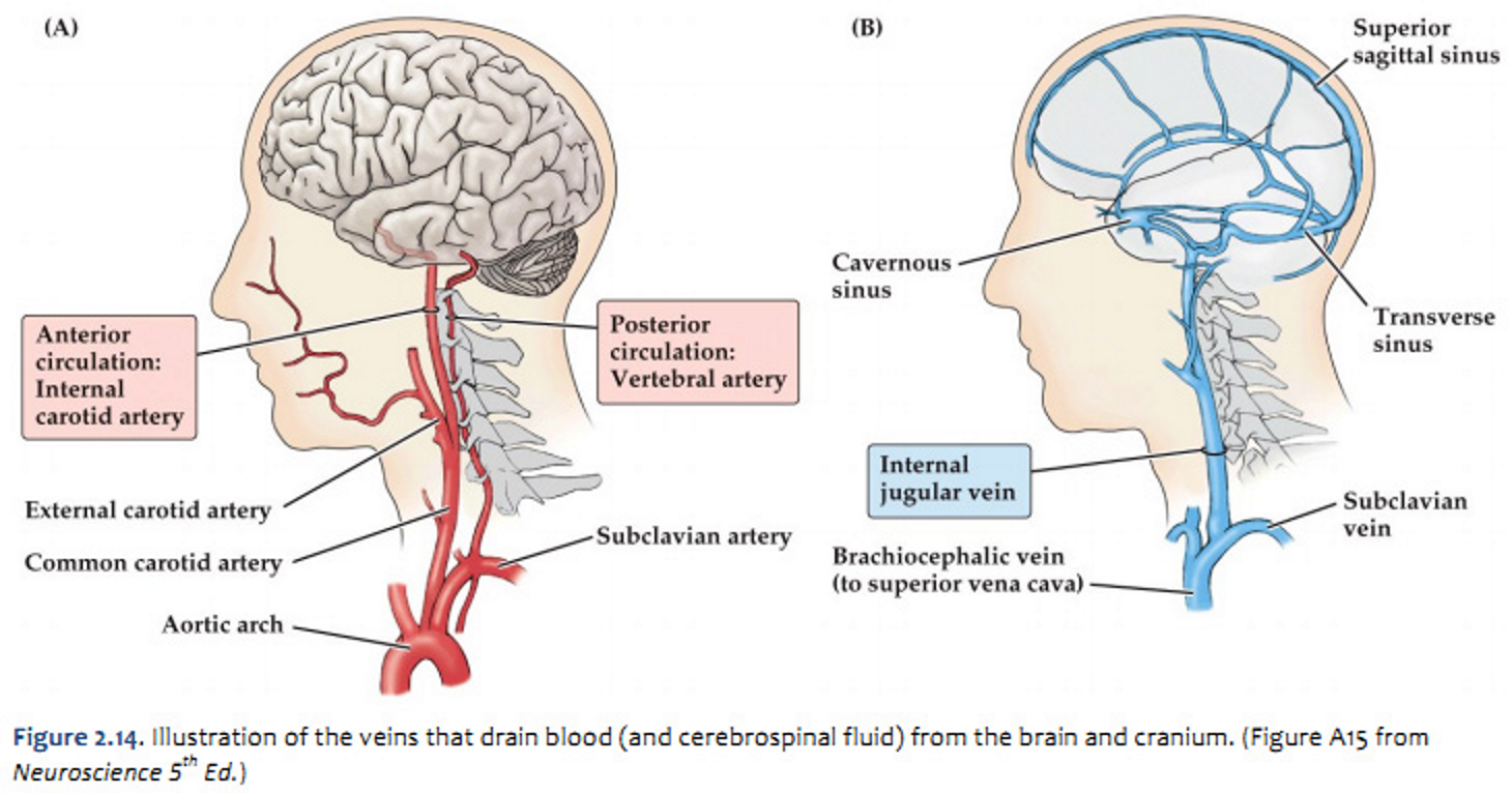
What is the blood supply like to the brain (2 points)?
Oxygenated (arterial) blood is distributed throughout the brain via arteries in the circle of Willis (shown in red).
Deoxygenated (venous) blood exists the cranium via the internal jugular veins, which drain blood from dural sinuses and vertebral veins (shown in blue).
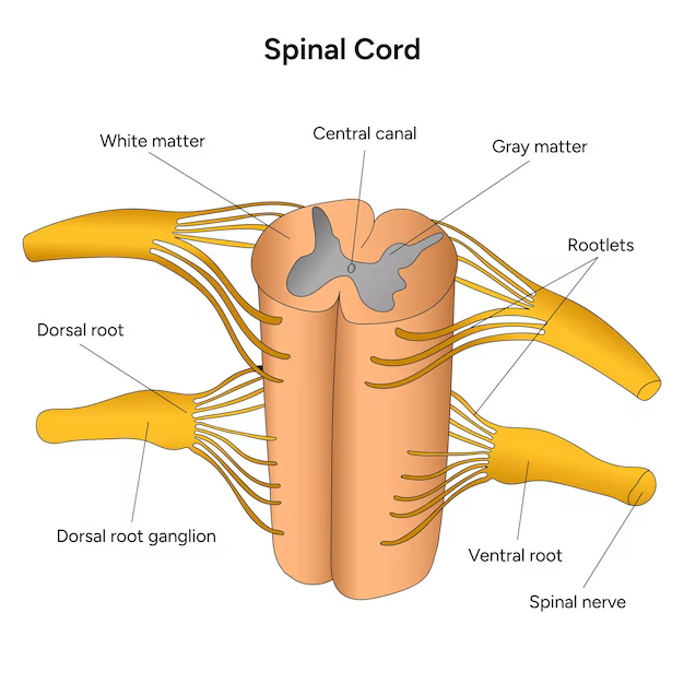
What is the function of three key structures in a spinal segment?
Dorsal root = Carries sensory (afferent) information into the spinal cord from the body
Ventral root = Carries motor (efferent) signals out of the spinal cord to muscles
Spinal nerve (mixed nerve) = Formed by dorsal + ventral roots joining which facilitate connection between the brain and various body parts.
What are the four plexuses?
Cervical plexus
Brachial plexus
Lumar plexus
Sacral plexus
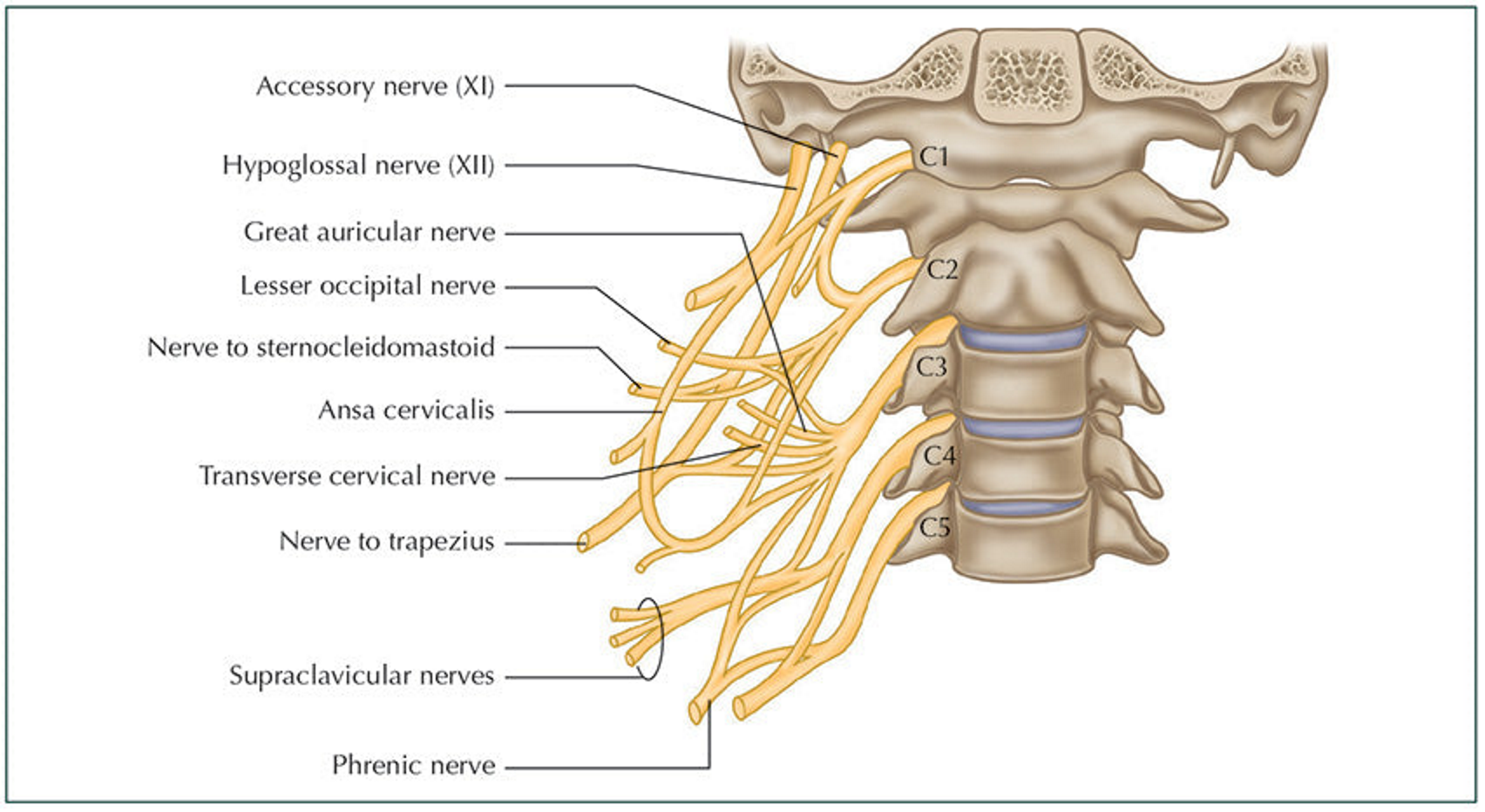
Cervical plexus and an example of a peripheral nerve
A plexus responsible for controlling the neck and diaphragm.
A peripheral nerve is phrenic.
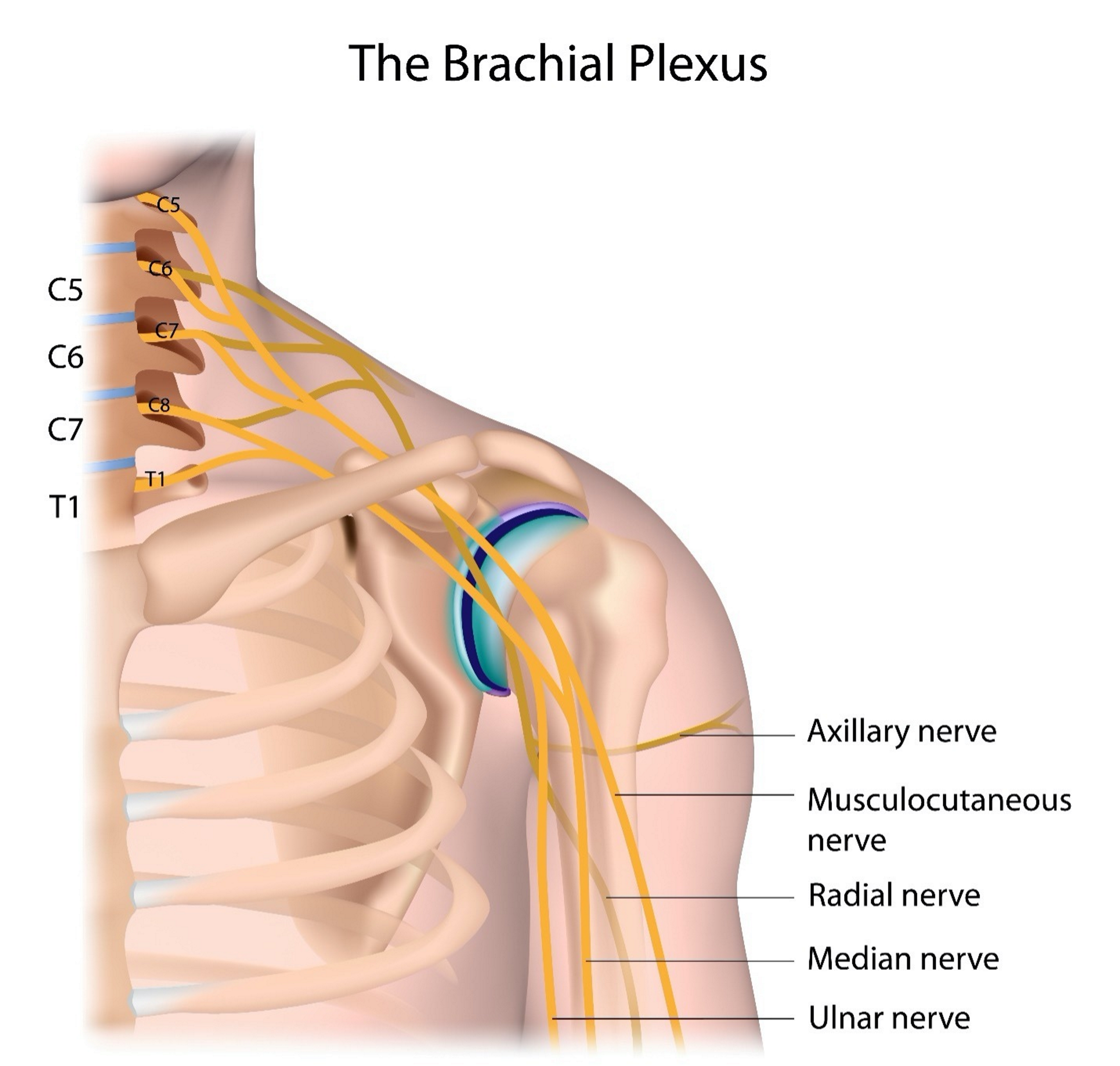
Brachial plexus and an example of a peripheral nerve.
A plexus responsible for controlling the pectoral and arm.
A peripheral nerve is musculocutaneous.
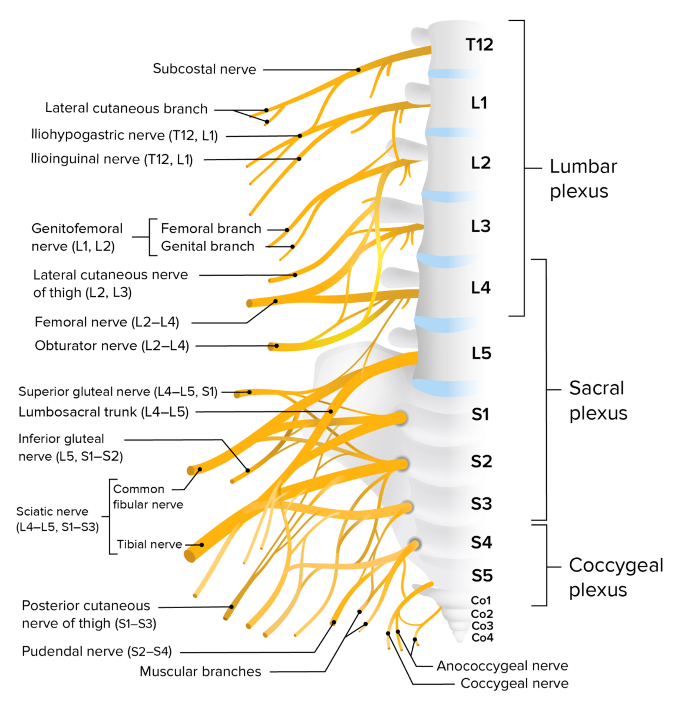
Lumar plexus and an example of a peripheral nerve
A plexus responsible for controlling abdomen, pelvis, and leg.
A peripheral nerve is femoral.
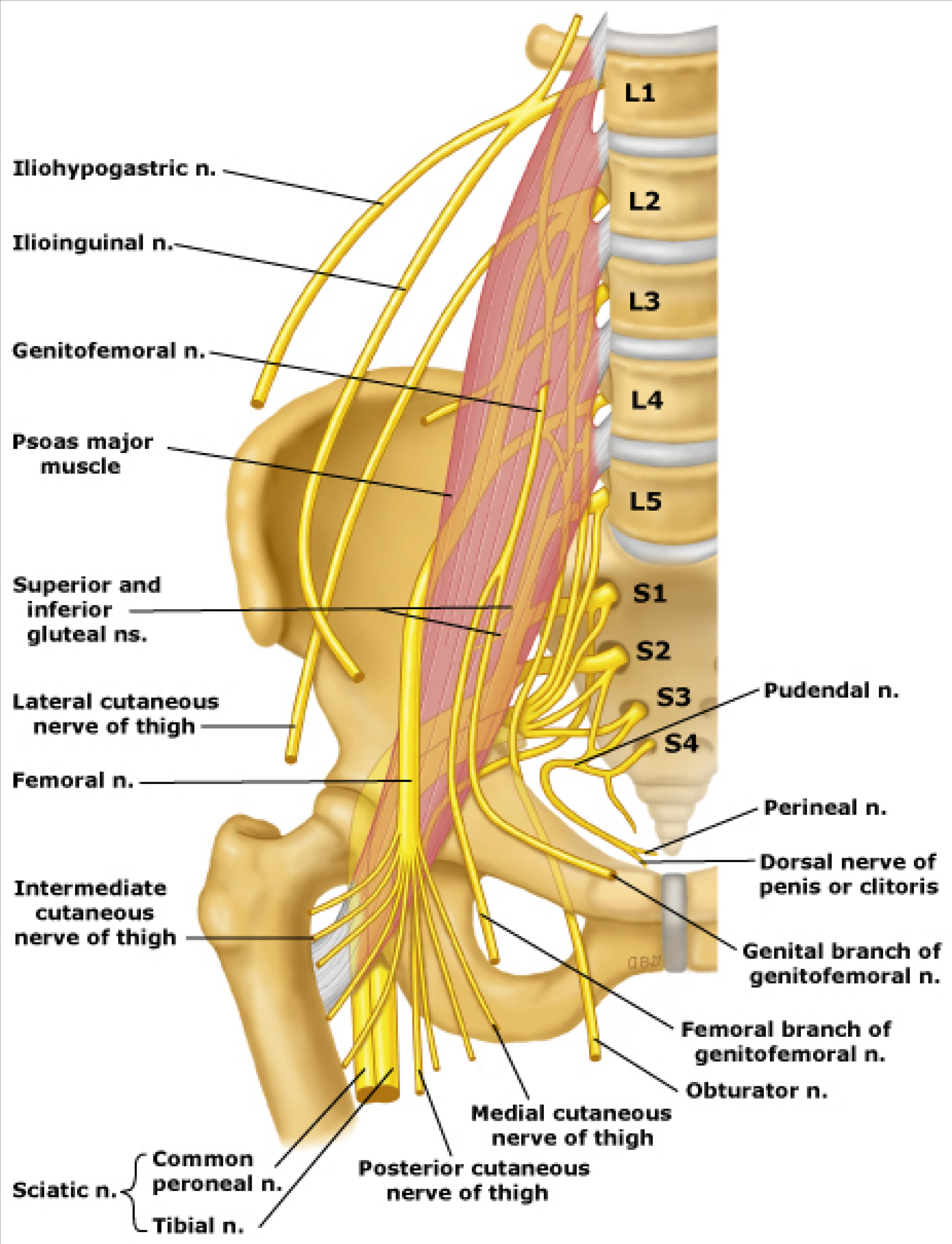
Sacral plexus and an example of a peripheral nerve
A plexus responsible for controlling pelvis and leg.
A peripheral nerve is sciatic.
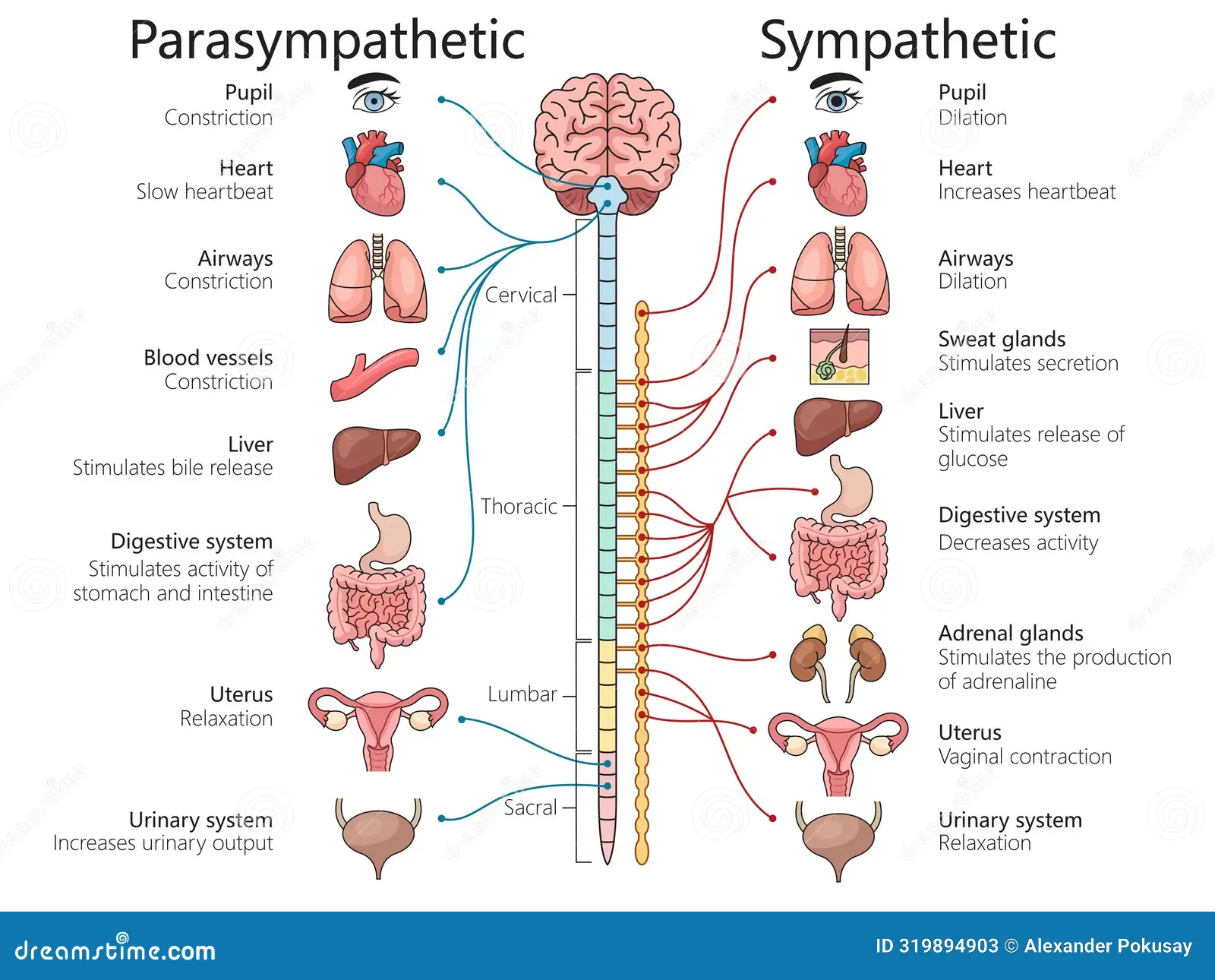
What are three divisions of the autonomic nervous system and functions?
Parasympathetic = Responsible for the rest-and-digest response.
Sympathetic = Responsible for the fight-or-flight response.
Enteric = Response for control of the gastrointestinal tract.