human a&p winter 2026 exam 1
1/94
There's no tags or description
Looks like no tags are added yet.
Name | Mastery | Learn | Test | Matching | Spaced | Call with Kai |
|---|
No study sessions yet.
95 Terms
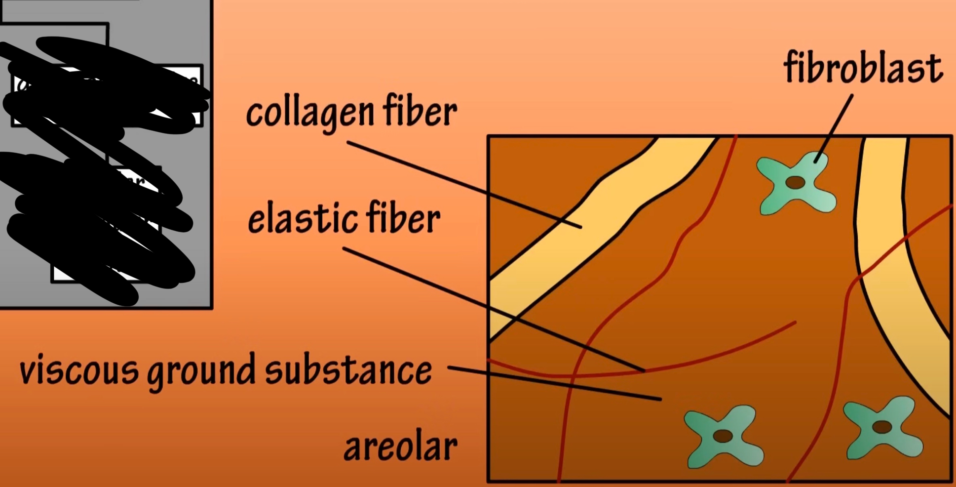
loose connective tissue areolar
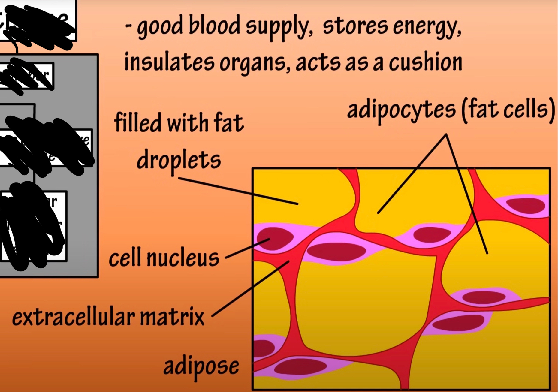
loose connective tissue adipose
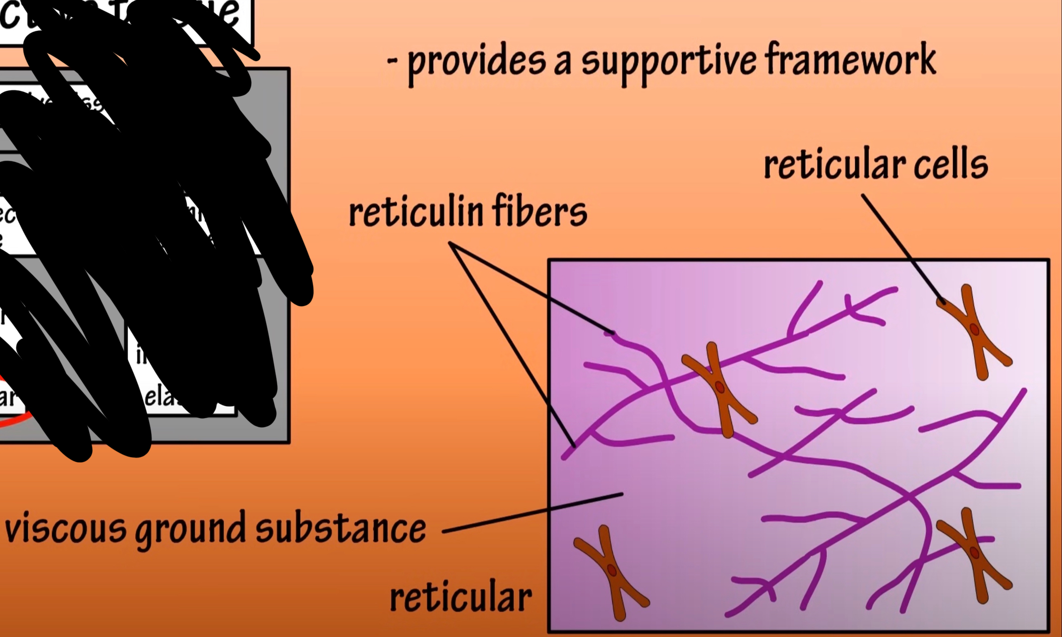
loose connective tissue reticular
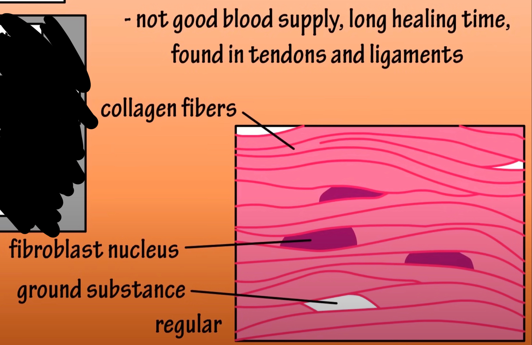
dense connective tissue regular
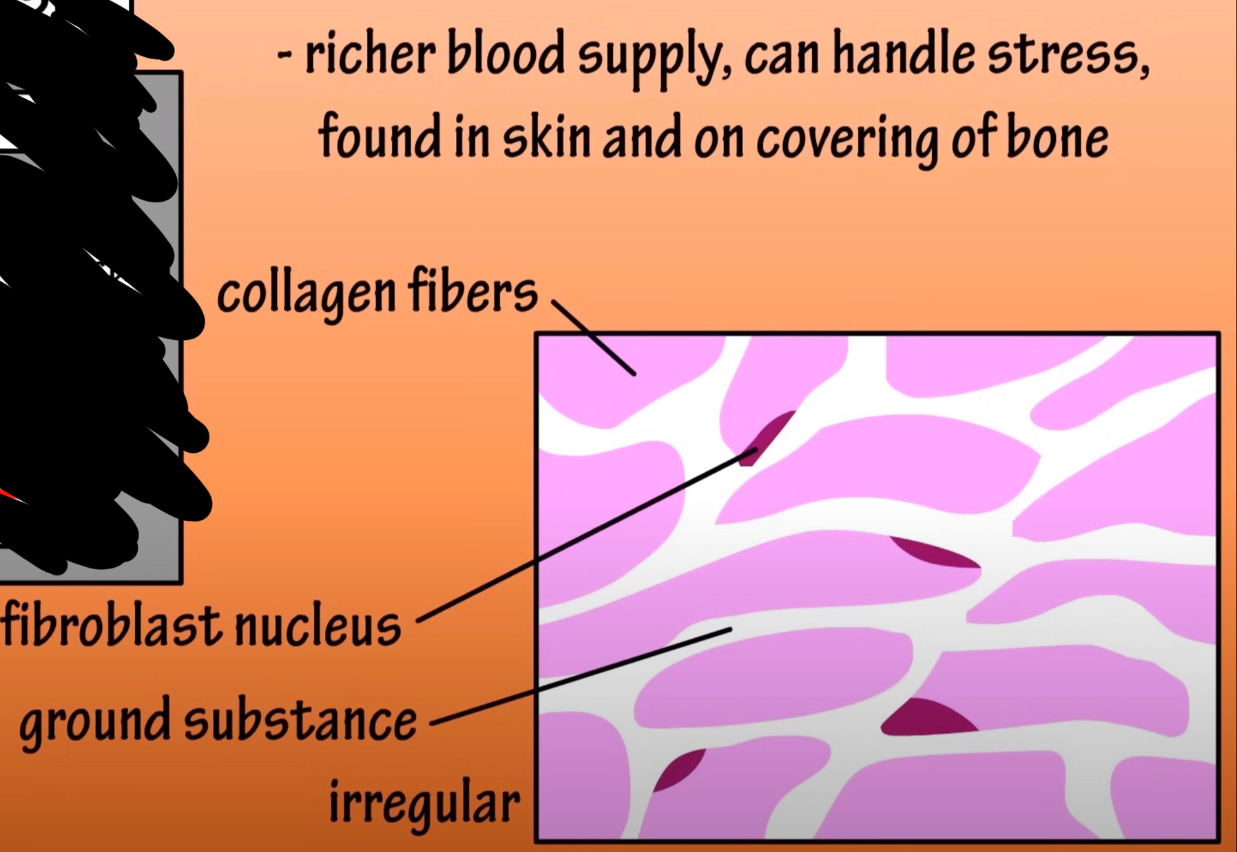
dense connective tissue irregular
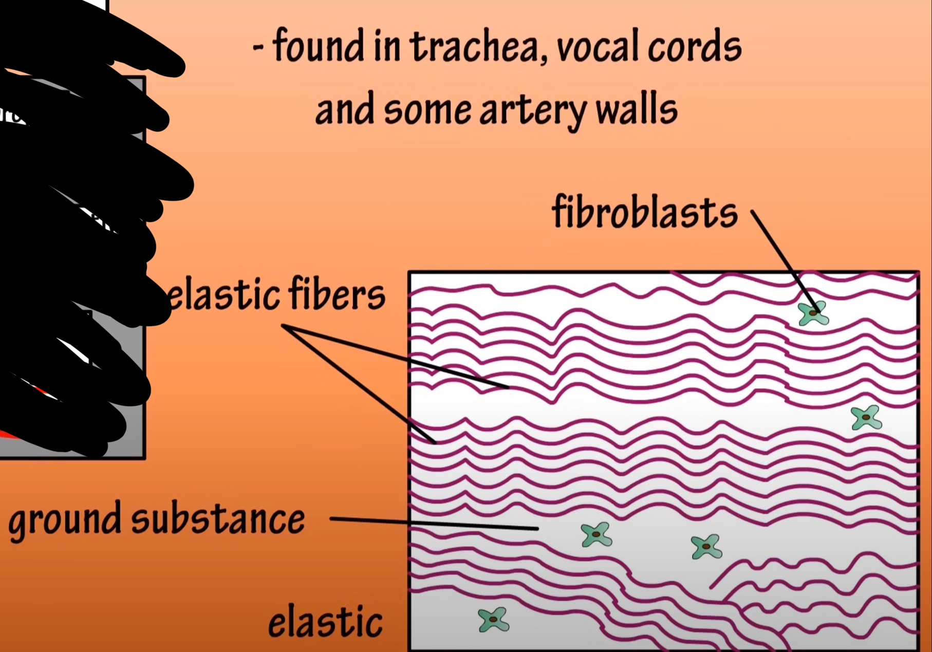
dense connective tissue elastic
When identifying the 3 general locations of epithelial tissue we find they form the blank, blank and blank of the body
coverings, linings and glands
list 3 general functions of epithelial tissue
protection, secretion & absorption
there are blood vessels present in epithelial tissue
false epithelial tissue is avascular
the cells in epithelial tissue have a lot of space between them
false they are tightly packed and have little space in between
there are nerve endings present in epithelial tissue
true
the free surface of epithelial tissue is known as the basement membrane
false the apical is the surface
to help epithelial tissues in their protective function, many epithelia produce secretions. identify 3 secretions produced by these tissues
sweat, mucous and tears
epithelial tissue on outermost layer of skin
stratified squamous
epithelial tissue lining of closed thoracic cavity
simple squamous
epithelial tissue covering the surface of the heart
simple squamous
epithelial tissue lining the closed abdominopelvic cavity
simple squamous
epithelial tissue lining of the mouth
stratified squamous
epithelial tissue forming glands and their ducts
simple cuboidal
epithelial tissue lining the trachea (respiratory passageway)
pseudostratified ciliated columnar
epithelial tissue covering the surface (outside) of the large intestine
simple squamous
epithelial tissue lining the anal canal (exit of the digestive tract)
stratified squamous
epithelial tissue lining the pharynx (throat) and esophaqus
stratified squamous
epithelial tissue lining the inside of the stomach
simple columnar
epithelial tissue lining the large intestine (colon)
simple columnar
epithelial tissue covering the ovary
simple cuboidal
epithelial tissue lining the inside of the uterus (female reproductive system)
simple columnar
epithelial tissue lining the inside of the urinary bladder
transitional
epithelial tissue lining the vagina (entrance to the female reproductive system)
stratified squamous
simple squamous
location - air sac of lungs, lining heart, blood vessels and lymphatic
functions - allows materials to pass through by diffusion & filtration & secretes lubricating substance
simple cuboidal
location - in ducts & secretory portions of small glands & in kidney tubules
function - secretes & absorbs
simple columnar
locations - ciliated tissues are in bronchi, uterine tubes & uterus ; smooth (non ciliated tissues) are in the digestive tract bladder
functions - absorbs also secretes mucous and enzymes
stratified squamous epithelium
location - lines the esophagus mouth and vagina
functions - protects against abrasion
pseudostratified ciliated columnar epithelium
location - ciliated tissue lines the trachea & much of the upper respiratory tract
function - secretes mucus ; ciliated tissue moves mucus
transitional epithelium
location - lines the bladder, uretha and ureters
function - allows the urinary organs to expand & stretch
areolar loose connective tissue
location - widely distributed under epithelia of body forms lamina propria of mucus membranes packages organ surrounds capillaries
function - wraps & cushion organs it macrophages phagocytize bacteria plays important role in inflammation holds & conveys tissue fluid
adipose tissue
location - under skin in the hypodermis around kidneys & eyeballs within abdomen & in breasts
function - provides reserve food fuels insulates against heat loss support and protect organs
reticular connective tissue
location - lymphoid organs (lymph nodes, bone marrow & spleen)
functions - fibers form a soft internal skeleton (stroma) that supports other cell types white blood cells, mast cell & macrophages
dense irregular connective tissue
location - fibrous capsules of organs & joints, dermis of the skin, submucosa of digestive tract
function - able to withstand tension exerted in many directions provides structural strength
dense regular connective tissue
location - tendons, most ligaments aponeuroses
function -attaches muscles to bones or to muscles
elastic connective tissue
location - walls of large arteries certain ligaments associate vertebral column; walls of bronchial tubes
function - allows recoil of tissue following stretching maintains pulsatile flow of blood through arteries aids recoil of lungs
hyaline
location - embryonic skeleton covers the end of the long bone in joint cavities costal cartilages ribs, nose, trachea & larynx
functions - supports & reinforces resilient cushioning properties resists compressive stress
fibrocartilage
location - intervertebral disc pubicsymphysis discs of knee joint
function - tensile strength with ability to absorb compressive shock
elastic cartilage
location - supports the external car
function - maintain the shape cartilage but more elastic fibers in matrix
bone
location - bone
function - supports & protects stores calcium & fat
blood
location - contained within blood vessels
function - transport oxygen, nutrients, hormones and waste products throughout the body while helping regulate temperature and fight infection
the skin is superficial to the bones
true
the ears are medial to the eyes
false the ears are lateral to eyes
the toes are posterior to the heel
false the toes are anterior to the heel
the small intestine is superior to the diaphragm
false is inferior to the diaphragm
the wrist is distal to the elbow
true
gallstones quadrant
RUQ
hepatitis quadrant
RUQ
stomach ulcer’s quadrant
LUQ
perforated intestine quadrant
any
ruptured spleen quadrant
LUQ
bladder infection quadrant
RLQ LLQ
appendicitis quadrant
RLQ
one effect of tobacco smoke is to destroy the cilia in the respiratory tract. what effect might this have on the respiratory function of the smoker?
prevents lungs from clearing mucus & debris is leading to mucus build up, coughing, higher risk of respiratory infection
tendons and ligaments are composed primarily of dense regular connective tissue. if a tendon or ligament is damaged, why does it take so long for it to heal?
poor blood supply
protraction
moving a body part forward
retraction
moving a body part downward
abduction
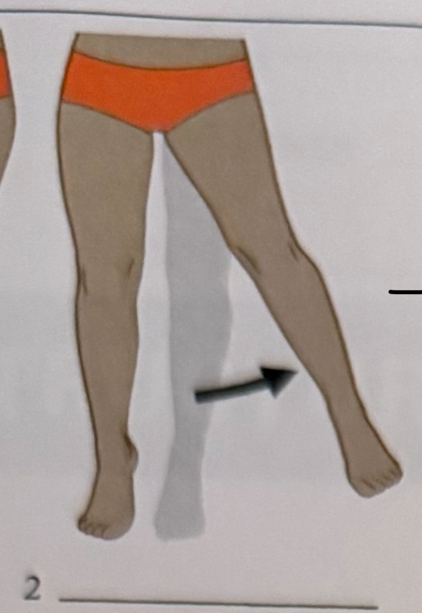
moving a body part away from the midline
adduction
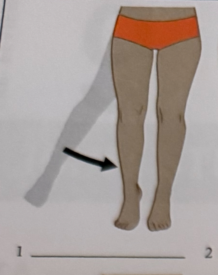
moving a body part toward the midline
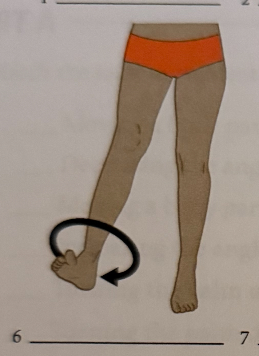
circumduction
moving a body part upward
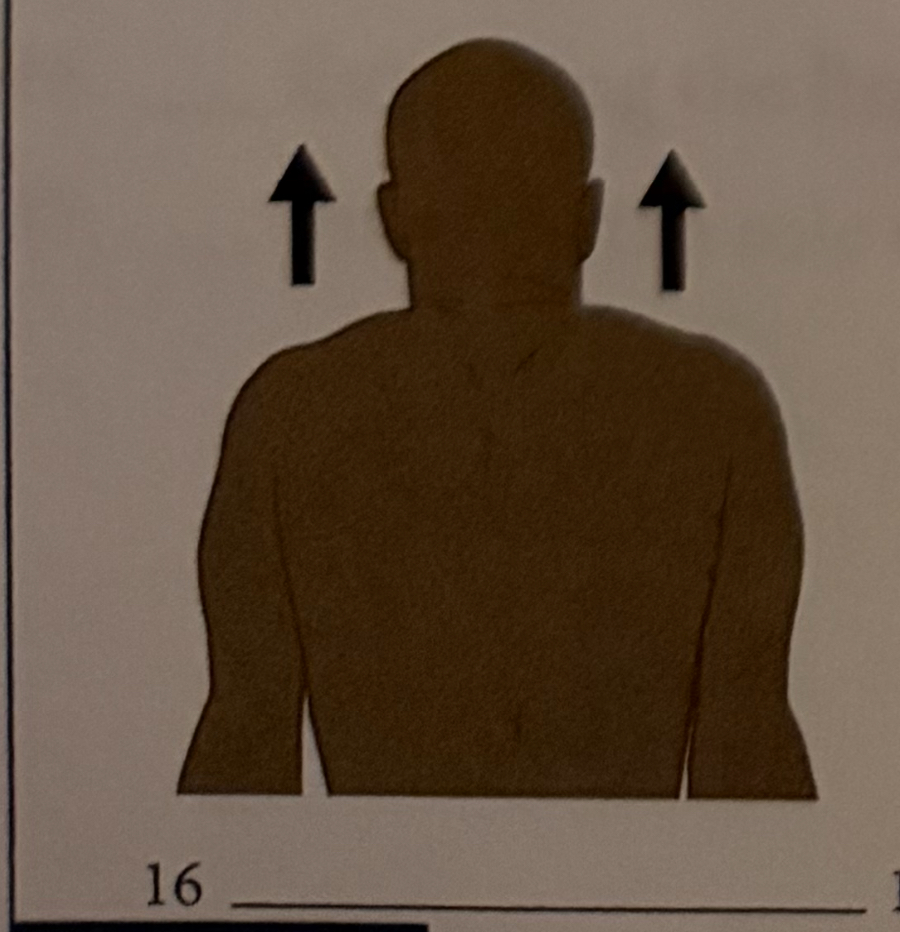
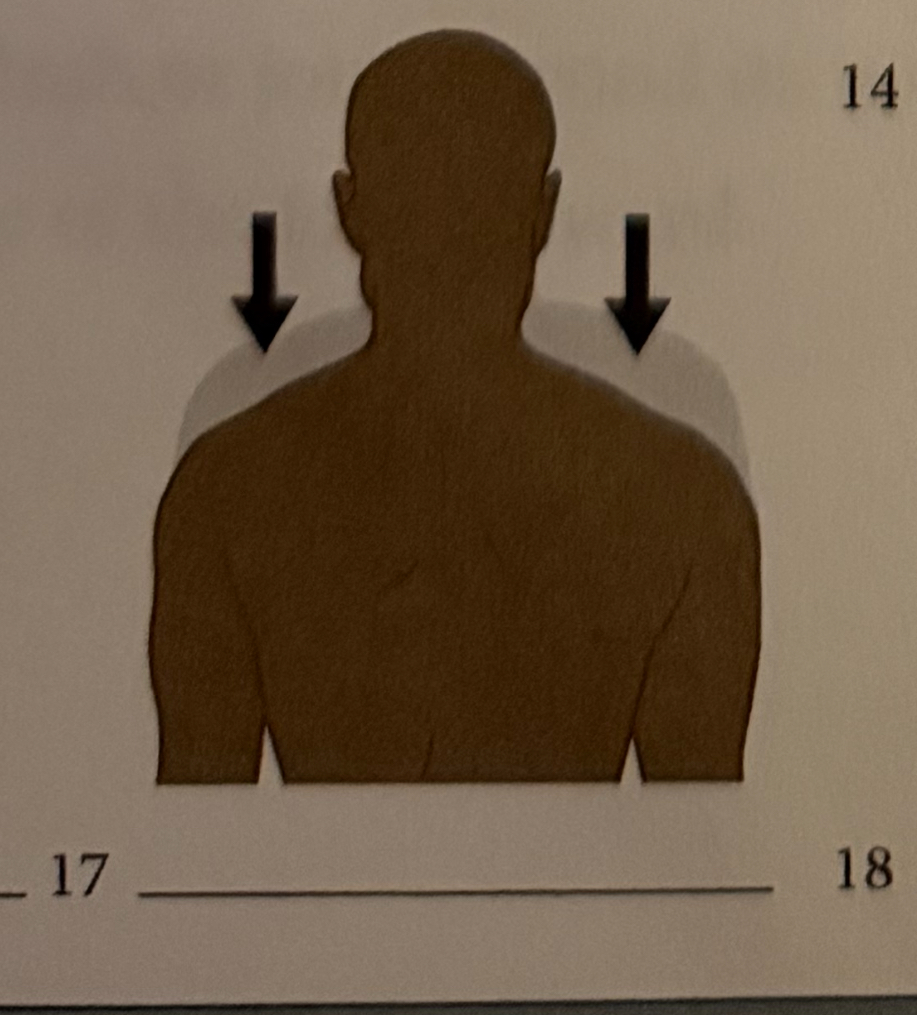
elevation
raising the heel of the foot
depression
moving a body part downward
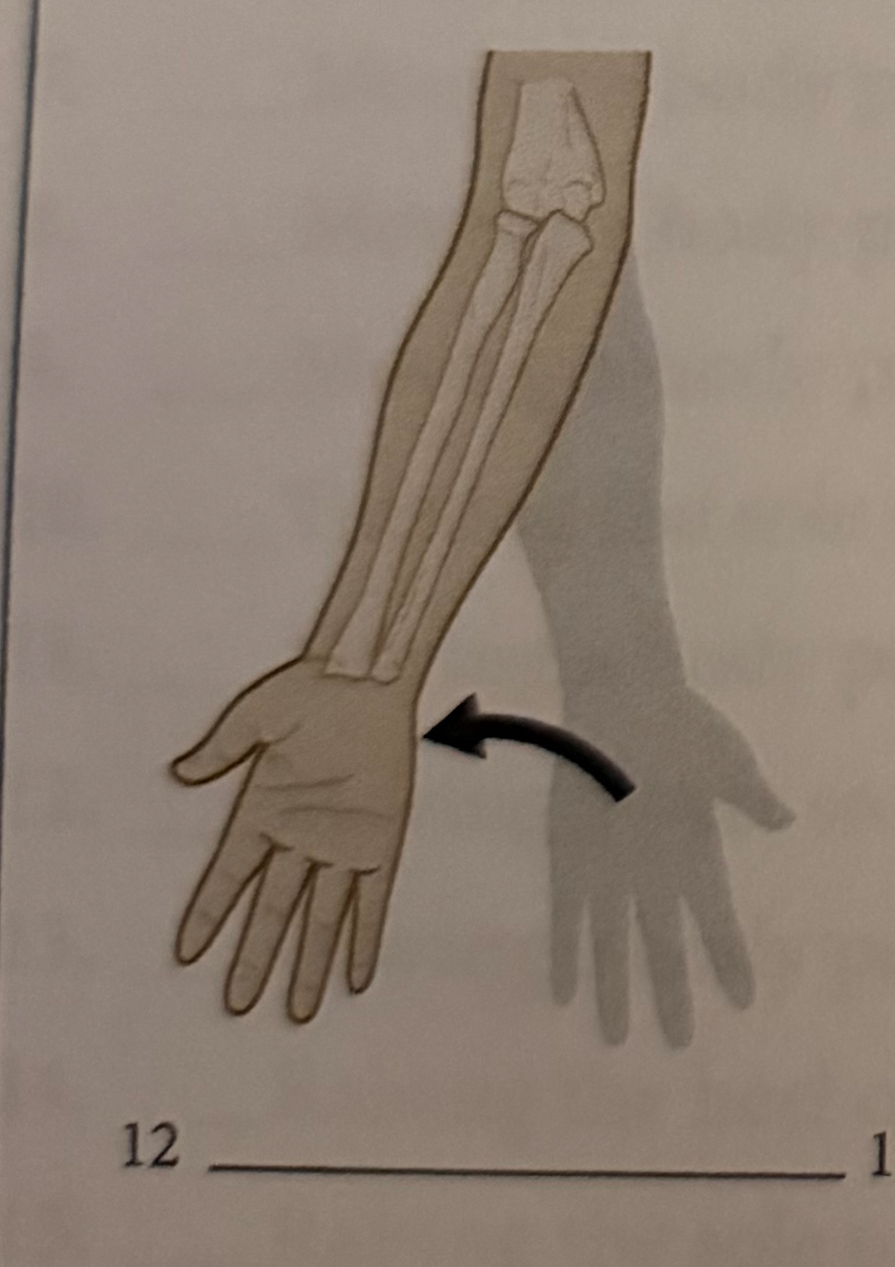
pronation
turning the palm downward
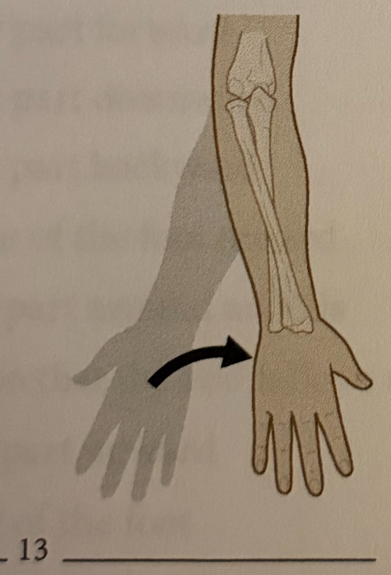
supination
turning the palm upward
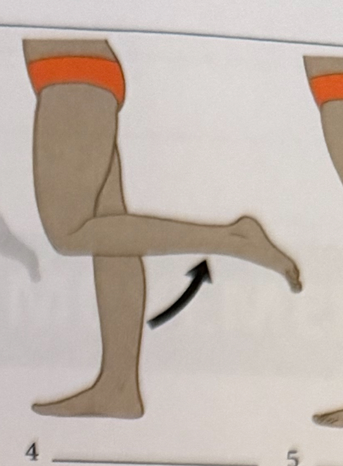
flexion
decreasing the angle of a joint

extension
increasing the angle of a joint
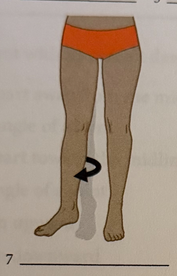
rotation
moving a body part around an axis
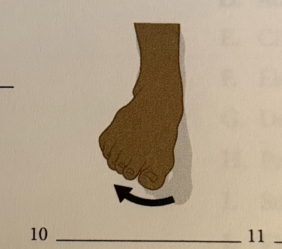
inversion
turning the sole of the food inward
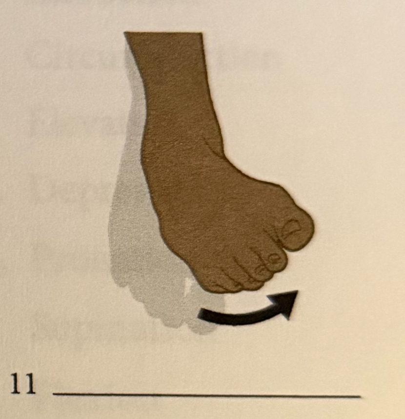
eversion
turning the sole of the foot outward
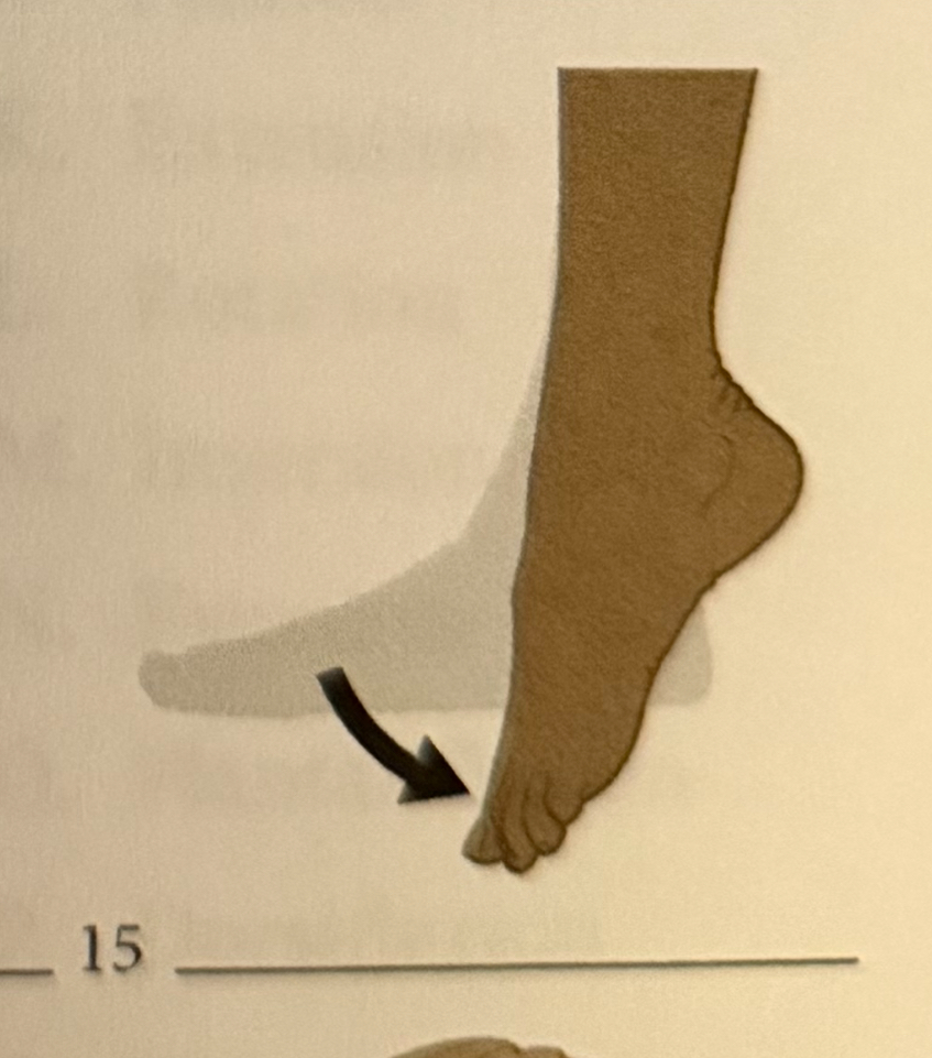
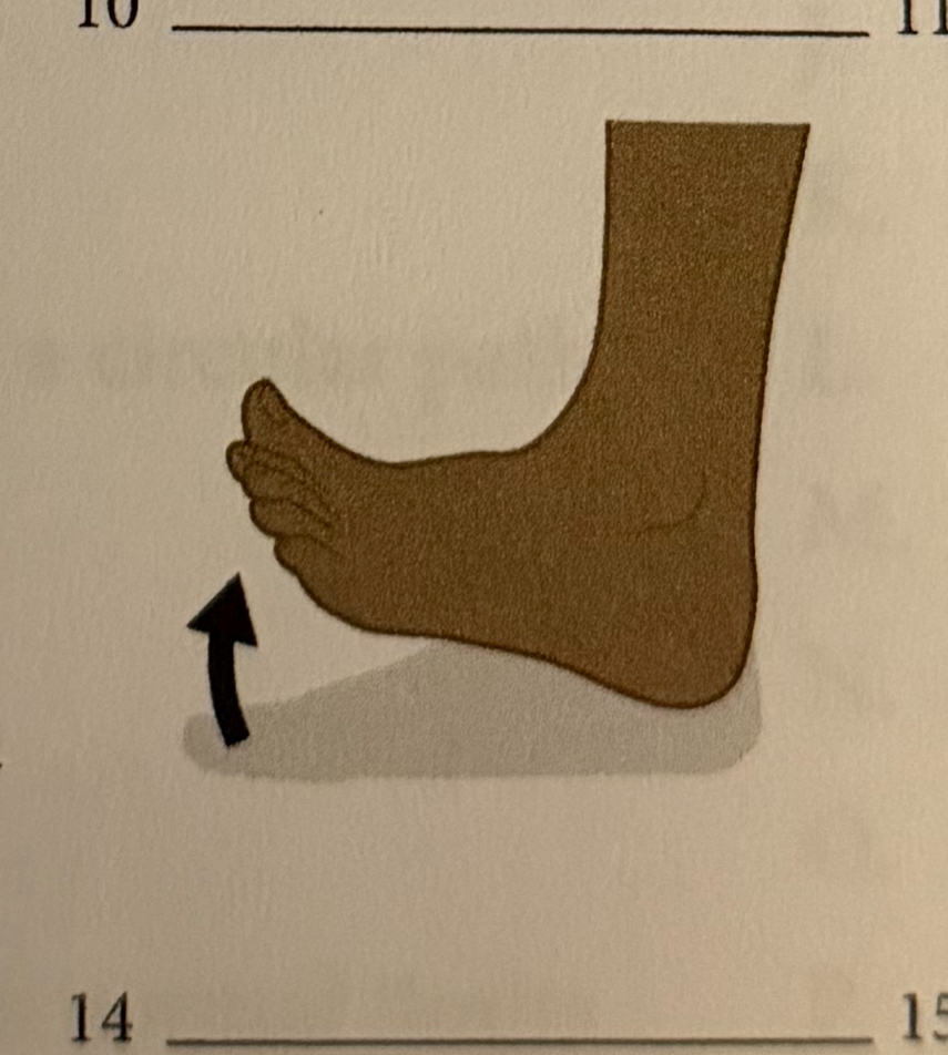
plantar flexion
raising the sole of the foot
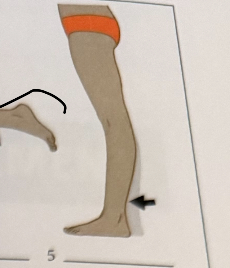
dorsiflexion
extending the limb or joint beyond its normal limits
hyperextension
moving a part so that the end follows a circular path
simple
cells that are thin and scale like
stratified
thin and branching these fibers form a supportive network for soft organs
pseudostratified
a single layer of cells that appear to be in several layers
columnar
cells arranged in a single layer
squamous
elongated, rectangular cells
cuboidal
cube shaped cells with a large central nucleus
collagen fibers
usually thick, these fibers provide strength and resiliency
elastic fibers
cells arranges in more than one layer
reticular fibers
usually thin these fibers allow a tissue to stretch and recoil to its original shape
matrix
material between connective tissue cells
fibroblasts
cells that secrete fibers
adipocytes
cells that store fat
chondrocytes
cartilage cells
lacuna
space within the matrix that contains a chondrocyte or osteocyte
osteocyte
bone cell
canaliculi
tiny channels in the bine tissue that connects one lacuna to another
lamellae
circular layers that compose an osteon
osteon
cylindrical unit of bone