Skin (cont.), Hair, Nails
 Call Kai
 Learn
Learn Practice Test
 Spaced Repetition
 Match
Match Flashcards
 Knowt Play
1/28

There's no tags or description
Looks like no tags are added yet.
Name | Mastery | Learn | Test | Matching | Spaced | Call with Kai | Chat |
|---|
No analytics yet
Send a link to your students to track their progress
29 Terms
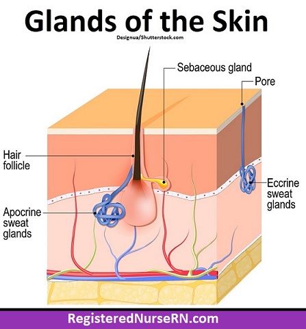
Sweat pore
the tiny surface opening of a sweat gland duct through which sweat is excreted onto the skin

Eccrine gland
secretes watery sweat for thermoregulation all over the body
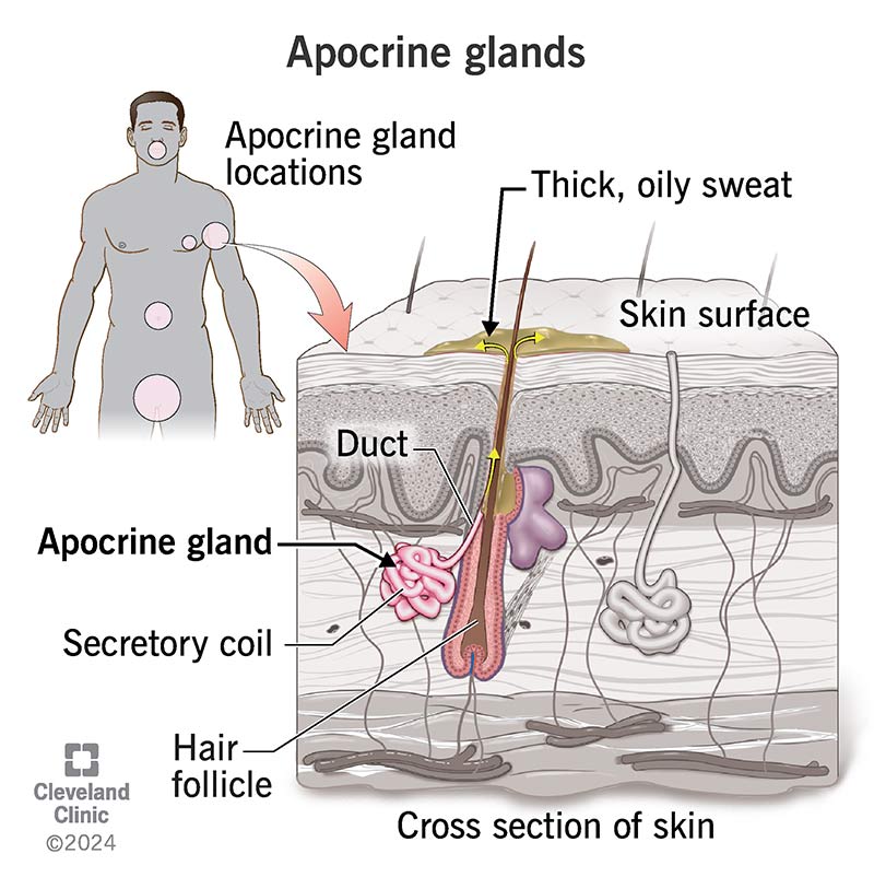
Apocrine gland
sweat gland located in axillary and anogenital areas that secretes a viscous, odor-producing fluid into hair follicles

Sebaceous gland
connected to hair follicles that secretes oily serum to lubricate skin and hair

Ceruminous gland
modified apocrine sweat gland in the ear canal that produces earwax (cerumen)
Sensory nerve
nerve fiber that transmits sensory signals from the skin to the CNS

Free nerve ending
unspecialized nerve endings in the epidermis and dermis that detect pain and temperature changes

Tactile corpuscle/Meissner’s corpuscle
encapsulated sensory receptor in the papillary dermis sensitive to light touch and low-frequency vibration

Lamellar corpuscle/Pacinian corpuscle
layered sensory receptor deep in the dermis/hypodermis sensitive to deep pressure and high-frequency vibration
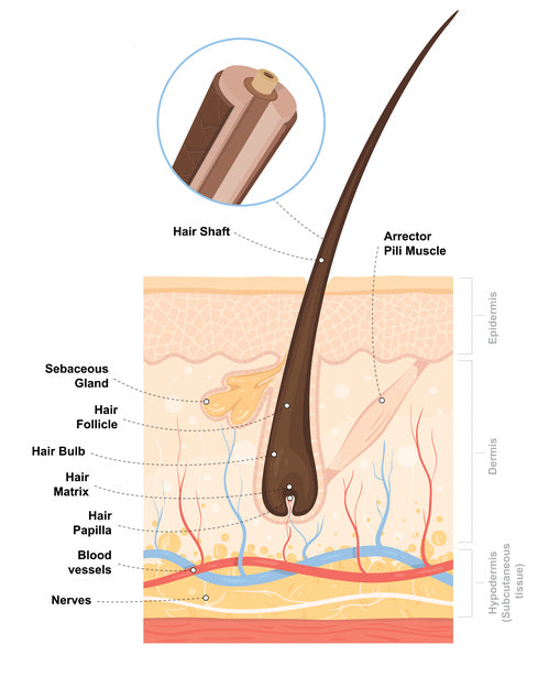
Hair shaft
portion of the hair that projects above the skin surface
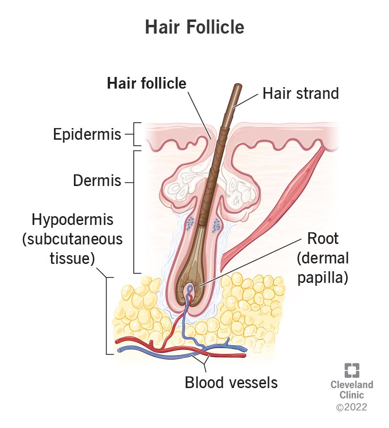
Hair root
portion of the hair embedded beneath the skin surface
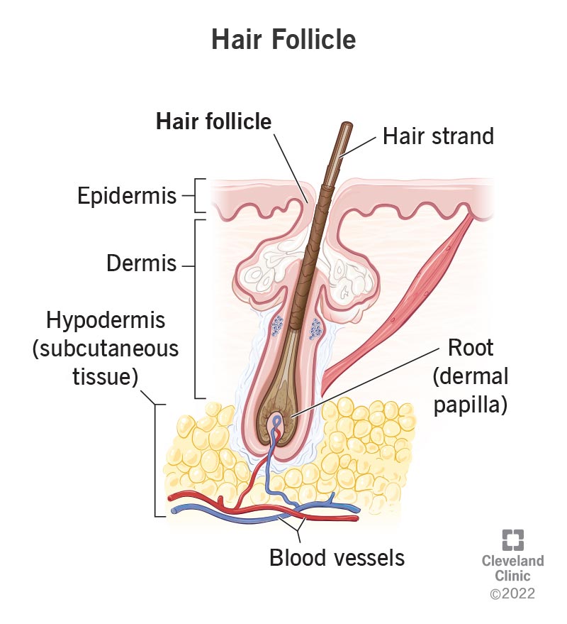
Hair follicle
tubular epithelial sheath that surrounds and produces the hair root
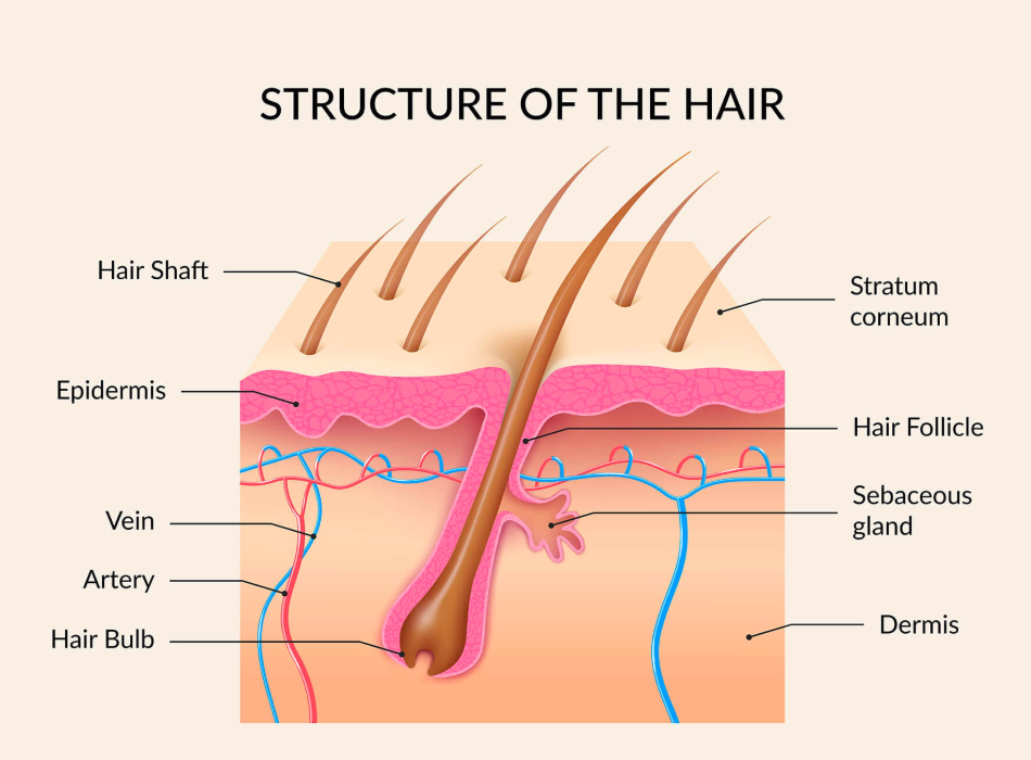
Hair bulb
expanded, bulbous base of the hair follicle containing dividing cells
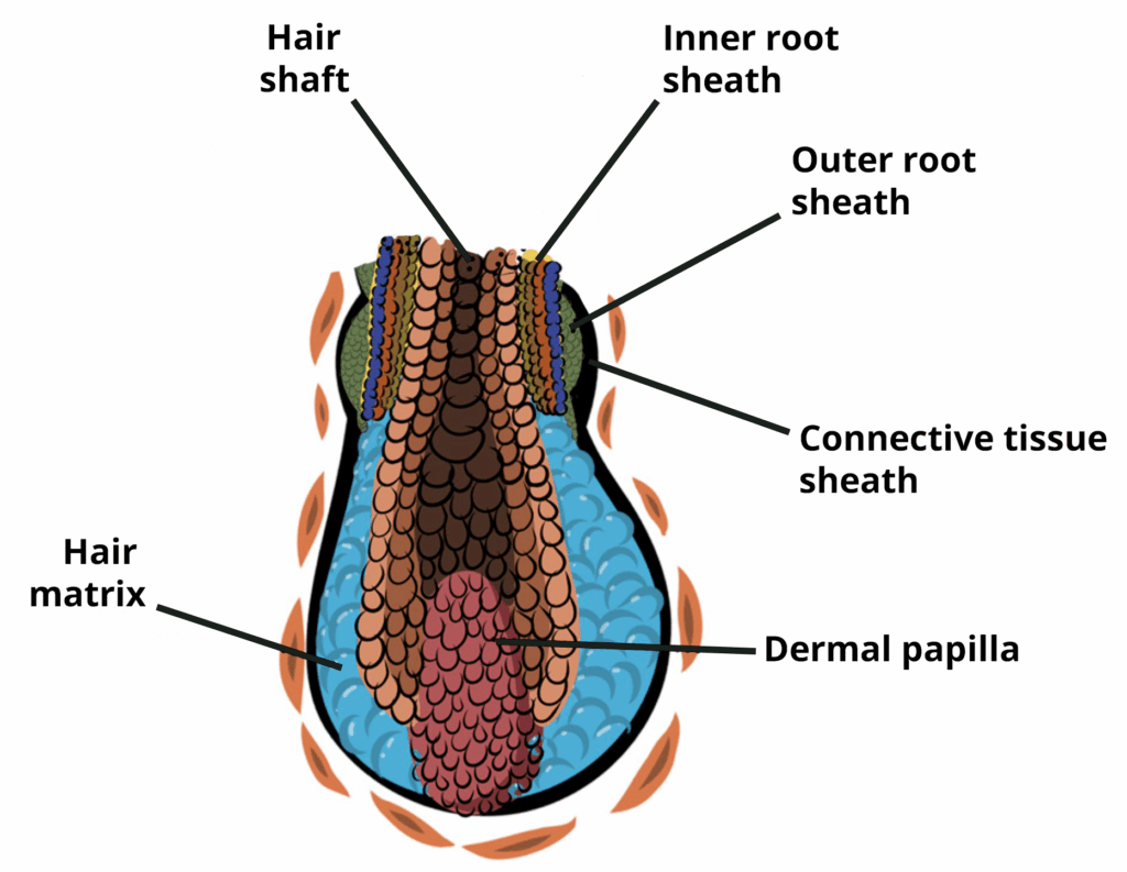
Papilla of hair
vascular connective tissue projection extending into the base of the hair bulb to supply nutrients

Hair matrix
layer of actively dividing germinal stem cells inside the hair bulb that produces the hair shaft

Arrector muscle of the hair/Arrector pili
smooth muscle attached to the hair follicle that contracts to pull the hair upright (goosebumps)
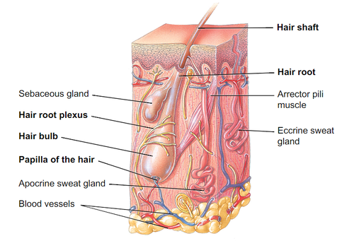
Hair root plexus
network of sensory nerve endings surround the hair follicle that detects hair movements
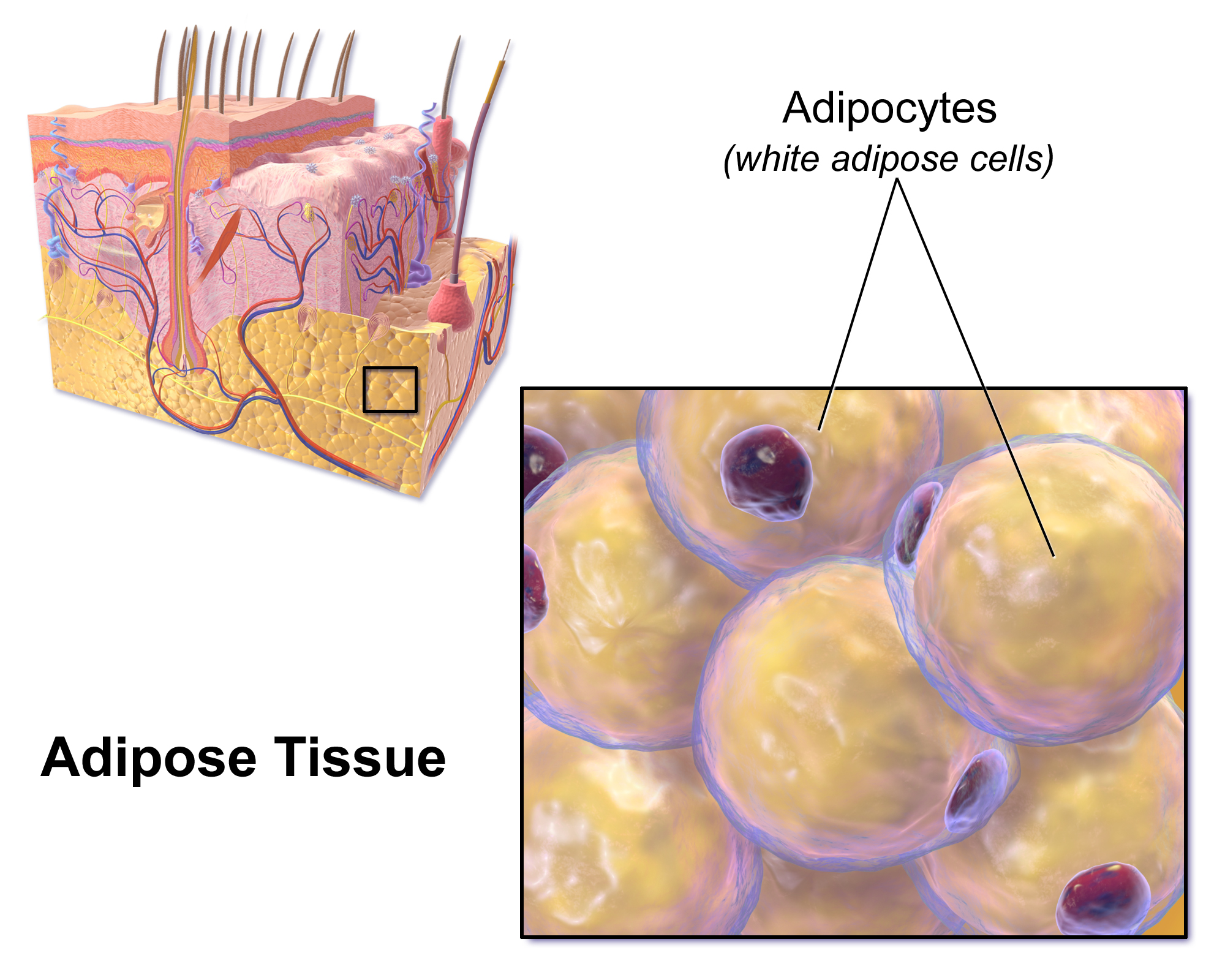
Adipose tissue
fat-storing connective tissue in the hypodermis that provides insulation, cushioning, and energy storage
Cutaneous vascular plexus
deep vascular network located at the boundary between the dermis and hypodermis
Papillary vascular plexus
superficial network of blood vessels located directly beneath the dermal papillae

Capillary loops
microscopic loops of blood vessels within the dermal papillae that nourish the epidermis
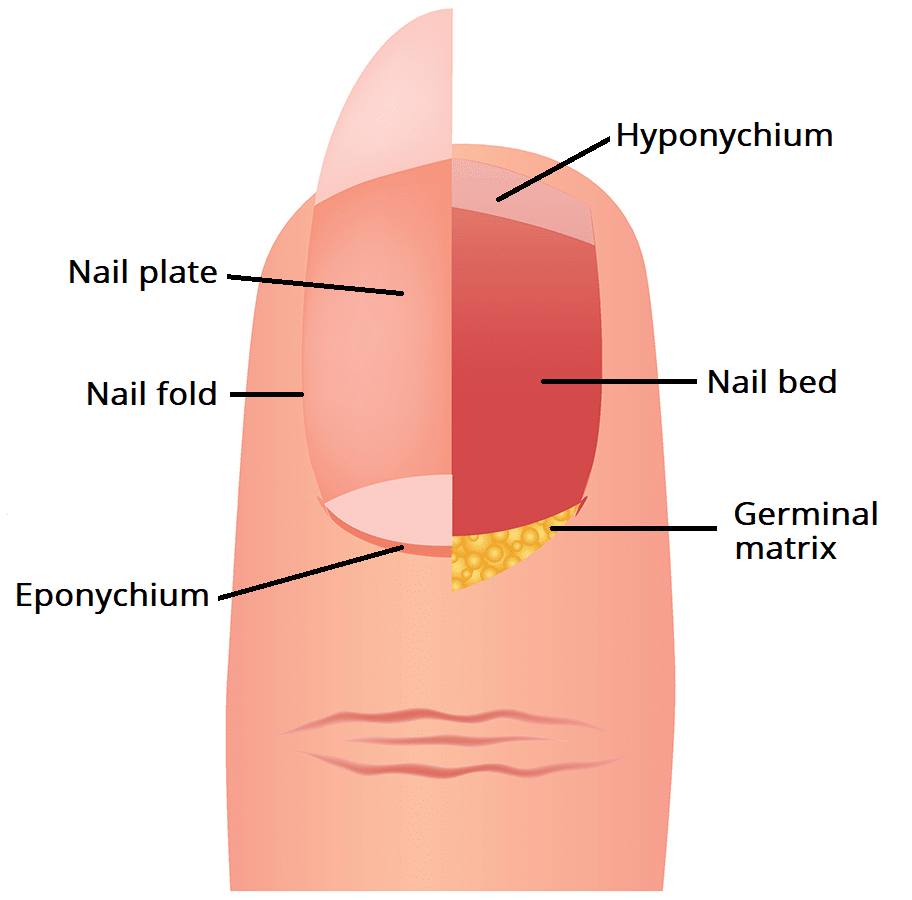
Nail plate
hard, exposed outer body of the nail composed of densely packed dead keratinized cells
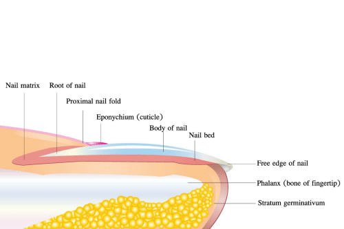
Nail root
hidden proximal portion of the nail embedded beneath the fold of skin at the base
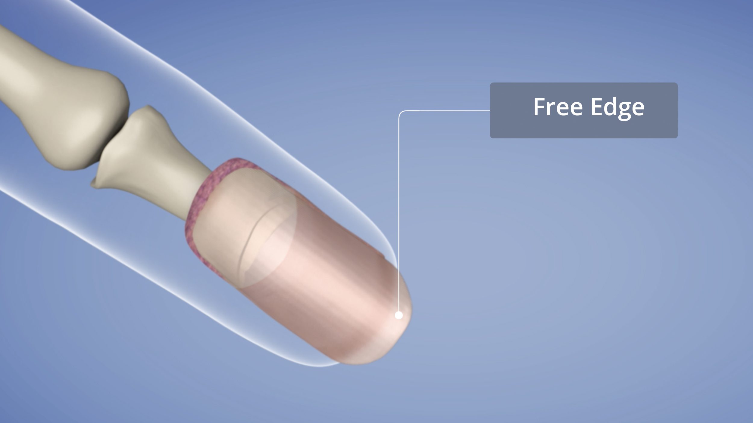
Free edge
the distal extension of the nail plate that overhands past the fingertip or toe

Nail matrix
actively dividing epithelial tissue at the proximal end under the nail root responsible for nail growth

Nail bed
layer of vascular epidermal tissue beneath the nail plate upon which the nail rests
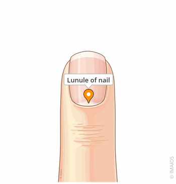
Lunule
pale, crescent-shaped area at the proximal base of the nail plate overlying the thick nail matrix
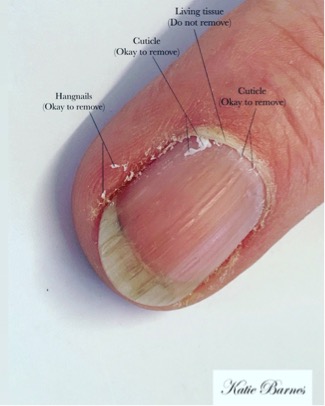
Eponychium/Cuticle
narrow fold of stratum corneum skin extending onto the base of the nail plate

Hyponychium
thickened region of stratum corneum beneath the free edge of the nail plate