2: Structure and Function of The Nervous System
1/68
There's no tags or description
Looks like no tags are added yet.
Name | Mastery | Learn | Test | Matching | Spaced |
|---|
No study sessions yet.
69 Terms
cells in the nervous system
neurons - transmit information;
glial cells - function depends on type (oligodendrocytes, Schwann cells, microglial cells, astrocytes);
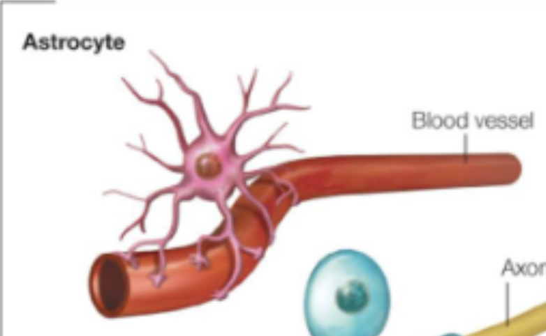
astrocytes
surround neurons, are in contact with blood vessels;
create the blood-brain barrier;
can modulate neuronal activity
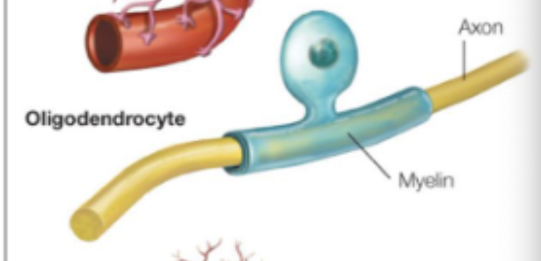
oligodendrocytes
CNS;
form myelin sheath around neuron’s axons: electrical insulation;
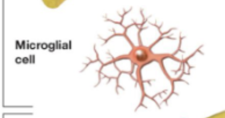
microglial cells
remove damaged cells;
Schwann cells
PNS;
form myelin sheath around neuron’s axons: electrical insulation;
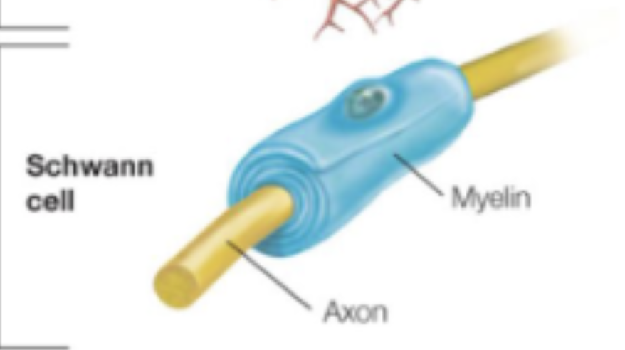
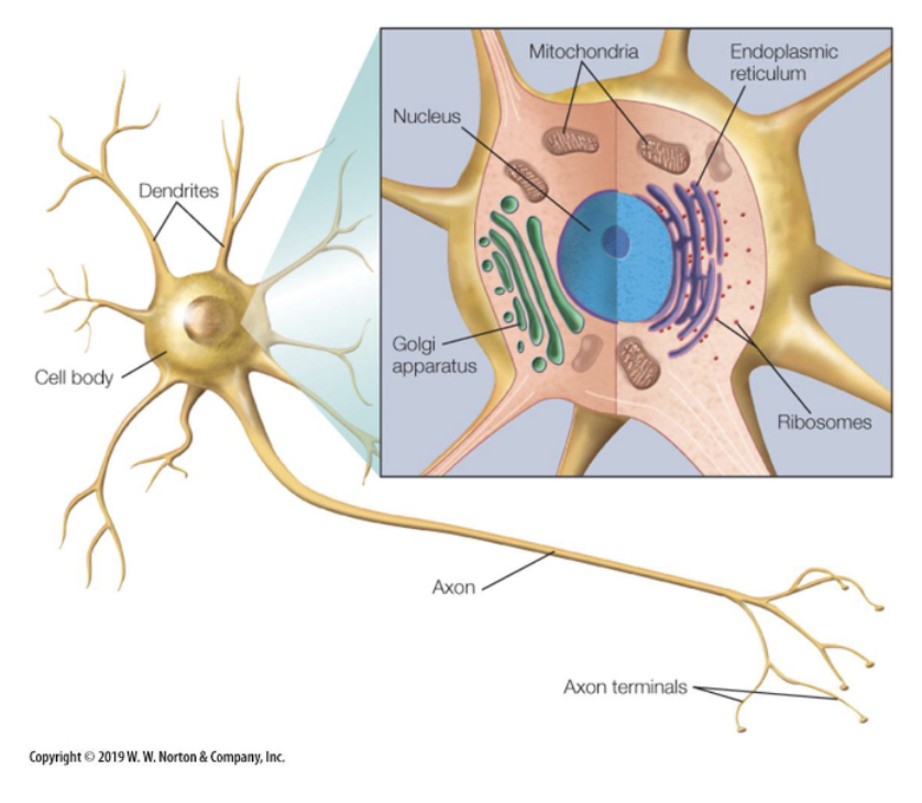
neuron
a typical eukaryotic cell with a cell membrane that encases a cell body.
The cell body (soma) is filled with cytoplasm, a salty intracellular fluid, that suspends the metabolic machinery that maintains the neuron (e.g., nucleus, ribosomes, Golgi apparatus).
In addition to a cell body, a neuron also has dendrites and an axon.
Dendrites receive input from other neurons (at the dendritic spines).
The axon outputs a signal to other neurons (at the axon terminals).
An electrical signal flows from the dendrites to the cell body to the axon.
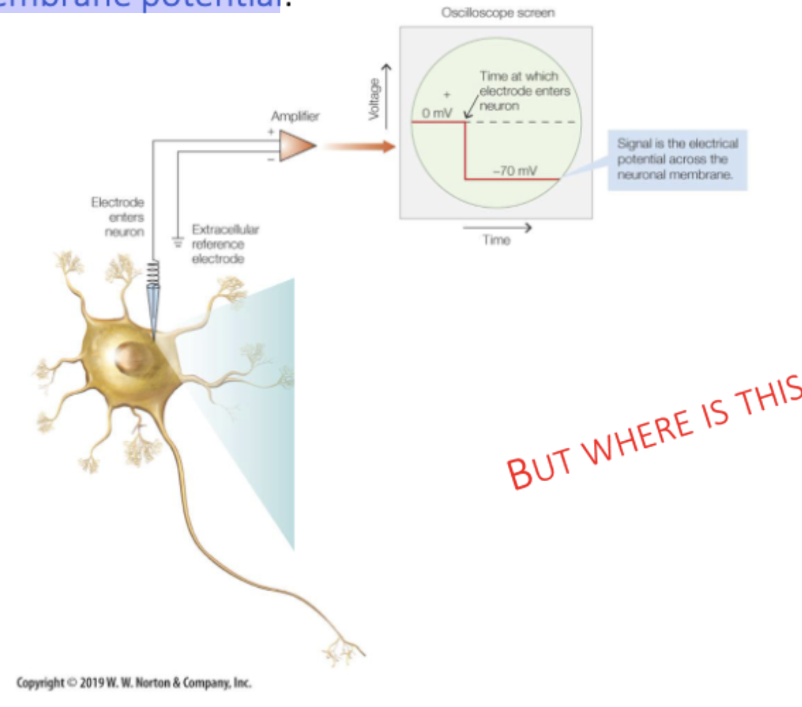
resting membrane potential
When a neuron is not sending a signal, it is at rest. If you stick an electrode in the neuron (with the extracellular space as a reference), you will measure an electrical voltage of -70mV.
ions
atoms or molecules that have either a positive or negative charge.
The intra- and extracellular fluid is made up of a combination of ions, predominantly:
Potassium (K+)
Sodium (Na+)
Calcium (Ca2+)
Chloride (Cl-)
Organic anions (A-)
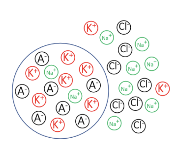
The intracellular fluid has more K+, the extracellular space has more Na+.
transmembrane proteins
in the cell membrane, allow the passing of ions.
ion channels: selectively permit one type of ion to pass. More potassium (K+) channels in cell membrane.
ion pumps: active transport proteins. Sodium-potassium pump pumps 3 sodium ions (Na+) out of the cell and 2 potassium ions into the cell. Requires energy!!
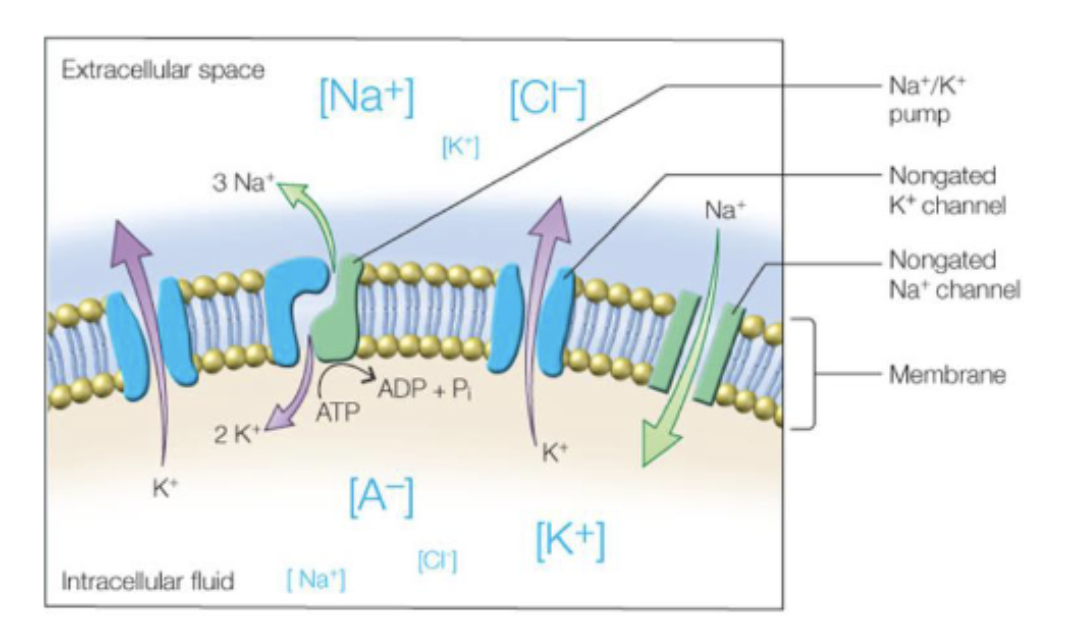
concentration gradient
Intracellular fluid consists mostly of: positively charged potassium ions (K+) and negatively charged organic anions (A-).
Extracellular fluid consists mostly of: positively charged sodium ions (Na+) and negatively charged chloride ions (Cl-).
The difference in the concentration of K+ and Na+ inside and outside the cell is caused by the Na+/K+ pump (an enzyme that actively ‘pumps’ Na+ out of the cell and pumps K+ inside the cell).
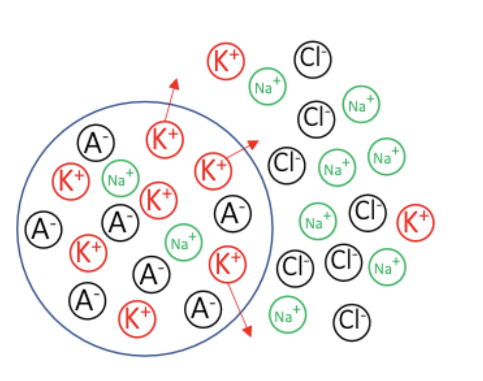
The cell membrane is mostly permeable to K+ (via potassium channels). The force caused by the concentration gradient wants to move K+ from intra- to extracellular space.
electrical gradient
The cell membrane is mostly permeable to K+ (via potassium channels). The force caused by the concentration gradient wants to move K+ from intra- to extracellular space.
Because the other ions can’t move, an electrical imbalance arises (more positively charged outside the cell)! Now there is an electrical gradient! The force of the electrical gradient wants to move K+ from extra- to intracellular space.
electrochemical equilibrium state
At some point, the two opposing forces (the concentration gradient and the electrical gradient) reach an electrochemical equilibrium state.
In the equilibrium state, the difference in electrical charge is -70 mV (= resting membrane potential).
action potential
the rapid depolarization and repolarization on the neuron’s output.
When a neuron is active (i.e., when it is passing information) the resting potential changes into an action potential.
action potential, beginning
A neuron receives a signal at the dendrites, more specifically on dendritic spines. Dendritic spines contain channels that open when a neurotransmitter binds to the channel receptor (ligand-gated channels).
Sodium (Na+) channel that opens when a specific neurotransmitter binds to its receptors.
Because the concentration of Na+ is smaller inside the cell compared to outside the cell, Na+ will flow in the cell.
Because Na+ is positively charged, there will be a local change
in the potential.
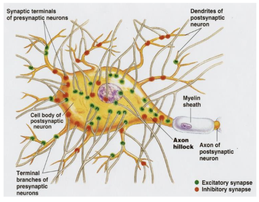
In this case the potential is getting more positive (an excitatory postsynaptic potential or EPSP). Other types of channels change the potential to a more negative state (inhibitory postsynaptic potential or IPSP). One neuron receives many, many signals from other neurons (on their dendrites but also directly on the soma).
The EPSP (or IPSP) spreads through the rest of the dendrites and towards the soma via electrotonic conduction. This happens very fast (instantaneously, because electrically), yet only over a short distance (depending on thickness of dendrite). Decremental conduction.
All these signals (both EPSPs and IPSPs) are integrated in the axon hillock. If there are sufficient excitatory signals the axon hillock will initiate an electrical signal that will be propagated to the axon (i.e., an action potential).
temporal/spatial summation
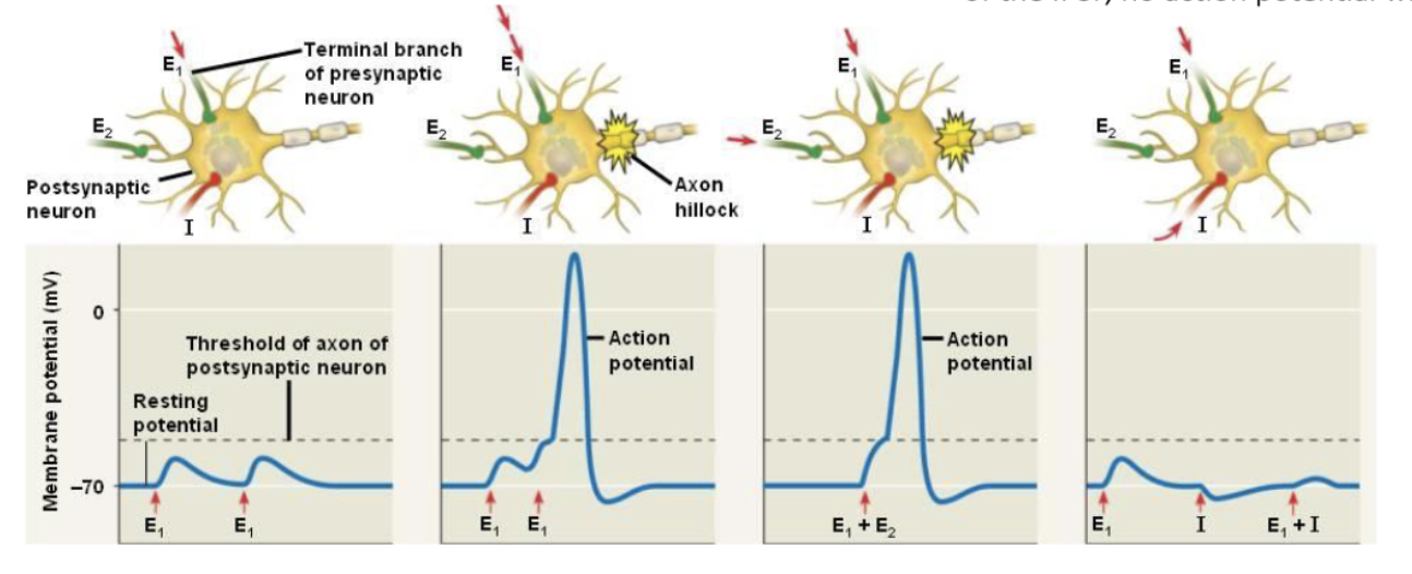
A single EPSP is not strong enough to elicit an action potential. The EPSP decays fast. Two EPSPs on the same location quickly after each other do!
Temporal summation: when the EPSPs follow in close succession, the integrated potential at the axon hillock will be large enough to trigger an action potential.
Spatial summation: when different EPSPs occur simultaneously at different locations, the integrated potential at the axon hillock will be large enough to trigger an action potential.
The axon hillock integrates all signals (both EPSPs and IPSPs). If the strength of the EPSP is as strong as that of the IPSP, no action potential will be initiated.
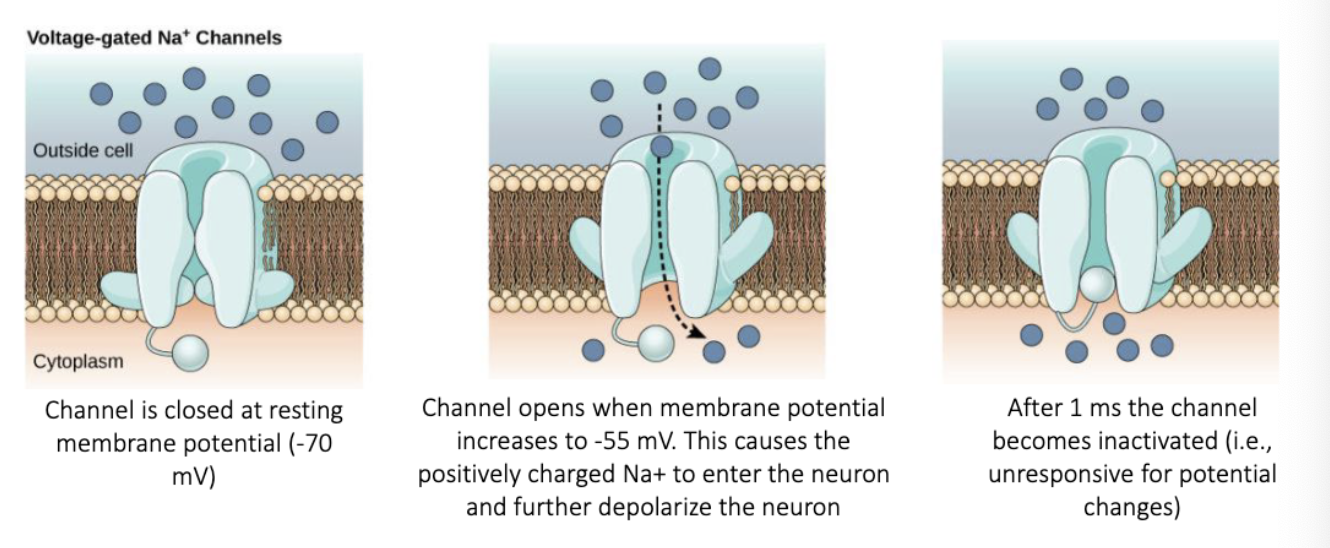
voltage-gated sodium channels
The axon hillock ‘produces’ an action potential because of voltage-gated sodium channels (that are present at the axon hillock). In contrast to ligand gated channels, these channels are triggered by changes in the membrane potential.
When the neuron returns to its rest membrane potential, it undershoots to a state of hyperpolarization before it returns to its resting membrane potential of -70 mV. This period is called the refractory period. During this period, the neuron can’t become active again.
ARP = Absolute Refractory Period. Na+ are still closed so a new action potential can not be generated.
RRP = Relative Refractory Period. An action potential can happen but only with larger-then-normal depolarizing currents.
electrotonic conduction through axon
After action potential begins activity at the axon hillock, this potential will ‘travel’ along the axon.
The strong depolarization spreads via electrotonic conduction to neighboring parts of the axon. There, voltage gated channels open as well, generating an action potential again. This process repeats along the axon, always producing a novel action potential.
Because of the refractory period, this neighboring action potential cannot spread back, only forward.
Because the action potential has to be generated by the opening of Na+ channels at every next site, the conduction velocity of the action potential is relatively slow compared to electrotonic conduction: 0.5 – 10 m/s depending on axonal diameter.
But the big advantage of action potentials over electrotonic conduction is that it can travel over much larger distances, in principle any length of axon, because it is generated over and over again.
Higher speeds are obtained by myelin sheets that act as insulators around the axons. Only at intervals of up to 1.5 mm (depending on axon diameter), small ‘naked’ sections are present: nodes of Ranvier.
Action potentials are only generated at the nodes of Ranvier. Between these nodes, the signal is passively spread very fast speeding up the conduction velocity to up to 150 m/s.
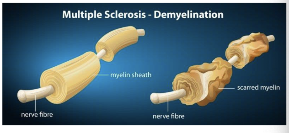
multiple sclerosis (MS)
In multiple sclerosis, the immune system destroys the myelin sheets of neurons. Many sensory and motor functions are impaired.
synaptic transmission
the transfer of a signal from one neuron to another neuron.
Two types of synapses: chemical and electrical. Predominantly chemical!
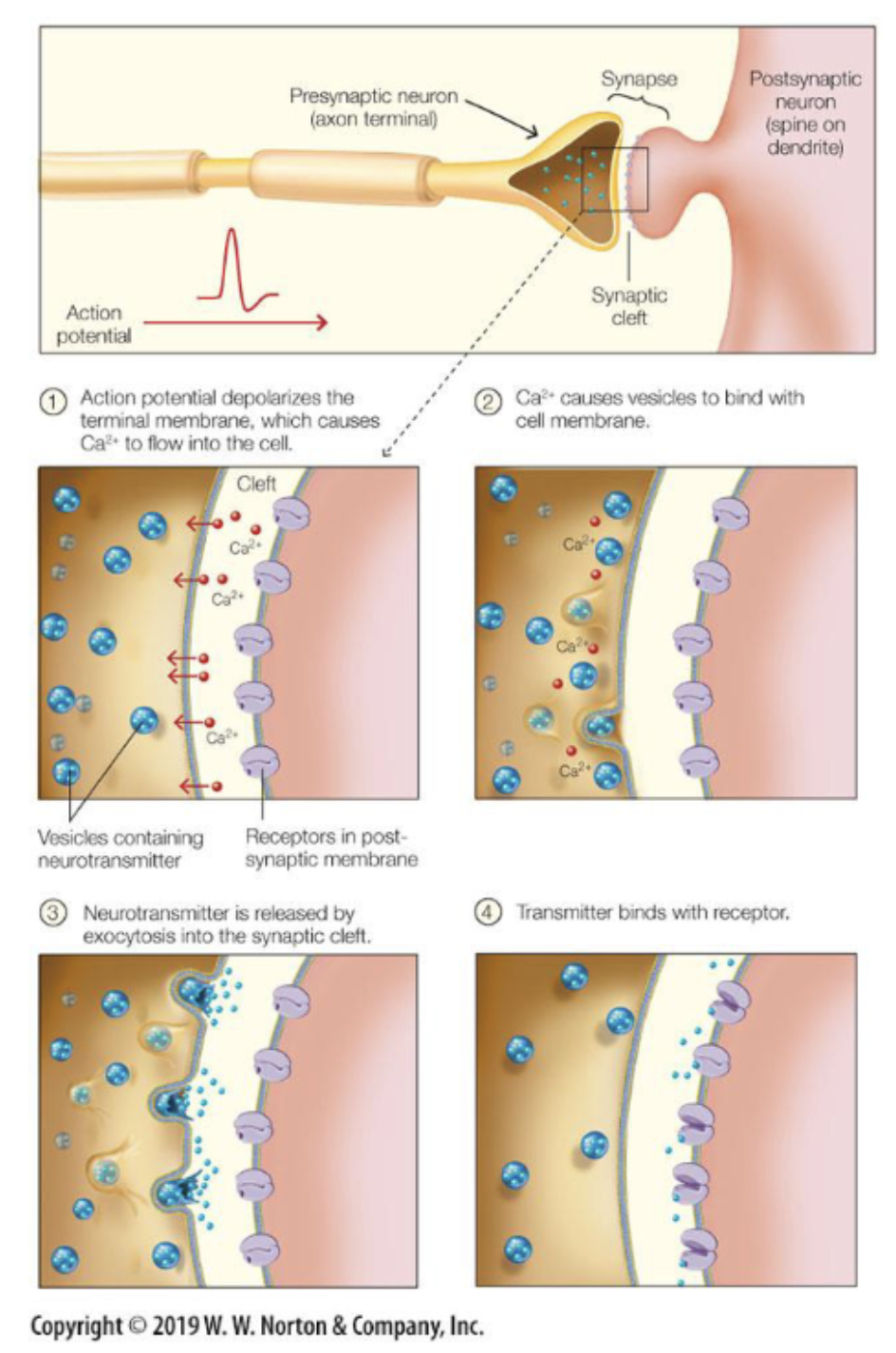
chemical synaptic transmissions
Signal between neurons, no physical contact.
Neurons communicate via chemical synapses: space between axon terminal of sending neuron and dendrite of receiving neuron is the synaptic cleft.
Presynaptic neuron releases neurotransmitters in synaptic cleft that can bind to specialized receptors of the postsynaptic neuron.
Axon terminal (the presynaptic terminal) contains vesicles filled with neurotransmitter molecules.
When an electrical current invades the presynaptic terminal, a series of events (the opening of the voltage-gated calcium (Ca2+) channels and the ensuing influx of Ca2+) leads to the fusion of the vesicles with the presynaptic membrane.
Neurotransmitter molecules are released in the synaptic cleft and can bind to postsynaptic receptors. This creates EPSPs and IPSPs that changes the cell’s polarization and could initiate an action potential.
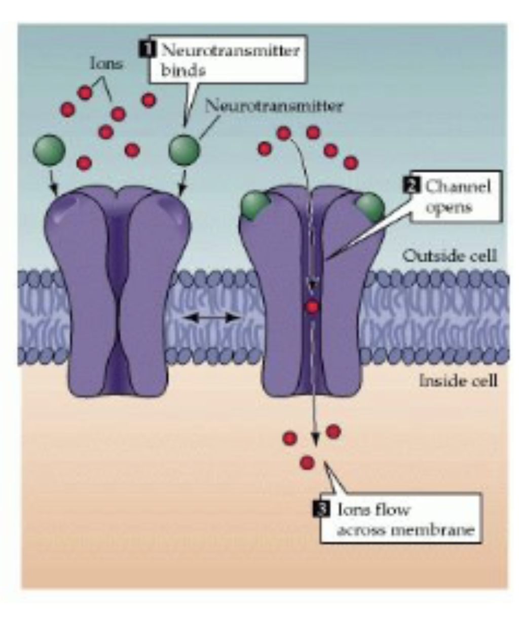
postsynaptic receptors
Different types (>100) of postsynaptic receptors. A receptor binds to a specific neurotransmitter.
ligand-gated ion channels: binding of neurotransmitter with receptor directly opens the ion channel; fast signalling; milliseconds.
G-protein-coupled receptors: indirect effect via a second messenger, slower signalling, hundreds of ms/s;
The effect of a neurotransmitter depends on the postsynaptic receptor: The same type of neurotransmitter can bind to different postsynaptic receptors that can have very different effects (increase or decrease firing).
glutamate
most prevalent;
excitatory;
gamma-aminobutyric acid (GABA)
second most prevalent;
inhibitory;
acethylcholine (ACh)
present in neuromuscular junction;
excites muscles;
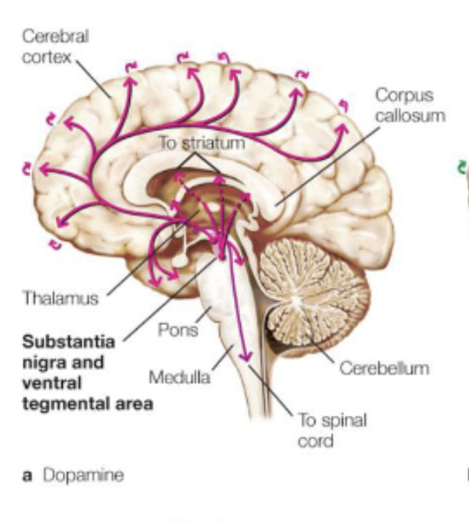
dopamine
motor control/cognition;
Substantia Nigra and Ventral Tegmental Area;
involved in Parkinson’s;
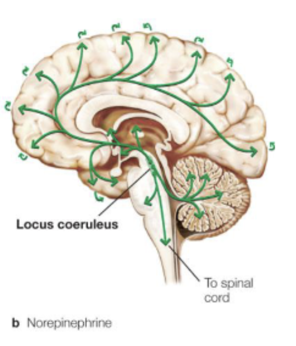
norepinephrine/noradrenaline
arousal;
Locus Coeruleus;
fight-or-flight response;
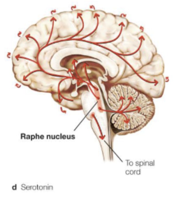
serotonin
mood/cognition;
Raphe Nucleus;
Depression;
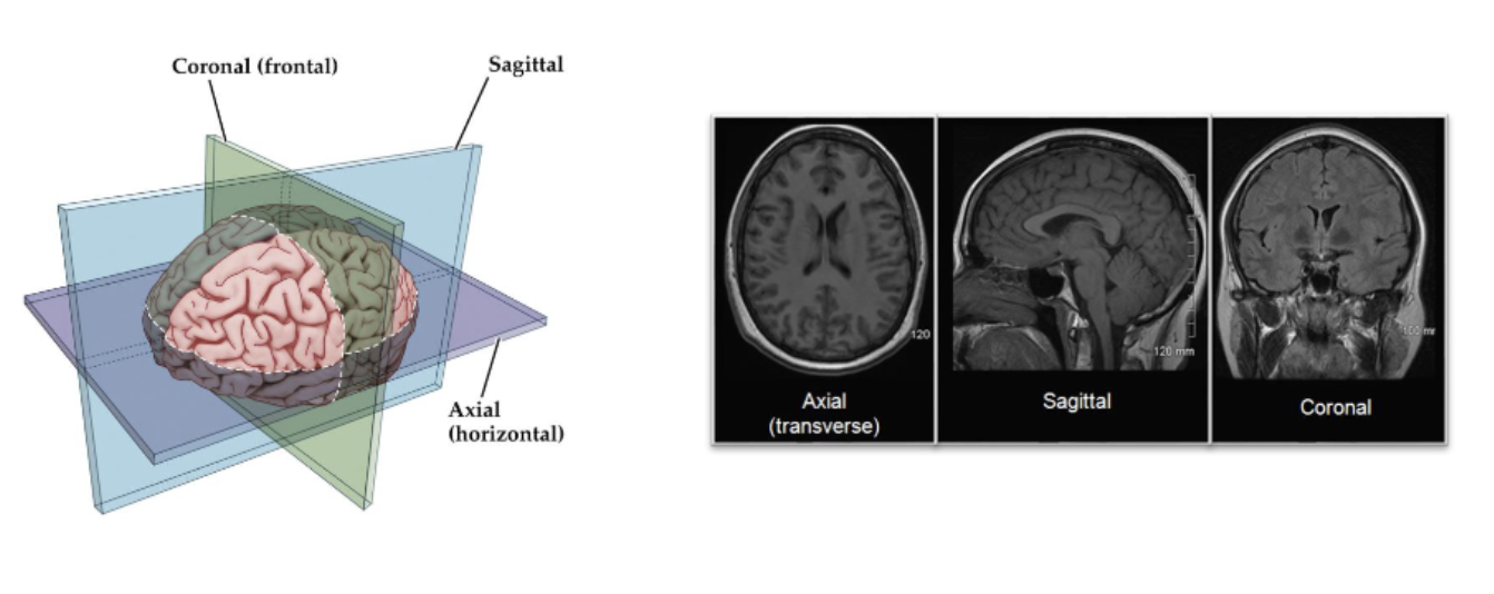
dorsal, ventral, rostral, caudal
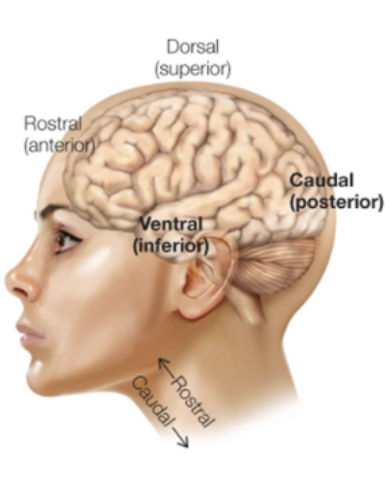
cross-sections of the brain
caudal/frontal = front to back/anterior to posterior
sagittal/longitudinal = left to right/lateral-medial-lateral
axial/horizontal/transverse = top to bottom/dorsal-ventral
nucleus
cluster of neurons in CNS
ganglion
cluster of neurons in PNS
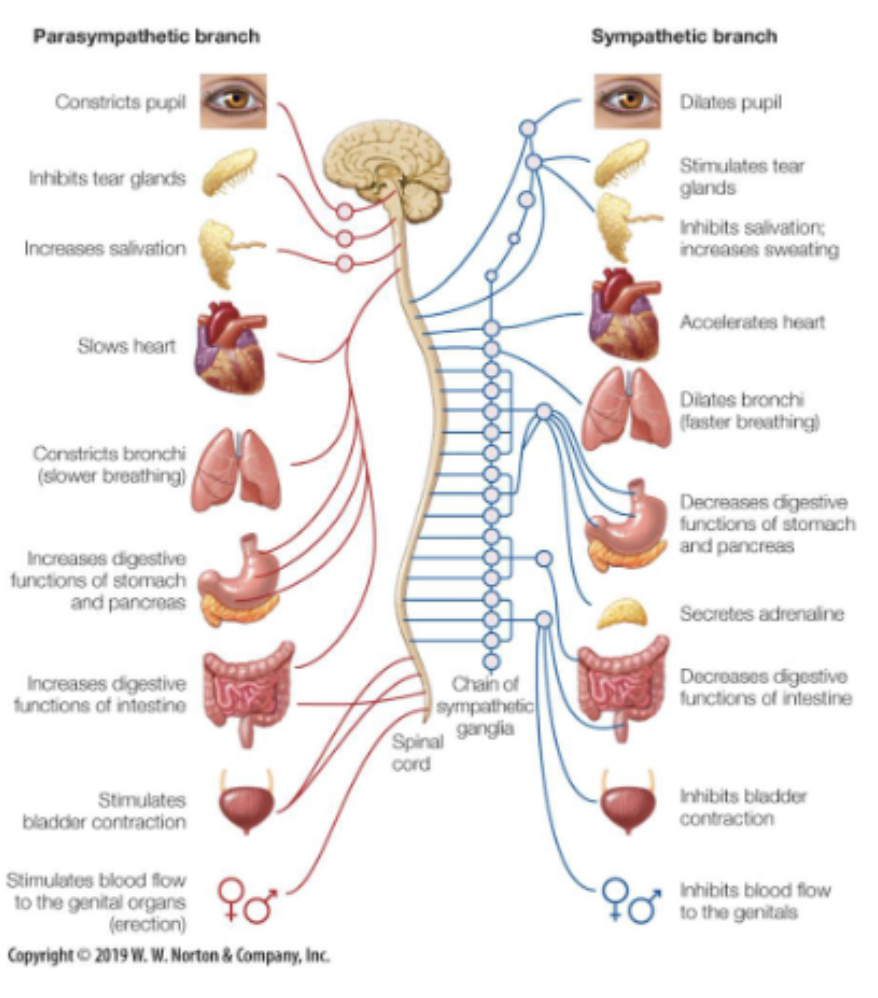
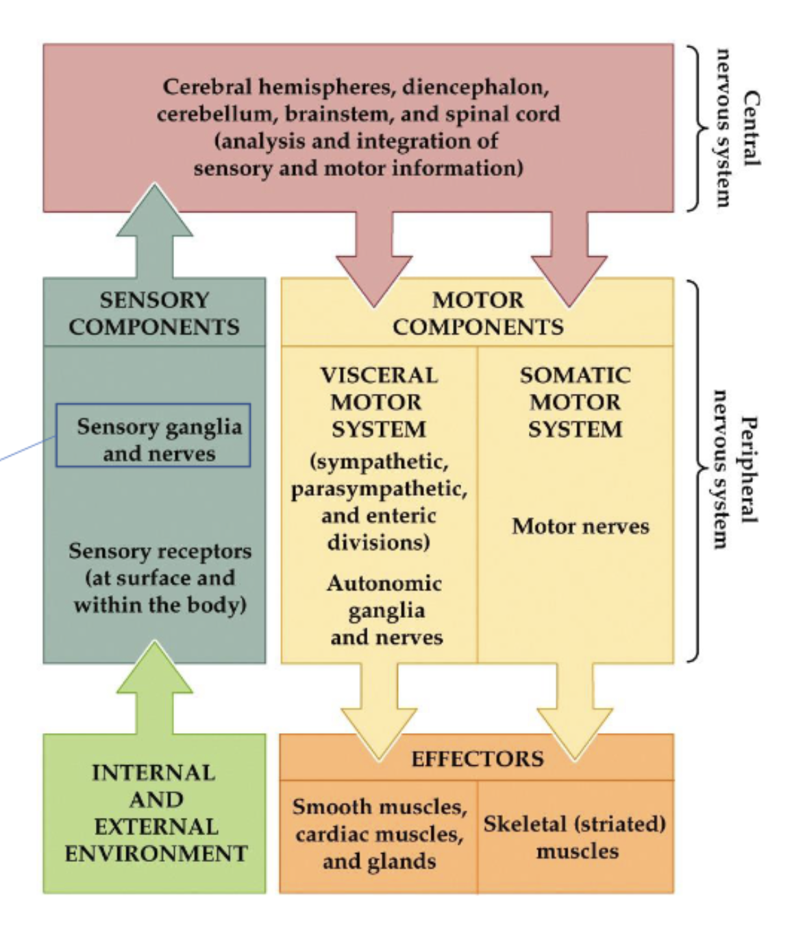
PNS
connects the CNS to the limbs and organs.
Consists of:
Somatic motor system: voluntary muscles
Autonomic motor system: involuntary muscles (reflexes) and organs (e.g., heart, bladder,...). This system is always active but can be in one of two states:
Sympathetic state: expending energy (‘fight or flight’)
Parasympathetic state: conserving energy (‘rest and digest’)
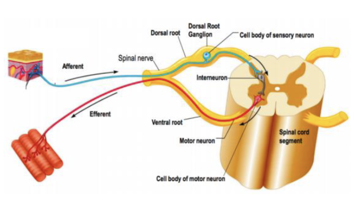
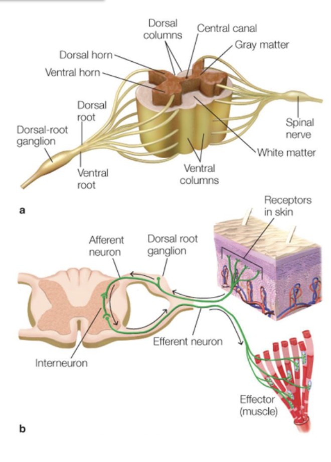
dorsal root ganglion
sensory neurons that transmit sensory information from the body to the central nervous system
information travelling through the nervous system
neuron locations
Neurons can be found in the thin sheet of tissue on the ‘outside’ of the brain (the grey matter) and in (mostly) subcortical nuclei.
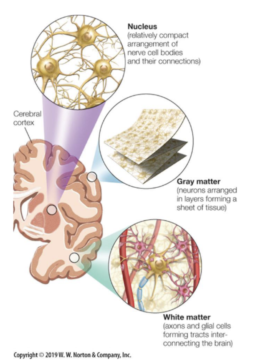
white matter
Neurons in distant brain areas are connected via axons with myelin sheets that form the white matter.
meninges
The CNS is covered with three protective membranes:
Dura Mater: thick membrane, closest to the skull
Arachnoid Mater. Not attached to the dura mater. Does not line the brain down to the sulci (except for the longitudinal fissure).
Pia Mater. Delicate membrane that firmly adheres to the surface of the brain. Can get infected (meningitis).
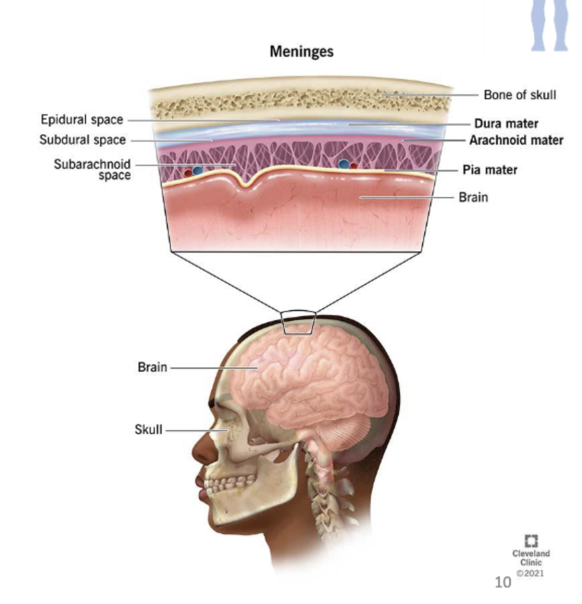
cerebrospinal fluid (CSF)
The fluid the brain floats in.
The area between the arachnoid membrane and the pia mater (the subarachnoid space) is filled with CSF.
protects the brain (reduces shock when hit), cleans the brain.
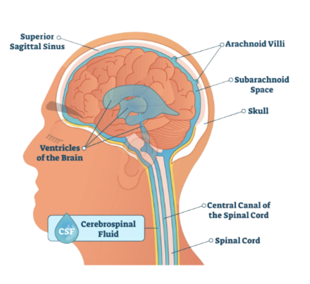
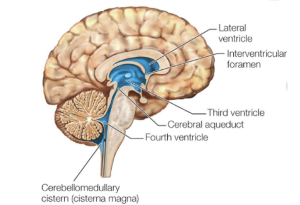
ventricles
cavities in the brain filled with CSF.
Four large connected ventricles:
left and right lateral ventricle: connect via the interventricular foramen to
third ventricle: connects via the cerebral aqueduct to
fourth ventricle
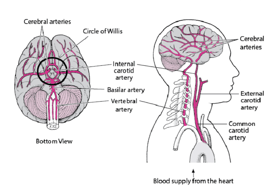
blood supply in the brain
Brain needs energy and oxygen, delivered via blood (the brain consumes about 20% of your blood supply).
Interruptions of blood supply to the brain (i.e., a stroke) can be caused by an obstruction of an artery or by hemorrhage from a blood vessel. This can cause damage to the brain (brain lesions).
main sources of blood in the brain
Two main sources:
Internal carotid arteries
Vertebral arteries
Left and right vertebral arteries merge at the level of the pons → basilar artery.
Basilar artery and internal carotid meet at circle of Willis.
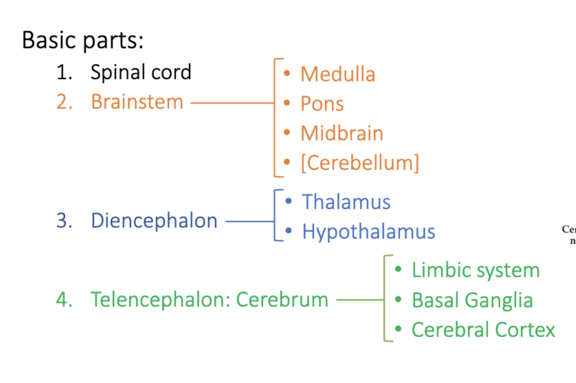
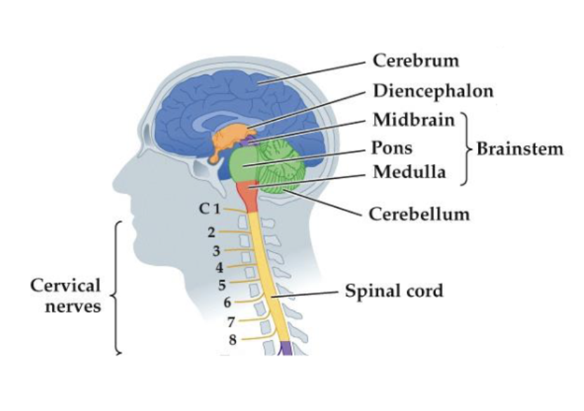
structure of the CNS
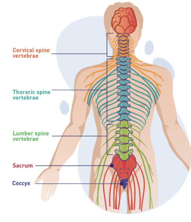
vertebral column
Spinal cord is enclosed by the bony vertebral column.
Vertebral column is divided into cervical, thoracic, lumber, sacral and coccygeal regions.
spinal cord
Each segment of the vertebral column gives rise to peripheral (spinal) nerves that innervate the body.
Sensory signal enter the spinal cord via the dorsal horn (afferent signals – towards the CNS) and leave it via the ventral horn (efferent signals – away from the CNS).
Some signals go directly from the dorsal to the ventral horn, via an
interneuron (e.g., spinal reflexes like the knee jerk).
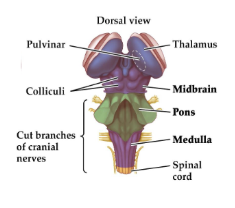
brainstem
Highly complex structure that houses the nuclei most essential for the maintenance of life. Damage to the brainstem is “very bad news”.
Three main parts: medulla (oblongata), pons, cerebellum, midbrain.
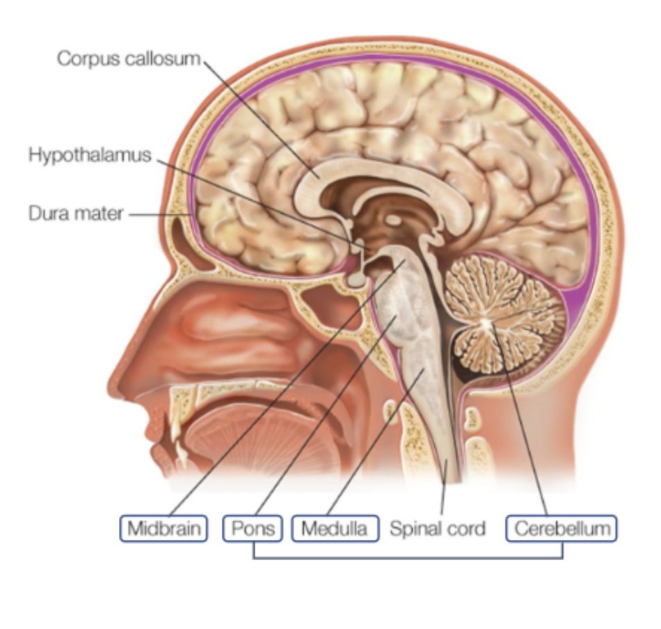
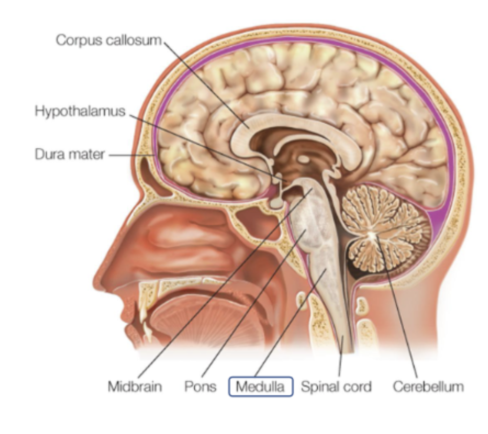
medulla (oblongata)
Cell bodies of many of the 12 cranial nerves (sensory and motor innervations to face, neck, throat).
Corticospinal motor axons (i.e., axons coming from the cortex to the spinal cord) cross here: motor neurons from left hemisphere cross here to control muscles from the right side of the body.
Responsible for autonomic (involuntary) functions such as heart rate, sneezing, blood pressure etc.
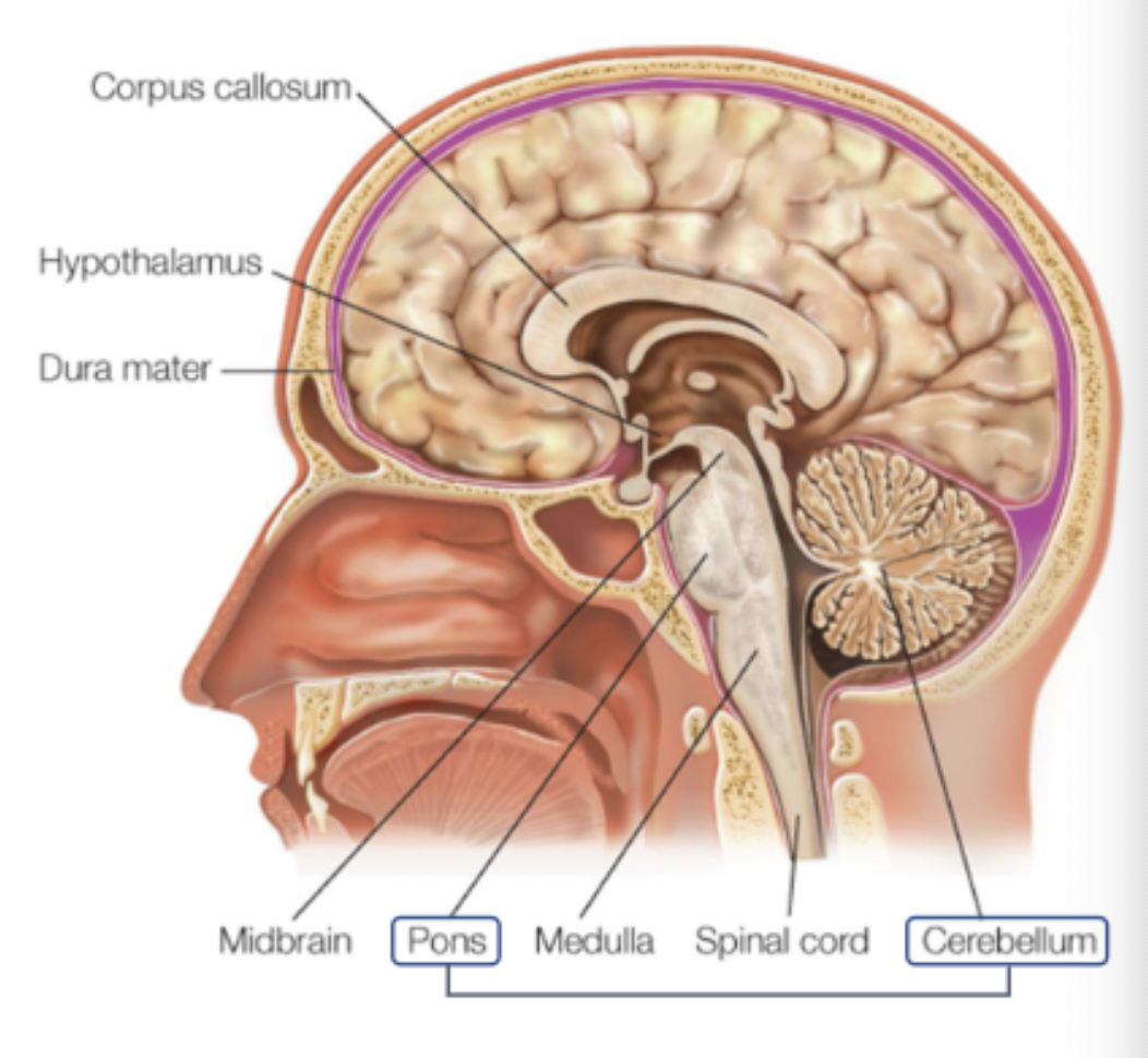
pons
“bridge”,
contains nuclei that connect the forebrain to the cerebellum
nuclei that deal with sleep, swallowing, facial expressions and more
related to sleep paralysis and generating REM sleep.

cerebellum
“little brain”
houses 69 billion neurons;
involvement in motor control and maintaining balance;
implicated in aspects of cognitive processing (language, attention)
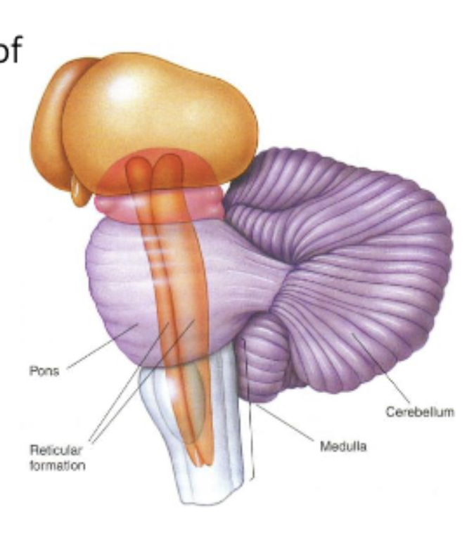
reticular formation
located throughout the brainstem;
crucially involved in arousal and attention. Damage to the reticular formation is likely to affect the state of consciousness (results in coma)
Home to the raphe nuclei (serotonin synthesis);
midbrain
mesencephalon
dorsal part: tectum
superior colliculus: reflections towards visual stimuli
inferior colliculus: reflections towards auditory stimuli
ventral part: tegmentum
Ventral Tegmental Area and Substantia Nigra - dopamine production;
periaqueductal grey: modulation of pain;
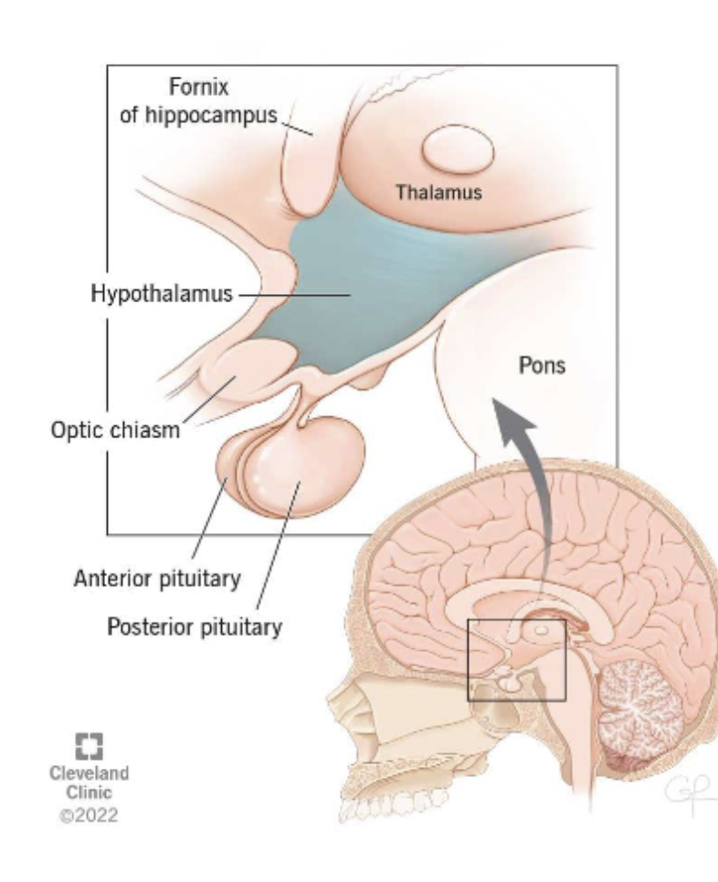
diencephalon
thalamus and hypothalamus
thalamus
Left and right thalamus, connected via the Massa Intermedia.
Switch board of the brain. Receives input from all sensory areas of the brain (except smell).
Consists of several nuclei that act as specific relays for incoming sensory information:
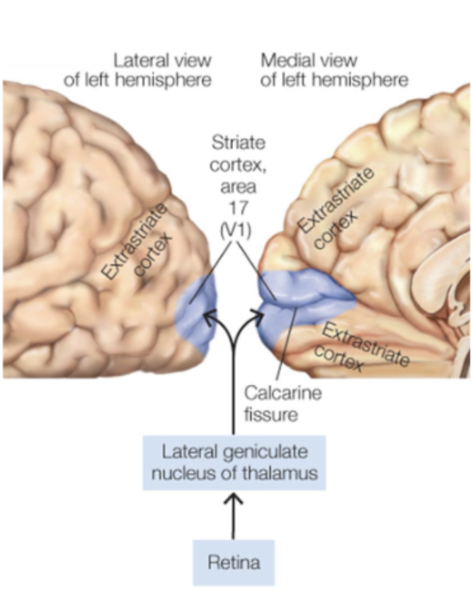
Lateral geniculate nucleus (LGN): visual information
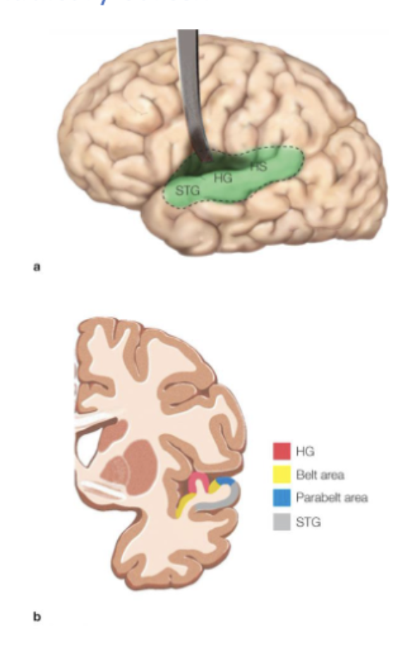
Medial geniculate nucleus: auditory information
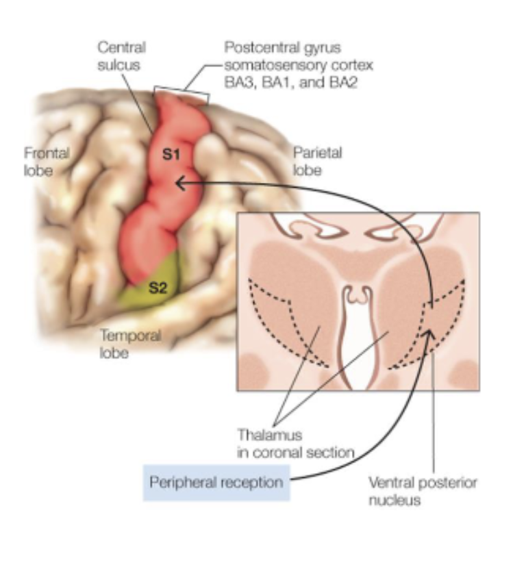
Ventral posterior nuclei: somatosensory information
Pulvinar: attention and integrative functions

hypothalamus
Controls the functions necessary for homeostasis (i.e., the normal state of the body): body temperature, metabolic rate, circadian rhythm.
Produces hormones but also regulates hormone production in other areas, for example in the pituitary gland (axonal connections to the posterior pituitary gland).
telencephalon (cerebrum)
limbic system, basal ganglia, olfactory bulbs, cerebral cortex
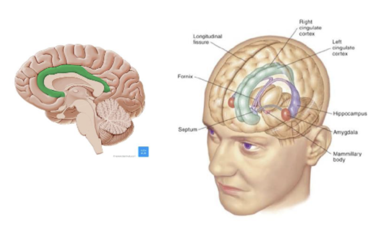
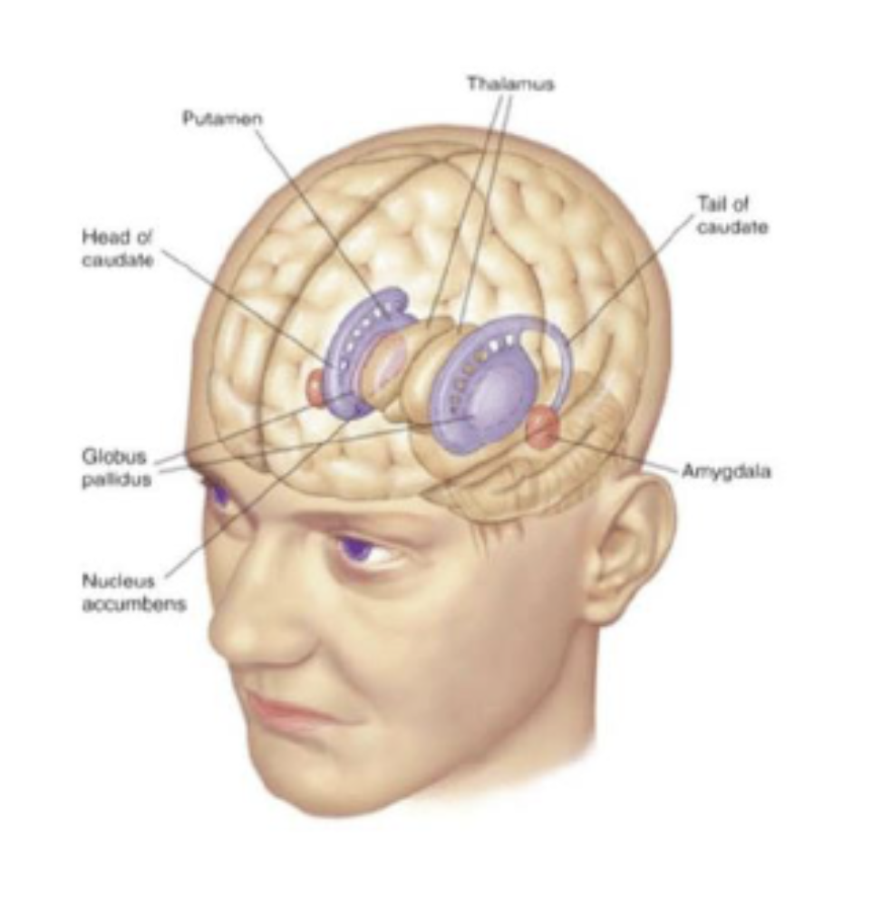
limbic system
cingulate cortex;
hippocampus: memory, spatial processing;
amygdala: emotional processing;
basal ganglia
striatum: putamen + caudate nucleus;
globus pallidus;
nucleus accumbens;
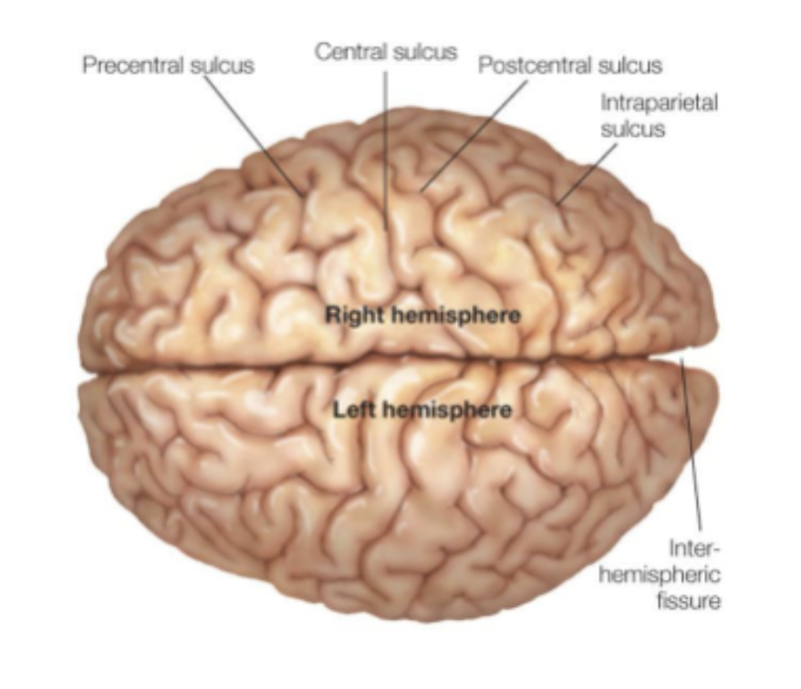
the cerebral cortex
Cortex is thin folded sheet of neurons and supporting cells. Folding creates convex convolutions (gyri) and concavities between the gyri (sulci).
Central sulcus is dividing concavity between frontal and parietal lobe.
The gyrus anterior of the central sulcus is the precentral gyrus; the gyrus posterior of the central sulcus is the postcentral gyrus.
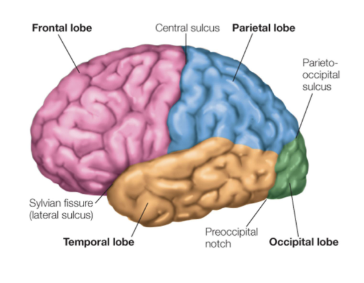
lobes
cytoarchitectonics
microanatomy, more subdivisions of the brain (Brodmann’s 52 areas).
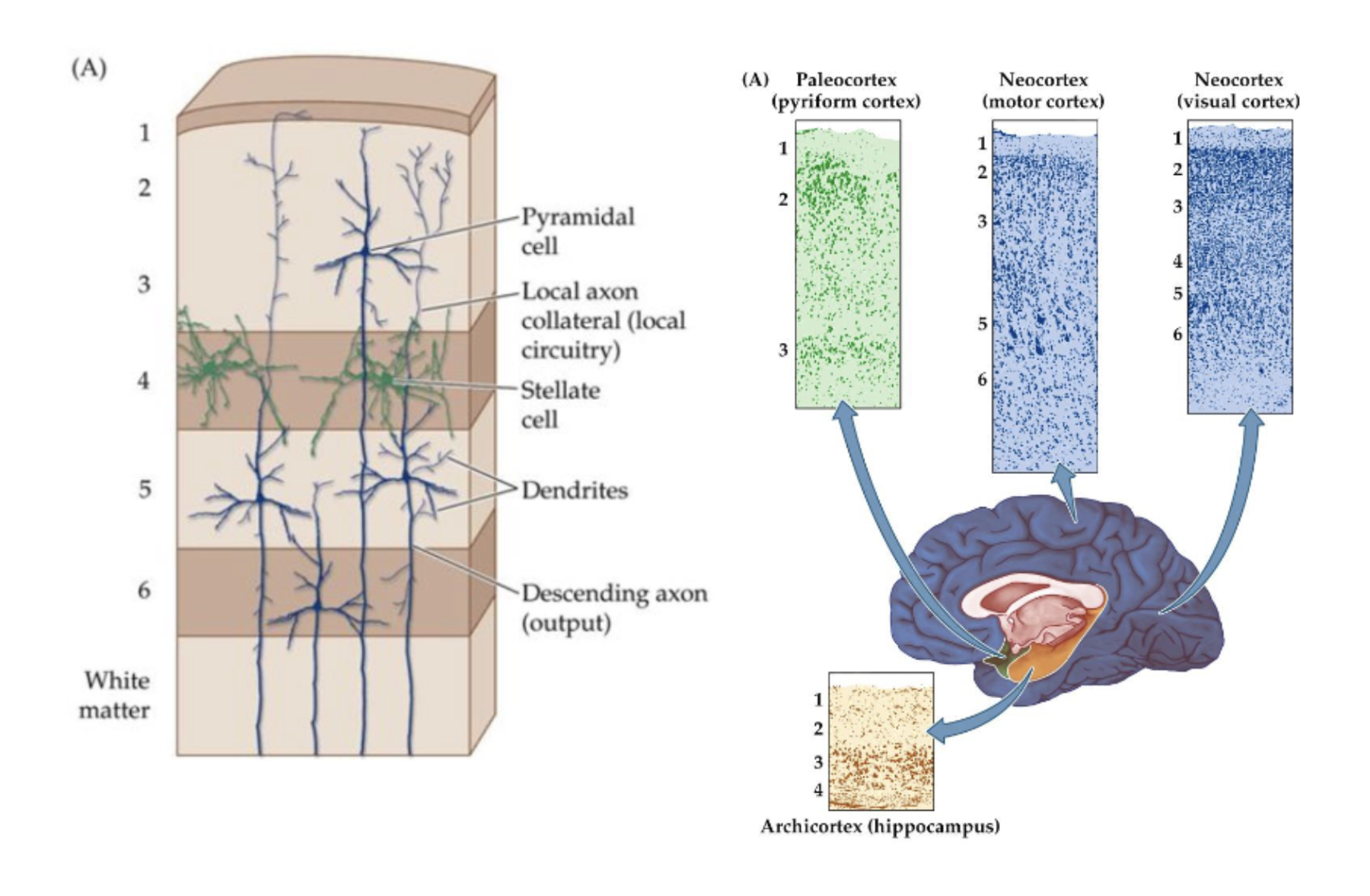
laminae
Most of the neocortex consist of 6 cellular layers (laminae). This is often called the grey matter.
Evolutionary older parts of the brain have less layers (e.g., paleocortex, archicortex).
Layers differ in cell densities, cell types, inputs, and outputs
1: no cell bodies, mostly axons and dendrites
2-3: pyramidal cells, locally connected to other cortical sites
4: stellate cells, receive input from thalamus
5-6: large pyramidal cells, output leaves the cortex
Arranged in a vertical structures called cortical columns of about 0.5 mm wide.
brain areas functional subtypes
primary sensory areas
primary motor areas
unimodal association areas (process one type of information);
multimodal association areas (integrate > one type of information);
paralimbic and limbic areas;
frontal lobe
motor cortex: in front of central sulcus
Primary motor cortex - generating movements
Premotor cortex - controlling movements
Supplemental motor cortex - planning movements
prefrontal cortex: anterior to the motor cortex
houses executive functions: planning, organising, controlling and executing behaviour;
last to develop!
evolutionarily the youngest;
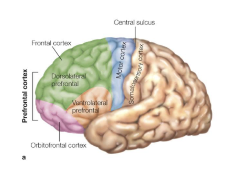
parietal lobe
somatosensory cortex: the postcentral sulcus
Primary somatosensory cortex (S1): receives input from thalamus about touch, pain, temperature, limb position;
Secondary somatosensory cortex (S2): unimodal association area that further processes sensory information;
occipital lobe
visual cortex
temporal lobe
auditory cortex
primate brains
Primates have a higher ratio of neuron number to brain size.
Off all primates, humans have the largest brains.
connectivity according to “small-world architecture”
Local ‘modules’ with many short connections
Small number of very long connections
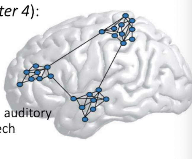
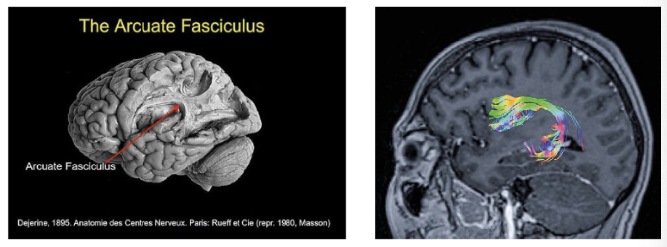
structural connectivity related to functional connectivity
In language processing, Wernicke’s area (related to interpreting an auditory code) is strongly connected to distant Broca’s area (related to speech production) via a dense bundle of axons, the Arcuate Fasciculus.
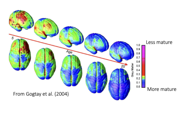
brain growth
The size of the brain quadruples from birth to adulthood. Not because of an increase in neurons, but because of synaptogenesis (the formation of synapses) and the growth of dendritic trees, the extension of axons, myelination and the proliferation of glial cells.
Synaptogenesis is followed by synaptic pruning (eliminating redundant synaptic connections). Use it or lose it!
Maturation of the cortex can be indicated by gray matter volume loss. When we mature, white matter increases linearly with age, but gray matter first increases and then decreases. The maturation of gray matter differs depending on the cortical region.