B&C - Methods in Cognitive Neuroscience
1/76
There's no tags or description
Looks like no tags are added yet.
Name | Mastery | Learn | Test | Matching | Spaced |
|---|
No study sessions yet.
77 Terms
What’s the key idea underlying cognitive psychology and behavioral methods?
• information enters brain → internal processes take place → behavior is put out
• use mental representations that undergo internal transformations to find out which internal processes take place
In which ways is information represented in cognitive psychology and behavioral methods?
• multiple ways → context often dictates which representation is accessed
What’s an example for mental representations that undergo internal transformations?
• participants need to decide whether two letters belong to same category (both vowels or both consonants) or to different categories (one consonant and one vowel)
• letters from same category could be physically identical (b and b), phonetically identical (b and B) or same category (b and f)
• participants need to respond as fast and accurate as possible → reaction time is measured
• although response in conditions “physical identity”, “phonetic identity” and “same category” is the same, big difference in reaction times
• reason (=internal process): we use different representations to solve task → physical representation (just looking at shape), letter identity representation, category representation (vowel or consonant)
What does it mean when it’s said that we “translate or transform representation” for action or behavior?/Which processes take place in the translation/transformation process?
• (see →) encode → compare → decide → respond or act
• that’s what we try to study with behavioral methods
What does the translation/transformation process look like, when developing a representation of having to buy coffee?
• when you noticed you had no coffee at home, smell of coffee later today will activate representation of coffee → representation will be used to decide on course of action
encode “coffee” as “things to buy” in memory
when smelling coffee, you compare activated representation of “coffee” to items in “things to buy” list
you decide if coffee is part of that list
you respond or act accordingly → go to shop
What kind of process is memory comparison?
• serial process → but not all processes are serial
How does cognitive psychology try to understand these different internal processes?
• design experiments specifically for that
What’s a serial comparison?
• compare one item we have encoded in memory from list at the time
What’s a parallel comparison?
• compare one item we have encoded in memory to all items at the same time
What kind of process is memory comparison?
• compelling evidence that it’s serial process → but not all processes serial
What’s the word superiority effect?
• Participants are more accurate in identifying the target vowel when it is embedded in a word → suggests that letter and word levels of representation are activated in parallel.
Ways of understanding how cognitive processes are related to brain (7)
• studying damaged brain
• methods to perturb neural function
• structural analysis of the brain
• methods to measure neural activity
• neuroimaging
• connectivity maps
• computational neuroscience
What’s the main idea behind investigating how cognition is affected in people with brain damage?
• brain injury disrupts/eliminates processing ability of affected brain structure
• for example: Broca’s patient “Tan”
What’s a single dissociation?
• When a lesion to brain area X impairs the ability of a patient to do task A but not task B, then we can say that brain area X and task A are associated, whereas brain area X and task B are dissociated
What’s a double dissociation?
• occurs when damage to area X impairs the ability to do task A but not task B, and damage to area Y impairs the ability to do task B but not task A. The two areas have complementary processing.
• strongest evidence comes from double dissociations
What are disadvantages of studying the damaged brain? (8)
• lesions never identical between patients
• can’t make our own lesions in humans
• animals need to be trained extensively to learn task
• animals cannot learn all complex behavior we can do as humans (e. g., reading)
• ethical issues in animal research
• problems of interpretation
• decline in cognitive function because of brain lesion could also indicate that other brain areas don’t receive input anymore and might not function properly anymore → diaschisis
• lesion might involve damage to cortical site but also to fiber tract that connects more distant areas
What is diaschisis?
• (changed) activity in surviving neurons after damage to other neurons
What are methods to perturb neural function? (4)
• pharmacological interventions
• genetic manipulations
• invasive stimulation methods
• non-invasive stimulation methods
What’s the underlying key idea of pharmacological interventions?
• signaling between neurons done by neurotransmitters
• interfering with neurotransmitters changes signalling → administration of agonists and antagonists
What are agonists?
• drugs that work like real neurotransmitters
What are antagonists?
• drug that blocks neurotransmission
What are disadvantages of pharmacological interventions?
• very unspecific → administering drug affects whole brain
• except in animas: there you can inject it directly in specific area
How are genetic manipulations used to perturb neural function?
• used in animal research
• use knock-out animals
What’s a knock-out animal?
• animal with specific genes manipulated so that they don’t express themselves
What’s a wild-type animal?
• normal animal (no manipulated genes)
How do you use invasive stimulation methods to perturb neural function and what are 2 examples?
• mostly done in animal research, but also in patients undergoing neurosurgery (e.g., epilepsy)
• for example by implanting electrodes that stimulate specific brain areas electrically
• examples: deep brain stimulation, optogenetics
What’s deep brain stimulation and what is it used for?
• electrode implanted in subthalamic nucleus
• used to alleviate symptoms related to Parkinson’s disease
How do optogenetics work?
• light-gated ion channels: channels that open or close in response to light → e. g. in green algae (called “channelrhodopsin”)
• genetic material responsible for making these channels is extracted and inserted in virus
• virus is injected in brain area (e. g. of rat) → will infect targeted neurons
• inserted genetic material will lead to production of channelrhodopsins in membranes of infected neurons
• by shining light on channelrhodopsins light-gated ions channels will open → every light pulse causes action potential
• effect for genetically modified neurons in rats’ amygdalae: rat behaves less anxious when stimulated
What happens in non-invasive stimulation methods?
• stimulating brain from outside skull
3 types of non-invasive stimulation methods
• transcranial magnetic stimulation (TMS)
• transcranial direct current stimulation (tDCS)
• transcranial alternating current stimulation (tACS)
What happens during transcranial magnetic stimulation (TMS)?
• strong current is sent through coil and produces strong magnetic field
• this field induces changing electrical field in underlying brain area and causes neuronal activity (e. g. motor activity)
What is single pulse TMS?
• during experiment, TMS pulse is delivered on each trial (e.g., when stimulus is presented)
What is repetitive TMS?
• before start of experiment, train of pulses is delivered that changes underlying brain area for longer duration
What is theta burst stimulation?
• form of repetitive TMS
• applies very-high-frequency stimulation for short period of time
• depresses activity for relatively long time (45-60 min)
What are effects of TMS and what do they depend on?
• depend on strength of pulse
• strong pulse causes temporary “lesions”
• weaker pulse can facilitate activation
What are advantages (1) and disadvantages (4) of TMS?
• advantage: high temporal resolution → pulse has immediate effect
• disadvantages:
• small spatial resolution → pulse affects relatively large brain area
• you can only target superficial brain regions → not possible to target structures deeper in brain
• pulse can cause contraction of scalp and head muscles → annoying for subjects, difficult to mimic in control condition
• might trigger epileptic seizures (rare)
What happens in transcranial direct current stimulation (tDCS) and what is it used for?
• small electrical current is directly applied to scalp
• effects can last for extended period
• used in research and to treat clinical problems (e. g. depression)
Disadvantages of tDCS?
• results hard to replicate
• some question if it can be strong enough to have any effect on brain at all
What is transcranial alternating current stimulation?
• similar to tDCS, but electrical current oscillates
Methods for structural analysis of the brain
• Computerized tomography (CT or CAT)
• Magnetic resonance imaging (MRI)
• Diffusion tensor imaging (DTI)
Computerized tomography
• first method for structural analysis of brain
• based on X-rays
• currently only used in medical imaging
Magnetic resonance imaging
• uses magnetic properties of atoms → most pervasive atom in brain is hydrogen
• provides structural image of brain at high spatial resolution
• variant of MRI is used for diffusion tensor imaging (DTI)
Mechanics behind magnetic resonance imaging
• in normal state, orientation of spinning protons is randomly distributed
• exposure to magnetic field of MRI scanner aligns orientation of protons
• when radio frequency pulse is applied, axes of protons are shifted in predictable manner and put protons in elevated energy state
• when pulse is turned off, protons release their energy as they spin back to orientation of magnetic field → MRI scanner reads out energy and produces image
Diffusion tensor imaging (DTI)
• creates image of structural connectivity of brain
• measures density and flow of water molecules in axons
What are methods to measure neural activity? (5)
• single-cell neurophysiology
• invasive neurophysiology in humans
• non-invasive electrical recording of neural activity
→ electroencephalography (EEG)
→ magnetoencephalography (MEG)
Which subjects is single-cell neurophysiology applied to?
• animals
What’s the receptive field?
• limited region of space
• if stimulus is presented in that region, visual neuron reacts to it
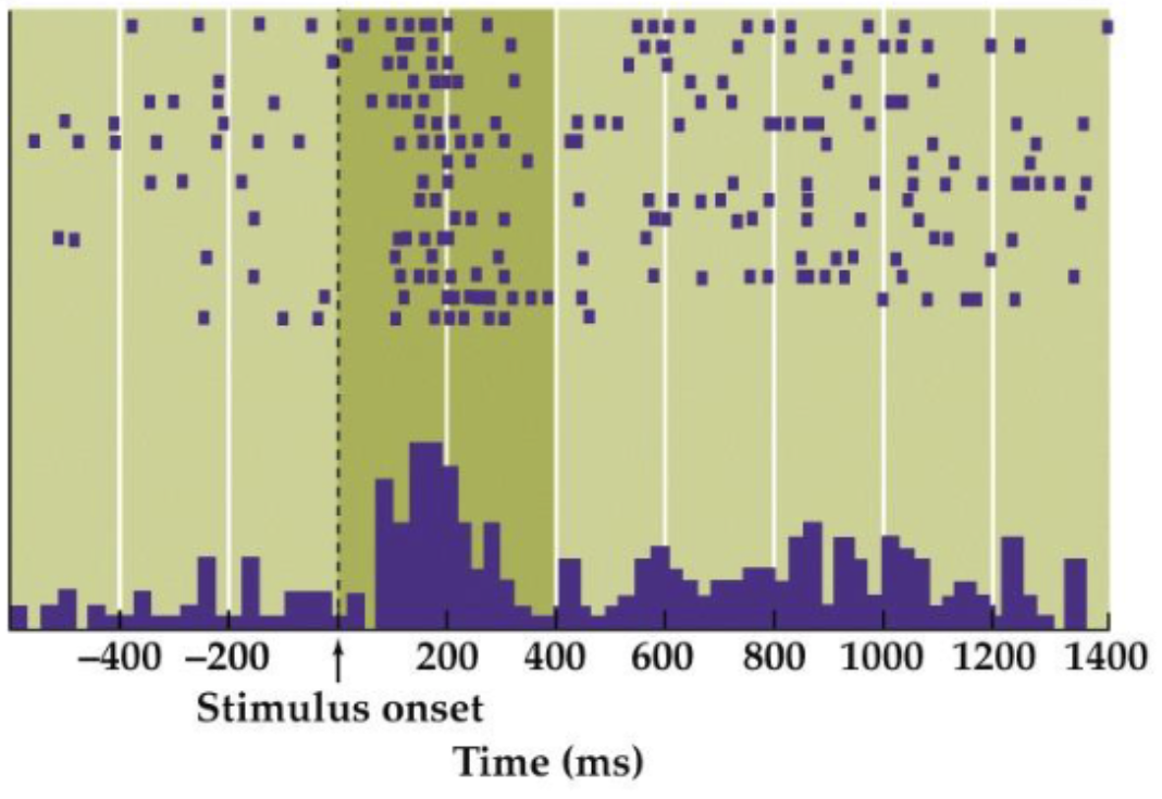
How does single-cell neurophysiology in animals work?
• visual stimulus is present in receptive field of neuron that we record with electrode that is inserted in brain
• trials are visualized
• “spikes” → action potential, every time neuron is active
• visualized as purple dots in raster plot
• to count spikes at all time points, all trials on presentation of stimulus are aligned in peristimulus histogram
When is invasive neurophysiology used in humans and what happens during it?
• when they undergo brain surgery
• Electrocorticography (ECoG) → grid of electrodes is placed directly on cortex
• neural activity often shows oscillatory activity → oscillations can have different frequencies and different amplitudes (power)
• changes of power of different frequency bands over time can be plotted in electrocorticogram (= time-frequency plot)
Which types of waves have which frequencies?
• 1-4 Hz: Delta
• 4-8 Hz: Theta
• 7.5-12 Hz: Alpha
• 13-30 Hz: Beta
• 30-70 Hz: Gamma
• >70 Hz: High Gamma
Electroencephalography (EEG)
• electrical recordings outside skull
• cap with electrodes (ranging between 20 to 256) is placed on subject’s head
• electrodes register changes in electrical potentials
Processing of data from EEGs
• oscillations: represent power of different frequency bands→ EEG data can be decomposed in sum of weighted sine waves → Fourier analysis
• event-related potentials (ERPs): small voltage differences in ongoing EEG triggered by sensory and cognitive events
Interpretation of data from EEGs
• EEG is recorded while a stimulus is repetitively presented
• EEG data is cut in portions (”epochs”) and aligned to onset of stimulus presentation
• epochs are averaged and signal that is most common in all epochs remains
How can you observe spatial-temporal patterns of the event-related potentials?
• topographic maps show these patterns
• → you can observe how activity at electrodes dynamically changes over time and space
Disadvantage of EEGs
• EEG tells you something about electrical potential at level of electrodes, not about electrical potentials at brain level
• inverse problem → pattern observed at sensors can be caused by infinite number of different brain activations
• low spatial resolution → low conductance of CSF → meninges, skull and scalp “blurs” electrical signal before it reaches electrodes
• that’s also why EEG can’t detect activity from deep brain structures (electrical signal has decayed before it reaches electrodes)
Magnetoencephalography
• in brain, electrical current produces magnetic field orthogonal to electrical field
• electrical currents run along axons of active neurons
• MEG is sensitive to neurons that are parallel to skull
Advantages of MEG
• magnetic field is not obstructed by skull etc.
• in theory, more possible to obtain signals from deep brain structures
• better spatial resolution compared to EEG
Disadvantages of MEG
• much more expensive than EEG
• participants’ movements are restricted
Methods of neuroimaging
• Positron emission tomography (PET)
• Functional Magnetic Resonance Imaging (fMRI)
Underlying key idea of neuroimaging
• indirect measure of neural activity → measuring metabolic activity related to activity
• hemodynamic response
• division of brain in voxels (small cubes) and see where change in metabolic activity takes place → about 100.000 voxels in fMRI image of brain
Hemodynamic response
• when brain area is active, blood flow to that region increases to supply that area with more energy → e.g. through oxygen, glucose, …
Key idea behind positron emission tomography
• radioactive substance (tracer) is injected into bloodstream of subjects (e.g. radioactive oxygen incorporated in water molecules)
• tracer becomes distributed in brain according to physiological needs → active brain area consumes more oxygen, so tracer will be more present in active brain areas
• because tracer is radioactive, series of events will cause it to emit radioactive beams that can be detected by specialized detectors
Disadvantages of positron emission tomography
• scary to subjects: radioactivity, needles, …
• very expensive → requires different facilities (cyclotron to make tracer, staff that can inject subjects, PET scanner)
• slow process: uptake in blood, transport to brain, time required to get clear signal → limitation in experimental designs that can be used, only “blocked” designs are possible
What are “blocked designs”?
• many trials from one condition are presented in a block
• allows integration of activity across time
• brain activity in one block is compared to activity of another block that has trials from another condition
• in PET, tracer needs to decay between different blocks → takes long time (10 min)
Use of PET today
• not used for functional imaging studies (because of fMRI)
• still useful to study some structural aspects
• e.g.: using Raclopride as tracer that binds to dopamine D2 receptors to detect their distribution (only in striatum)
Underlying key idea of functional Magnetic Resonance Imaging (fMRI)
• blood with oxygen (oxyhemoglobin) has different magnetic properties than blood without oxygen (deoxyhemoglobin)
• active brain areas use more oxygen → results in local increase in oxyhemoglobin
• small changes in magnetic properties of blood “Blood Oxygenation Level-Dependent signals” (BOLD) are detected by fMRI scanner
• supply of oxyhemoglobin is relatively fast
• if brain area is not active anymore, increase in blood oxyhemoglobin is stopped very fast, directed to other active brain areas
• BOLD signal still slow, but response to event takes only ~12sec
Event-related designs
• fMRI allows that
• change conditions on trial-by-trial level
Analysis techniques of fMRI
• still advancing
• Multivoxel Pattern Analysis (MVPA) uses machine learning techniques to recognize activity patterns
Scanning protocols of fMRI
• still advancing
• Magnetic Resonance Spectroscopy → gives information about chemical composition of tissue (e.g. measure concentration of neurotransmitters in different brain areas) → low temporal and spatial resolution (→ large voxels, long scan time)
Limitations of fMRI
• relatively low temporal resolution
• brains of different participants need to be “normalized” to common space → standardized template of brain
• stats require correction methods
Connectivity maps
• study of how brain is connected
• “modern type of phrenology”
• → brains regions don’t work in isolation, but as complex, interconnected network
Construction of connectivity maps
• define network nodes → e.g. brain areas discovered through fMRI
• measure correlation between e.g. brain signals from different network nodes
• put all pairwise correlations in association matrix
• visualize correlations in connectivity map
Use of connectivity maps
• to spot individual differences and relate them to cognition
• see how they change within individual (e.g. through learning)
Computational neuroscience
• simulate cognitive processes in computer model
• makes predictions by asking or trying what will happen if we lesion specific part of model
• contain neural networks → information processing is distributed over units whose inputs and outputs represent specific features
Converging methods
• integrates different methods
• use method to generate hypothesis to test with different methods
• important aspects: convergence, complementarity
Convergence
• study a theoretical concept with different paradigms
• hypothesis that is tested with different experimental designs that all give the same results (”converge”) provides very strong empirical evidence
• Combining information from multiple studies can be done by performing a meta-analysis
Complementarity
• different methods provide different sorts of information → some methods have high temporal resolution, others have high spatial resolution