Lab abnormalities, Diagnostic Imaging, Kidney Biopsy and Interventional Neprhology
 Call Kai
 Learn
Learn Practice Test
 Spaced Repetition
 Match
Match Flashcards
 Knowt Play
1/16

Earn XP
Description and Tags
Don't just scan the chapter—CT it through, ultrasound your weak spots, and biopsy every concept until the diagnosis is PASS!
Name | Mastery | Learn | Test | Matching | Spaced | Call with Kai | Chat |
|---|
No analytics yet
Send a link to your students to track their progress
17 Terms
Which of the following is a characteristic of an ideal glomerular filtration marker?
a. Extensively reabsorbed and metabolized in the proximal tubule.
b. High degree of binding to plasma proteins.
c. Freely filtered in the glomerulus with minimal tubular secretion or reabsorption.
d. Fully secreted in the distal tubule.
Answer: c
Reference: Chapter 23, Laboratory Assessment of Kidney Disease, Page 734
A 73-year-old woman with a monoclonal spike on electrophoresis has a urine dipstick showing 1+ protein. However, her quantitative protein-to-creatinine ratio (uPCR) is significantly higher than her albumin-to-creatinine ratio (uACR). Which statement explains this discrepancy?
a. The patient has severe glomerular injury causing massive albumin leak.
b. The dipstick is a sensitive marker for overflow proteinuria.
c. The dipstick reliably detects albumin but poorly detects monoclonal light chains.
d. The higher uPCR is a result of laboratory error in creatinine measurement.
Answer: c
Reference: Chapter 23, Laboratory Assessment of Kidney Disease, Page 757.e6-7
Explanation: Option c is correct because dipsticks use a colorimetric indicator specific for albumin; in cast nephropathy, light chains (non-albumin proteins) are detected by uPCR but missed by the dipstick and uACR
Guidelines often prefer the measurement of urinary Albumin-to-Creatinine Ratio (ACR) over Protein-to-Creatinine Ratio (PCR) primarily because:
a. PCR is only accurate in pediatric populations.
b. ACR allows for better standardization and albumin is the predominant protein in most kidney diseases.
c. ACR is linear across all ranges of proteinuria while PCR is not.
d. ACR is superior in predicting doubling of serum creatinine.
Answer: b
Reference: Chapter 23, Laboratory Assessment of Kidney Disease, Page 750
Explanation: Option b is correct; ACR is easier to standardize and albumin is typically the most important protein lost. Option a is wrong as PCR is used in adults too. Option c is incorrect; both have variability, and the relationship between them is actually nonlinear. Option d is false for this reason—both are equal
A 66-year-old male with diabetic nephropathy presents with fatigue and weight loss. His creatinine has risen from 180 to 370 μmol/L in 6 months. Urinalysis shows 3+ RBCs and microscopy reveals red cell casts. What is the most appropriate interpretation?
a. This is the natural progression of diabetic nephropathy.
b. The rise in creatinine is caused by his unintentional weight loss.
c. He likely has a superimposed rapidly progressive glomerulonephritis (RPGN).
d. The result is a laboratory error and should be repeated before acting.
Answer: c
Reference: Chapter 23, Laboratory Assessment of Kidney Disease, Page 757.e6-7
Explanation: Option c is correct because red cell casts are highly pathologic and strongly suggest active glomerular bleeding/proliferative GN, which is not typical for pure diabetic nephropathy
Microscopic hematuria is clinically defined as the presence of how many erythrocytes per high-power field (hpf) on repeated urine samples?
a. ≥ 1
b. ≥ 3
c. ≥ 5
d. ≥ 10
Answer: b
Reference: Chapter 23, Laboratory Assessment of Kidney Disease, Page 752
Dysmorphic red blood cells (e.g., acanthocytes) found in the urinary sediment are most likely to originate from which anatomical site?
a. Glomerular capillary
b. Ureteral mucosa
c. Bladder wall
d. Prostatic urethra
Answer: a
Reference: Chapter 23, Laboratory Assessment of Kidney Disease, Page 752
Explanation: Option a is correct; dysmorphic cells are damaged by pH and osmolality changes as they travel through the tubule after glomerular filtration
Which statement regarding the timing of urine collection for protein assessment is true? a. Random daytime samples are the most accurate for predicting 24-hour excretion.
b. First void samples are preferred because protein excretion is lower and less variable overnight.
c. Samples must be collected after high-intensity exercise to ensure maximum sensitivity.
d. Diurnal variation is negligible in patients with established CKD.
Answer: b
Reference: Chapter 23, Laboratory Assessment of Kidney Disease, Pages 750-751
Explanation: Option b is correct as first void specimens have less intraindividual variability and are recommended by guidelines. Option a is wrong as random samples are influenced by hydration and posture. Option c is incorrect because exercise causes transient, non-pathologic proteinuria. Option d is false as diurnal variation occurs in both healthy and CKD patients
Which of the following patient scenarios is most likely to show a transient, non-pathologic increase in urinary albumin excretion?
a. A healthy 22-year-old male who just finished a marathon.
b. A 60-year-old female with a 20-year history of Type 1 diabetes.
c. A patient with persistent red cell casts on microscopy.
d. A sedentary patient with a baseline eGFR of 35 mL/min/1.73 m2.
Answer: a
Reference: Chapter 23, Laboratory Assessment of Kidney Disease, Pages 746-747, 751
Explanation: high-intensity exercise can cause transient proteinuria lasting 24-48 hours.
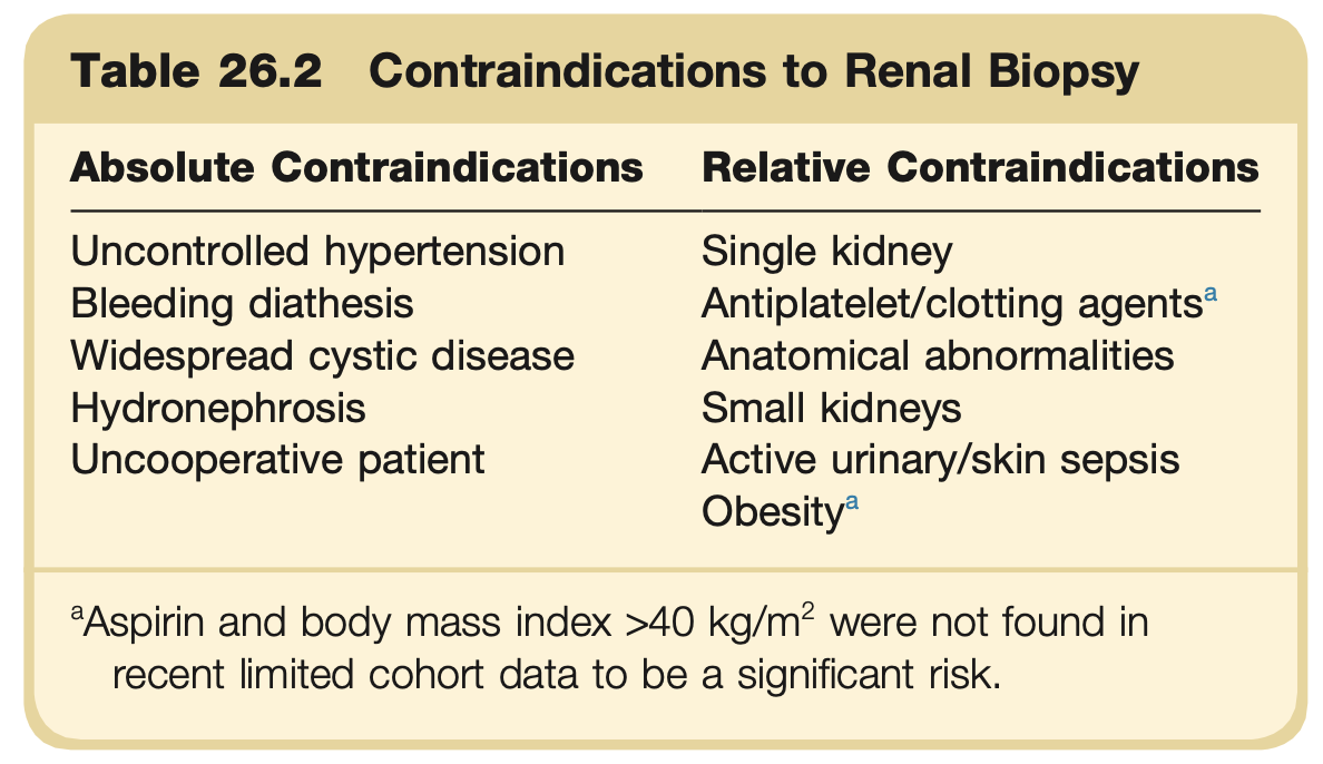
Which of the following is considered an absolute contraindication to a percutaneous renal biopsy?
a. Single functioning kidney.
b. Presence of a renal transplant.
c. Uncontrolled hypertension.
d. eGFR < 30 mL/min/1.73 m2.
Answer: c
Reference: Chapter 26, The Renal Biopsy, Page 864
For which of the following conditions is Electron Microscopy (EM) considered essential for reaching a definitive diagnosis?
a. IgA nephropathy
b. Acute tubulointerstitial nephritis
c. Thin basement membrane lesion
d. Myeloma cast nephropathy
Answer: c
Reference: Chapter 26, The Renal Biopsy, Pages 869, 871.e2
Explanation: Option c is correct because measuring the thickness of the GBM requires the high resolution of EM
What is the first-line imaging examination for azotemic patients to assess renal size and check for obstruction?
a. Noncontrast Computed Tomography (CT)
b. Ultrasonography (US)
c. Intravenous Urography (IVU)
d. Magnetic Resonance Renography
Answer: b
Reference: Chapter 25, Diagnostic Kidney Imaging, Page 797
Explanation: Ultrasonography is the most frequently used diagnostic examination for the evaluation of the kidneys and urinary tract. It is noninvasive, uses no ionizing radiation, and requires minimal patient preparation. It is the first-line examination in azotemic patients to assess renal size and the presence or absence of hydronephrosis and obstruction. It is used to assess the vasculature of native and transplanted kidneys. US is also used to evaluate renal structure and to characterize renal masses. It is the primary modality of imaging for evaluation of a transplanted kidney. It is also the most commonly used modality for imaging guidance for a kidney biopsy.
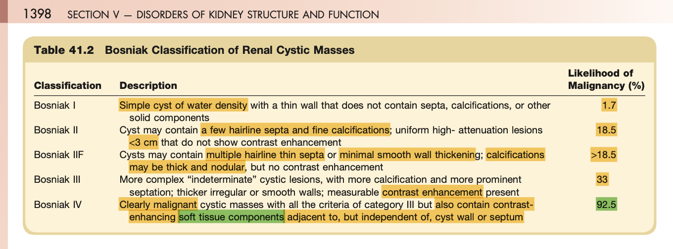
A 58-year-old woman is found to have a complex renal cyst. Contrast-enhanced ultrasound (CEUS) shows flow within a solid nodular component, and CT shows heterogeneous enhancement. This cyst is most appropriately classified as:
a. Bosniak Category I
b. Bosniak Category II
c. Bosniak Category III
d. Bosniak Category IV
Answer: d
Reference: Chapter 25, Diagnostic Kidney Imaging, 838
Explanation: In 1986, Bosniak developed a classification system based on CT imaging characteristics to help guide the clinical management of cystic renal masses.256–263 Category I lesions are simple, benign cysts (see Fig. 25.56). Category II cysts are benign with thin septa, fine rim-like calcification, or they are uniform high-density cysts less than 3 cm in diameter that do not enhance (Fig. 25.65). Category IIF represents more indeterminate category II lesions that necessitate follow-up, usually at 6 to 12 months, to prove benignity (Fig. 25.66).262 These cystic lesions may have multiple septa, or an area of thick or nodular calcification, or they may be high-density cysts larger than 3 cm in diameter. Category III cystic lesions have thickened, irregular walls, which demonstrate some enhancement. Dense irregular calcification may also be visible. In these cases, clinical history may be helpful in determining whether they are renal abscesses or infected cysts. Although some of these lesions are benign, surgery may be necessary for diagnosis and treatment.263 Biopsy has been advocated by some authorities.264–267 Category IV cystic masses are clearly malignant and demonstrate distinct enhanced soft tissue masses or nodules within the cyst (Fig. 25.67).268 Nephrectomy is required for these lesions, although if they are not larger than 5 to 6 cm and are in proper locations, a nephron-sparing procedure may be performed.
A CT scan of an incidental 3-cm renal mass shows an area with an attenuation of -25 Hounsfield Units (HU). This finding is virtually diagnostic for which tumor?
a. Angiomyolipoma (AML)
b. Renal cell carcinoma (RCC)
c. Oncocytoma
d. Transitional cell carcinoma
Answer: a
Reference: Chapter 25, Diagnostic Kidney Imaging, Page 840
Explanation: Renal hamartomas, known as angiomyolipomas (AMLs), are benign renal tumors composed of different tissues, including fat, muscle, vascular elements, and even cartilage. It is the fat component that makes AML distinguishable radiologically. On US the mass is solid and hyperechoic due to the presence of fat. On CT the diagnosis of AML can be made with ease, because most AML have a large amount of fat that exhibits low attenuation (<–10 HU). In uncommon cases, only a minimal amount of fat is present, and it must be searched for diligently. MRI with fat-suppressed and opposed-phase chemical shift sequences can be used to make an accurate diagnosis
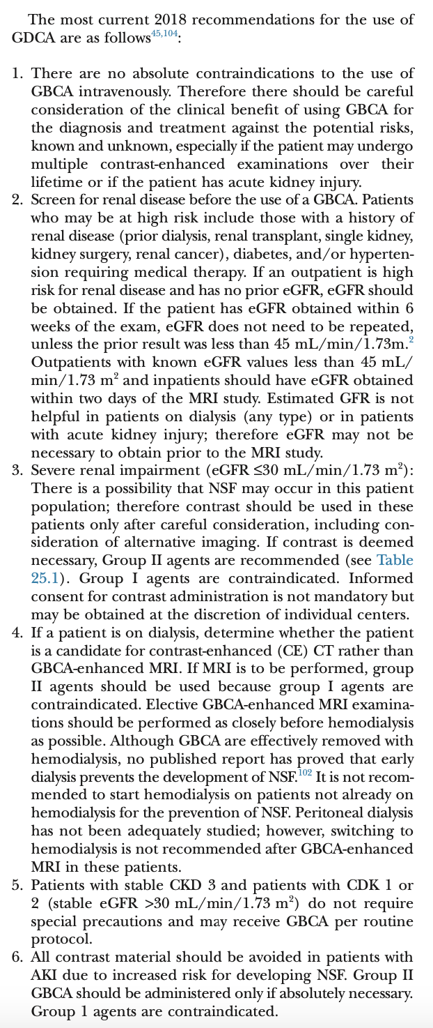
A patient with an eGFR of 25 mL/min/1.73 m2 needs an MRI. According to the ACR manual, which strategy should be followed regarding gadolinium-based contrast agents (GBCA)?
a. All GBCAs are absolutely contraindicated.
b. Use of Group II agents is preferred.
c. The patient must be started on hemodialysis immediately after the MRI to prevent NSF.
d. Group I agents should be used because they are more effectively filtered.
Answer: b
Reference: Chapter 25, Diagnostic Kidney Imaging, Pages 808
What is the most common minor complication resulting from a percutaneous kidney biopsy?
a. Acute kidney injury
b. Sepsis
c. Hematoma or gross hematuria
d. Arteriovenous fistula
Answer: c
Reference: Chapter 26, The Renal Biopsy, Page 863
A patient with a tunneled hemodialysis catheter that was previously functioning well presents for their session. The dialysis nurse is unable to consistently achieve a blood flow greater than 250 mL/min, with multiple alarms despite patient repositioning. Blood flow was decreased to 180ml/min during the session. On post access care, there resistance on flushing. What is the most appropriate initial empirical treatment for this catheter dysfunction?
a. Double anticoagulation with heparin dose during next dialysis session and monitor flow.
b. Instill a thrombolytic agent (e.g., t-PA) into the catheter lumens.
c. Refer the patient for a surgical catheter exchange.
d. Insert temporary access and start apixaban.
Answer: b
Reference: Chapter 68, Interventional Nephrology, p 2203
Explanation: There are two major indications for catheter exchange: dysfunction and infection. Catheter dysfunction is diagnosed when blood cannot be aspirated from the catheter lumen at the time of dialysis initiation, or more commonly, if it is not possible to consistently achieve a blood flow required to yield sufficient urea and other solute clearance (>250 mL/min). In catheters that were previously delivering an adequate blood flow intraluminal thrombus is the most common etiology for dysfunction, although a fibrin sheath may be the culprit in some patients. This problem is usually treated empirically in the dialysis unit by instilling t-PA into the catheter lumens.222 Tissue plasminogen activator (tPA) instillation is successful in about 70% to 80% of catheters, but problems with poor flow frequently recur within 2 to 3 weeks. If the thrombolytic agent does not improve the catheter flow, the patient is referred for catheter exchange.
A 55-year-old patient with a left arm AV fistula presents with severe, persistent left arm edema that is interfering with their quality of life. They have undergone multiple PTAs and one stent placement for left subclavian vein stenosis, but the symptoms recurred within weeks of each procedure. Which of the following is the most appropriate next step to resolve the edema? (Problem Solving)
a. Perform a fourth PTA with a higher-pressure balloon.
b. Place a second, larger stent inside the existing one.
c. Transition the patient to a tunneled femoral catheter.
d. Ligate the left arm vascular access.
Answer: d
Reference: Chapter 68, Interventional Nephrology, p 2196-2198
Explanation: Central vein stenosis is a frequent occurrence in patients on hemodialysis. Acute or chronic trauma of the central vessels caused by either temporary or permanent dialysis catheters is the major cause. Stenosis leads to impairment of venous return on the ipsilateral extremity, and may in turn result in malfunction or thrombosis of the vascular access. Although it may be asymptomatic, patients with central vein stenosis most commonly present with ipsilateral upper extremity edema. In some patients, a previously unappreciated central vein stenosis becomes evident clinically following creation of an ipsilateral AV fistula or graft. The diagnosis can be confirmed by angiography, ultrasound, or magnetic resonance venography.
The most commonly encountered location of central vein stenosis is at the junction of the cephalic vein with the subclavian vein (not catheter injury related). Other central veins that may be affected (often related to injury from previous catheters) include the subclavian vein, brachiocephalic vein, and superior vena cava. In patients with tunneled femoral catheters, central vein stenosis may occur in the external iliac vein, common iliac vein or inferior vena cava, resulting in ipsilateral lower extremity edema. The stenotic lesion is an aggressive neointimal proliferation, or clot and fibrin sheath formed around an indwelling dialysis catheter that becomes organized and incorporated into the vessel wall. Stenoses may progress over time to complete occlusion of the venous circulation. If left untreated, central vein stenosis will cause increased retrograde pressure and formation of venous collaterals. In some patients, the collaterals are sufficiently well developed to permit adequate venous drainage that prevents formation of edema.
The treatment of choice of symptomatic central vein stenosis is PTA of the stenotic lesion.198–205 Unfortunately, the long-term success of PTA of central venous stensosis is quite poor, due to a combination of elastic recoil and aggressive neointimal hyperplasia. In one study, the primary patency was substantially shorter after angioplasty of central vein stenosis, as compared to stenoses at more peripheral locations. As a result, patients with central vein stenosis may require multiple angioplasties to treat recurrent lesions.
Stent placement has been attempted in the management of refractory central vein stenosis due to elastic recoil. Several small series have reported the outcomes of stent placement for refractory central venous stenotic lesions. These studies have been limited by their retrospective study design, the small numbers of patients, and the absence of a control group. In two uncontrolled series, the primary patency following stent deployment for central vein stenosis was 42% to 50% at 6 months, and only 14% to 17% at 1 year. Although there are no published randomized studies comparing stent deployment with angioplasty of central vein stenosis, the primary patency utilizing stents appears no better than that achieved with angioplasty alone. In patients with ipsilateral vascular access and persistent upper extremity edema despite attempted angioplasty, the only recourse may be ligation of the vascular access, creation or placement of a contralateral arteriovenous access, transition to peritoneal dialysis, or urgent kidney transplantation.