Nervous System
 Call Kai
 Learn
Learn Practice Test
 Spaced Repetition
 Match
Match Flashcards
 Knowt Play
1/123

There's no tags or description
Looks like no tags are added yet.
Name | Mastery | Learn | Test | Matching | Spaced | Call with Kai | Chat |
|---|
No analytics yet
Send a link to your students to track their progress
124 Terms
Anatomy of a neuron
Some
Dendrites
Initial segment
Axon colateral
Axon
Axon terminal

initial segment of axon
Area of the axon having the lowest threshold for stimulation, so the action potentials begin at this point
Axon colateral
a branch of an axon from a single neuron
Can axons differentiate?
No, they are terminally differentiated cells
Amniotic cell
A cell that cannot divide
- neurons!
Nuclei
clusters of neuron cell bodies in CNS
Ganglia
clusters of cell bodies in the PNS
What kind of metabolic need to neurons have?
High metabolic need
Ratio of neurons to glia
1:1
Types of glial cells
astrocytes, microglia, oligodendrocytes/schwann cells, ependymal cells
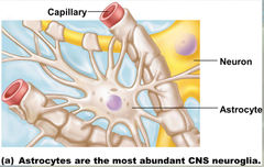
Astrocytes
Provide structural and metabolic support for neurons.
Form blood brain barrier
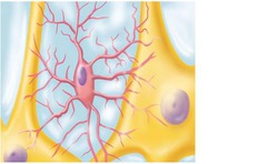
Microglia
Act as phagocytes, eating damaged cells and bacteria, act as the brains immune system
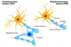
Oligodendrocytes and Schwann cells
form myelin sheath
- Oligodendrocytes in CNS
- Schwann cells in PNS
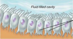
Ependymal cells
produce cerebrospinal fluid
How are neurotransmitters and other materials transported from the soma to the axon terminal?
Transported on microtubule tracks by kinesins and dyneins
Kinesin
motor proteins in anterograde transport
- to periphery of cell
- towards POSITIVE end of microtubule
Dynein
motor proteins in retrograde transport
- to nucleus of cell
- towards NEGATIVE end of microtubules
Classes of neurons
afferent, efferent, interneurons
Interneurons
Central nervous system neurons that internally communicate and intervene between the sensory inputs and motor outputs
Afferent neurons
Nerve cells that carry impulses towards the central nervous system from the PNS
- "sensory neurons"
Efferent neurons
Nerve cells that conduct impulses away from the central nervous system to the PNS
- "motor neurons"
Divisions of the PNS
Afferent and efferent
Divisions of afferent PNS
somatic sensory, visceral sensory, special sensory
somatic sensory division
carries signals from receptors in the skin, muscles, bones, and joints
- helps us sense touch, pain, temperature, vibration, etc
Visceral sensory division
carries signals from the viscera (heart, lungs, stomach, and urinary bladder)
- organ senses
Special sensory division
vision, hearing, taste, smell, balance
Divisions of the efferent PNS
somatic and autonomic
somatic motor division
carries signals to skeletal muscles
Divisions of autonomic nervous system
sympathetic, parasympathetic, enteric
Brain anatomy
Brain Lobes
-Frontal
-Parietal
-Temporal
-Occipital
Cerebellum
Brain stem

Frontal lobe function
involved in motor function, problem solving, memory, judgment, impulse control
Parietal lobe function
somatosensation and spatial processing
Temporal lobe function
Hearing, memory, language comprehension.
Occipital lobe function
visual processing
Cerebellum function
Balance and coordination
Parts of the brainstem
midbrain, pons, medulla oblongata, reticular formation
Forebrain
largest part of the brain, containing the cerebral cortex and the diencephalon
Diencephalon
thalamus and hypothalamus
Motor cortex
an area at the rear of the frontal lobes that controls voluntary movements

Somatosensory cortex
area at the front of the parietal lobes that registers and processes body touch and movement sensations

Hypothalamus
Master coordinator of the nervous and endocrine systems, responsible for controlling eating, drinking, reproduction, etc
Brainstem function
responsible for automatic survival functions
- all nerve fibers that relay signals go through the brainstem!
reticular formation function
essential for life
recieves and integrates information from across entire CNS
regulates breathing, sleep, attention, circulation, movement
Integrating centers
neuron clusters in the brainstem that control GI, respiration, cardiovascular function, and more
What protects the spinal cord?
The vertebral column (spine)
What part of the spinal cord recieves incoming senory information?
The posterior (dorsal) horn.
What part of the spinal cord sends out motor signals?
The anterior (ventral) horn.
Fiber tracts
in the spinal cord that transmit signals between different regions of the central nervous system.
run longitudinally through spinal cord
How many total nerve pairs are there in the PNS?
43 total
12 pairs of cranial nerves
31 pairs of spinal nerves
cervical
thoracic
lumbar
sacral
coccygeal
How many cervical nerves are there?
8
How many thoracic nerves are there?
12
How many lumbar nerves are there?
5
How many sacral nerves are there?
5
How many coccygeal nerves are there?
1
Function of cervical nerves
Control muscles and glands. Receive sensory input from head, neck, shoulders, arms hands
Function of thoracic nerves
Associated with chest and upper abdomen
Function of lumbar nerves
Associated with lower abdomen, hips, and legs
Function of sacral nerves
Associated with lower digestive tract and genitals
Are spinal nerves afferent or efferent?
Both!
Function of cranial nerves
special sensory function, balance, motor function of head, neck, facial muscles
Are cranial nerves afferent or efferent?
Some are purely afferent, some are purely efferent. Most are both.
Structure of somatic neurons
SINGLE neuron between CNS and skeletal muscles
Are somatic neurons inhibitory or excitatory?
Excitatory
Structure of autonomic neurons
Two neuron chain between CNS and affector organ/gland
What do autonomic neurons innervate?
smooth muscle, cardiac muscle, glands, GI organs
NO SKELETAL MUSCLE
Are autonomic neurons excitatory or inhibitory?
Either or
Where do sympathetic neurons originate from?
Thoracolumbar region
Sympathetic neuron structure
Short pre-ganglionic axon and long post-ganglionic axons
What neurotransmitter(s) do sympathetic neurons use?
Acetylcholine for pre-ganglionic synapses
Epinephrine/norepinephrine for post-ganglionic synapses
Where do parasympathetic neurons originate from?
Craniosacral region
Parasympathetic neuron structure
Long pre-ganglionic axons, short post-ganglionic axons
What neurotransmitter(s) do parasympathetic neurons use?
Acetylcholine used for both pre- and post-ganglionic synapses
Structure of adrenal medulla
A set sympathetic post-ganglionic neurons
What does the adrenal medulla release?
Catecholamines
80% epinephrine
20% norepinephrine
Relationship between sympathetic and parasympathetic nervous systems
Antagonistic—never activated together!
Physical support structures for the CNS
cranium, meninges, CSF
Meninges
Series of membranes between the brain and skull that protect the brain, blood vessels, contain CSF, and form partitions
What are the meninge layers?
Dura Mater
Arachnoid Mater
Pia Mater
Dura Mater
Outermost layer of meninges
Arachnoid mater
2nd layers in the meninges.
Subarachnoid space is filled with CSF and houses the largest blood vessels that supply the brain
Pia mater
The meninge layer closest to the brain
clings to the brain EXTREMELY tightly
most delicate
contains a network of blood vessels
Cerebrospinal fluid
Extracellular fluid of the CNS that provides cushioning and maintains a stable a optimal environment
Where is CSF made?
In the choroid plexus by ependymal cells
Where is CSF found?
Circulates through the subarachnoid space and the ventricles
How much volume of CSF do we have in our body?
~125-150 mL
Recycling of CSF
CSF is reabsorbed by arachnoid villi. CSF is recycled 3 times a day!
Blood Brain Barrier
A layer formed of impermeable brain capillaries, thick surrounding basal lamina, and astrocytes which helps maintain a stable brain environment
Permeability of BBB capillaries
Most impermeable capillaries in the entire body
ONLY lipid-soluble compounds can easily cross the BBB
What factors affect electrostatic force strength?
Quantity of charge and distance between charges
Coloumb’s law
Resting membrane potential
Charge formed by the separation of ions across a membrane
Why do different cells have different resting membrane potentials?
It comes down to differences in membrane permeability!
RMP for neurons
-70 mV
Concentration of Na+ intracellularly vs extracellularly
Na+ higher extracellularly
Concentration of K+ intracellularly vs extracellularly
K+ higher intracellularly
Concentration of Ca2+ intracellularly vs extracellularly
Ca2+ higher extracellularly
How to membrane potentials change?
Movement of ions across the membrane
Nernst Equation
Tells us what the membrane potential would be if the membrane was ONLY permeable to the ion of interest

Goldman-Hodgkins-Katz equation
Allows us to calculate RMP by looking at individual ion contribution

What establishes the RMP?
Sodium-potassium ATPase
3 Na+ out, 2 K+ in
Uses ATP
Depolarization
moving from RMP to more positive