A&P 1 ch. 10 The Muscular System
 Call Kai
 Learn
Learn Practice Test
 Spaced Repetition
 Match
Match Flashcards
 Knowt Play
1/505

There's no tags or description
Looks like no tags are added yet.
Name | Mastery | Learn | Test | Matching | Spaced | Call with Kai | Chat |
|---|
No analytics yet
Send a link to your students to track their progress
506 Terms
What is the fundamental purpose of all muscle tissue?
To convert the chemical energy of ATP into the mechanical energy of motion.
What are the three types of muscle tissue?
Skeletal, cardiac, and smooth muscle.
What are the 6 collective functions of muscle tissue?
Movement, stability, control of body openings/passages, heat production, hormone secretion, and glycemic control.
Term used for muscles that resist the pull of gravity and prevent us from falling or slumping over
Antigravity muscles
What percentage of body heat do skeletal muscles produce at rest versus during exercise?
20% to 30% at rest, and up to 85% during exercise.
What are the hormones secreted by exercised muscles called, and what do they do?
Myokines; they stimulate glucose synthesis by the liver and the breakdown of visceral fat.
How do skeletal muscles contribute to glycemic control?
They regulate blood glucose concentration by absorbing, storing, and using a large share of the body's glucose.
The term "muscular system" refers strictly to which type of muscle?
Skeletal muscle only
Myology
The study of skeletal muscles
Approximately how many muscles are in the human muscular system?
About 600 muscles
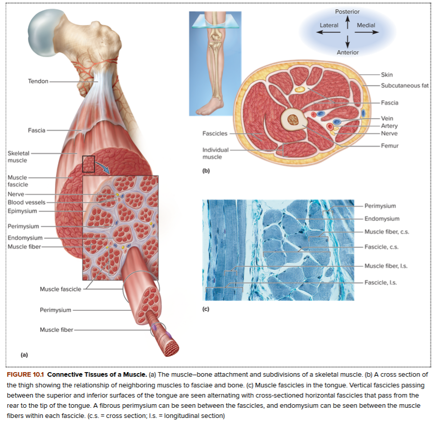
connective tissues of a muscle (image 1)
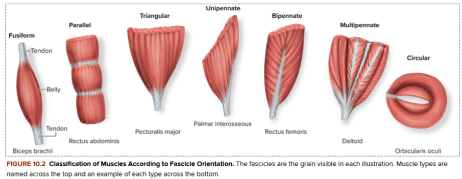
classification of muscles according to fascicle orientation (image 2)
Other three components make up a skeletal muscle
Connective tissue, nerves, and blood vessels
What is the order of skeletal muscle connective tissue components from smallest to largest (deep to superficial)?
Endomysium (deepest)
Perimysium (middle)
Epimysium (most superficial)
Fascia
Endomysium
A thin sleeve of loose connective tissue that surrounds each individual muscle fiber.
What vital functional role does the endomysium play regarding nourishment and stimulation?
It creates room for blood capillaries and nerve fibers to reach every individual muscle fiber.
The excitation of a muscle fiber relies on the exchange of which three ions between the endomysial fluid and the fibers?
Calcium, sodium, and potassium ions.
Perimysium
A thicker connective tissue sheath that wraps muscle fibers together into bundles called fascicles.
What is a fascicle, and approximately how many muscle fibers does it contain?
A bundle of muscle fibers wrapped by perimysium; it typically contains 20 to 60 muscle fibers.
Which connective tissue layer carries larger nerves, blood vessels, and stretch receptors called muscle spindles?
The perimysium
Epimysium
A fibrous sheath that surrounds the entire muscle.
Fascia
A sheet of connective tissue that separates neighboring muscles or muscle groups from each other and from the subcutaneous tissue.
What structural feature determines both the strength of a muscle and the direction in which it pulls?
The orientation of its fascicles
What are the structural features of a fusiform muscle?
They are thick in the middle with fascicles converging on each tapered end.
What are two major anatomical examples of fusiform muscles?
The biceps brachii of the arm and the gastrocnemius of the calf.
Muscle strength is directly proportional to what physical characteristic?
The diameter of a muscle at its thickest point
What are the structural characteristics of parallel muscles?
They have a fairly uniform width and parallel fascicles
What are three examples of parallel muscles that are elongated straps?
The rectus abdominis of the abdomen, sartorius of the thigh, and zygomaticus major of the face.
What term is used for squarish, four-sided parallel muscles, and what is an example?
Quadrilateral muscles; an example is the masseter of the jaw.
What is a functional advantage and a functional disadvantage of parallel muscles?
Advantage: They can shorten more than other muscle types.
Disadvantage: Having fewer muscle fibers than a fusiform muscle of the same mass, they produce less force.
What are the structural features of triangular (convergent) muscles?
They are fan-shaped—broad at one end with fascicles converging on the other, narrower end.
Two (2) anatomical examples of triangular muscles
The pectoralis major in the chest and the temporalis on the side of the head.
Why are triangular muscles relatively strong despite having small localized insertions on a bone?
Because they contain a large number of fibers in the wider part of the muscle.
What shape do pennate muscles resemble, and how do their fascicles insert?
They are feather-shaped; their fascicles insert obliquely on a tendon that runs the length of the muscle (like the shaft of a feather).
What is a unipennate muscle, and what are two examples?
A muscle where all fascicles approach the tendon from one side; examples include the palmar interosseous muscles of the hand and the semimembranosus of the thigh.
What is a bipennate muscle, and what is an example?
A muscle where fascicles approach the tendon from both sides; an example is the rectus femoris of the thigh.
What is a multipennate muscle, and what is an example?
A muscle shaped like a bunch of feathers with their quills converging on a single point; an example is the deltoid of the shoulder.
Why do pennate muscles generate more force than parallel or fusiform muscles?
Because they fit more muscle fibers into a given length of muscle.
What is another name for circular muscles, and what is their structural arrangement? Sphincters
Sphincters; they have fascicles arranged in rings around certain body openings and passages.
What happens when circular muscles contract?
They constrict the opening and tend to prevent the passage of material through it.
What are two examples of skeletal circular muscles?
The orbicularis oculi of the eyelids and the external urethral and an@l sphincters.
What type of muscle tissue can also form sphincters, and what are three examples?
Smooth muscle; examples include the pyloric valve (stomach to small intestine), some sphincters of the urinary tract, and the anal canal.
Muscle compartment
A package of groups of functionally related muscles, along with their supplying nerves and blood vessels, enclosed by fasciae.
Where in the body does muscular compartmentalization occur?
In the thoracic and abdominal walls, pelvic floor, and limbs.
Intermuscular septa
particularly thick fasciae that separate muscle compartments
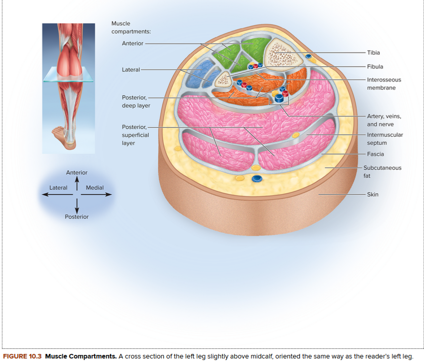
Muscle compartment (cross section) - Image 3
What causes compartment syndrome, and what is its primary danger?
It is caused by blood and fluid accumulation inside an inelastic muscle compartment. The resulting pressure obstructs arteries, causing ischemia (poor blood flow) that can lead to permanent muscle necrosis.
reversible DAMAGE? Nerves can regenerate after the pressure is relieved, but muscle necrosis (death) is irreversible.
What are two key clinical signs of compartment syndrome?
Severe tissue pressure/ischemia and myoglobinuria (dark-colored urine caused by myoglobin released from broken-down muscle).
How is compartment syndrome treated?
By immobilizing and resting the limb, or by performing a fasciotomy (an incision into the fascia to relieve pressure).
What protein is released into the blood during muscle breakdown?
Myoglobin
What is myoglobinuria, and what is its clinical appearance?
The presence of myoglobin in the urine; it gives the urine a dark color and is a key sign of compartment syndrome or degenerative muscle disorders.
Fasciotomy
An incision made into the fascia to relieve pressure inside a muscle compartment.
How is strong structural continuity created between a muscle and a bone?
Collagenous fibers emerge from the muscle's connective tissue, continue into the tendon or attachment, and then merge into the periosteum and matrix of the bone.
Three (3) other structures where muscles might insert
The fascia or tendon of another muscle, or the collagen fibers of the dermis (skin).
Where does the distal tendon of the biceps brachii partly insert?
On the fascia of the forearm.
What allows facial muscles to produce expressions like a smile or a wink?
They insert directly into the skin of the lips, eyelids, and other facial areas.
Two (2) forms of muscle attachment to bones?
Direct and indirect attachments
Direct (fleshy) attachment
An attachment where there is so little separation between muscle and bone that the red muscular tissue seems to emerge directly from the bone to the naked eye.
Two (2) anatomical examples of muscles with direct attachments
The brachialis and the lateral head of the triceps brachii.
What actually spans the gap between muscle and bone at a microscopic level in a direct attachment?
Collagen fibers (the muscle fibers themselves stop slightly short of the bone).
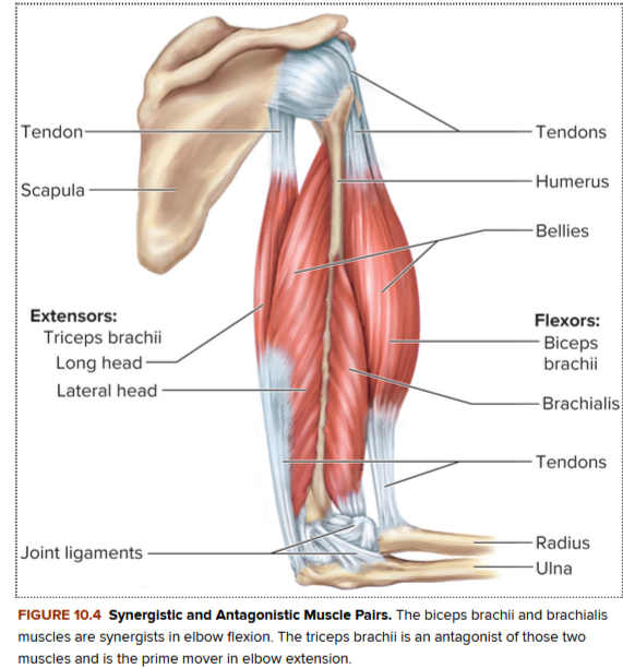
Synergistic and antagonistic muscle pairs (image 4)
Indirect attachment
An attachment where the muscle ends visibly short of its bony destination, and the gap is bridged by a fibrous cord or band called a tendon (Example; The biceps brachii)
What tendon can be easily palpated just above your heel?
The calcaneal or Achilles tendon
Two (2) tendons that can be felt on the anterior side of the wrist
The tendons of the palmaris longus and flexor carpi radialis muscles.
Aponeurosis
A broad, sheet-like tendon
Example: the palmar aponeurosis, located beneath the skin of the palm.
Retinaculum
A band of connective tissue under which groups of tendons from separate muscles pass (like a bracelet over the surface of the wrist).
The origin
attachment at the stationary end of a muscle
The insertion
attachment at the moving end of a muscle
How do the relative movements of the origin and insertion of the quadriceps femoris reverse between kicking a soccer ball and sitting down in a chair?
When kicking, the tibia moves more than the femur (tibia is insertion, femur is origin). When sitting down, the femur moves more than the tibia (femur becomes insertion, tibia becomes origin).
What alternative descriptive terms do some authorities use instead of origin and insertion?
Proximal and distal, medial and lateral, or superior and inferior muscle attachments.
authorities largely abandoned the terms "origin" and "insertion because they are imperfect and misleading; the moving and nonmoving ends of a muscle can be reversed depending on the action being performed.
They use Proximal and distal, medial and lateral, or superior and inferior muscle attachments.
How do the relative movements of the origin and insertion of the biceps brachii reverse between lifting weights and doing chin-ups?
In weight lifting, the arm is stationary (origin) and the forearm moves (insertion). In chin-ups, the forearm is stationary and the arm moves to lift the body, reversing the origin and insertion.
How do the relative movements of the origin and insertion of the quadriceps femoris reverse between kicking a soccer ball and sitting down in a chair?
When kicking, the tibia moves more than the femur (tibia is insertion, femur is origin). When sitting down, the femur moves more than the tibia (femur becomes insertion, tibia becomes origin).
Intrinsic muscle
A muscle that is contained entirely within a region of interest (such as the small muscles located entirely within the hand).
Extrinsic muscle
A muscle that acts upon a designated organ or region but arises from another region (such as forearm muscles that connect to the hand via long tendons).
Besides the hand, what are four other anatomical structures or regions where the intrinsic-extrinsic distinction applies?
The tongue, larynx, back, and foot.
"Action" of a muscle
The effect produced by a muscle, whether it is to produce or prevent a movement.
How can the action of the gastrocnemius change depending on what the quadriceps muscle is doing?
The gastrocnemius usually flexes the knee, but if the quadriceps prevents knee flexion, the gastrocnemius flexes the ankle instead (causing plantar flexion).
Prime mover (agonist)
The muscle that produces most of the force during a particular joint action.
What muscle serves as the prime mover during flexion of the elbow?
The brachialis
Synergist
A muscle that aids the prime mover
Example: the biceps brachii working with the brachialis to flex the elbow
What are two ways a synergist can optimize a movement besides just adding power?
It can stabilize a joint to restrict undesirable movements (like rotation) or modify the direction of a movement so the action is more coordinated.
Antagonist
A muscle that opposes the prime mover
What are the protective functional roles of an antagonist maintaining tension on a joint during an action?
It limits the speed or range of the prime mover, preventing excessive movement, joint injury, or inappropriate actions.
What muscle acts as the antagonist to slow down and stop elbow extension when you reach out to pick up a cup of tea?
The brachialis (while the triceps brachii serves as the prime mover).
Antagonistic pair of muscles
A pair of muscles that act on opposite sides of a joint because a single muscle can only pull, not push.
Fixator
A muscle that prevents a bone from moving by holding it steady, allowing another muscle attached to it to pull on something else.
What muscles act as fixators to hold the scapula firmly in place when the biceps brachii contracts?
The rhomboids
Innervation
The identity of the nerve that supplies a given muscle.
What is the functional consequence if a muscle's nerve is severed?
The muscle is paralyzed
From where do spinal nerves arise, and how do they emerge from the vertebral column?
They arise from the spinal cord and emerge through the intervertebral foramina.
What regions of the body do spinal nerves supply?
Muscles below the neck
Immediately after emerging from an intervertebral foramen, each spinal nerve branches into what two structures?
A posterior and an anterior ramus
Plexus in relation to spinal nerves
Weblike networks of spinal nerves adjacent to the vertebral column.
From where do cranial nerves arise, and how do they emerge from the skull?
They arise from the base of the brain and emerge through the skull foramina.
What regions of the body do cranial nerves innervate?
Muscles of the head and neck
How are cranial nerves uniquely identified or labeled?
By roman numerals (CN I to CN XII) and by names.
How much of the body's total blood flow does the muscular system receive at rest versus during exercise?
About one-quarter (1.24 L/min) at rest, and up to three-quarters (11.6 L/min) during exercise.
What structural adaptation prevents the capillaries of skeletal muscle from breaking when a muscle lengthens?
The capillaries undulate or coil when the muscle is contracted, giving them enough slack to stretch out straight without breaking.
How do Latin roots help translate the muscle name depressor labii inferioris?
It is a muscle that lowers (depresses) the bottom (inferior) lip (labium).
How do Latin roots help translate the muscle name flexor digiti minimi brevis?
It is a short (brevis) muscle that flexes the smallest (minimi) finger (digit).