6.2
 Call Kai
 Learn
Learn Practice Test
 Spaced Repetition
 Match
Match Flashcards
 Knowt Play
1/29

There's no tags or description
Looks like no tags are added yet.
Name | Mastery | Learn | Test | Matching | Spaced | Call with Kai | Chat |
|---|
No analytics yet
Send a link to your students to track their progress
30 Terms
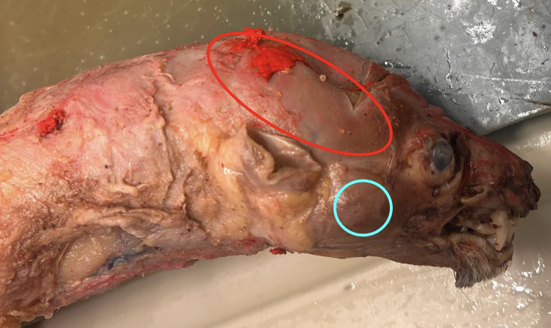
A= red circle B= blue circle
A: Temporalis
B: Masseter

mylohyoid

digastric
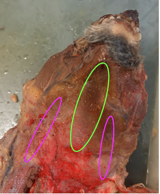
A= green circle B= pink circle
A: mylohyoid
B: digastric
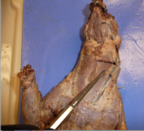
sternomastoid
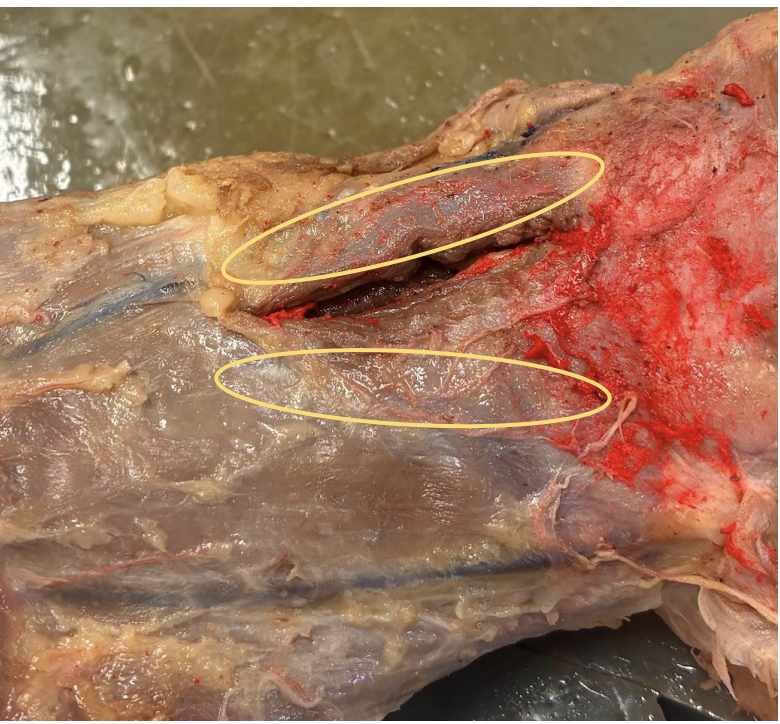
sternomastoid
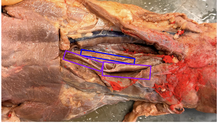
A= purple box B= blue box
A: sternohyoid
B: sternothyroid
how are the sternothyroid and sternohyoid anatomically positioned?
Neck muscles viewed ventrally
The sternohyoid is superficial, and the sternothyroid is deep
Both are thin and stuck together
sternohyoid goes up to the hyoid bone (top of the neck), whereas the sternothyroid cuts off less cranialy
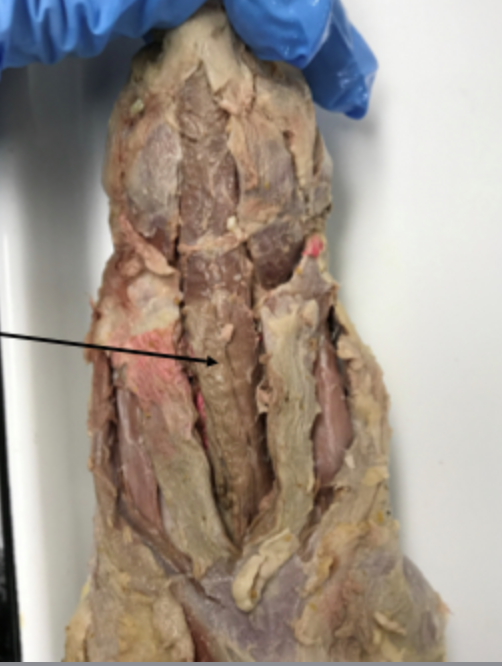
sternohyoid
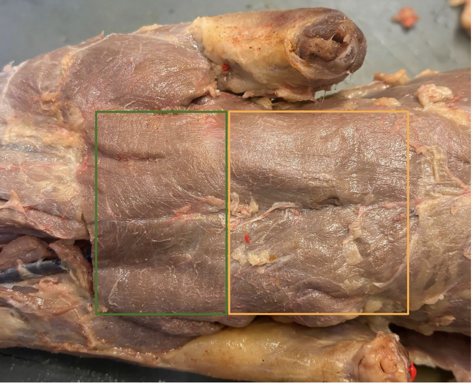
A= green box B= yellow box
A: pectoralis majors
B: pectoralis minors
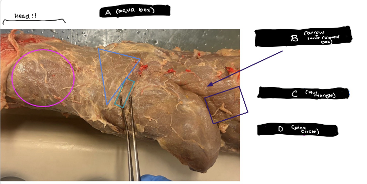
A: spinodeltoid
B: latissiumus dorsi
C: acromiotrapezius
D: clavotrapezius

spinodeltoid
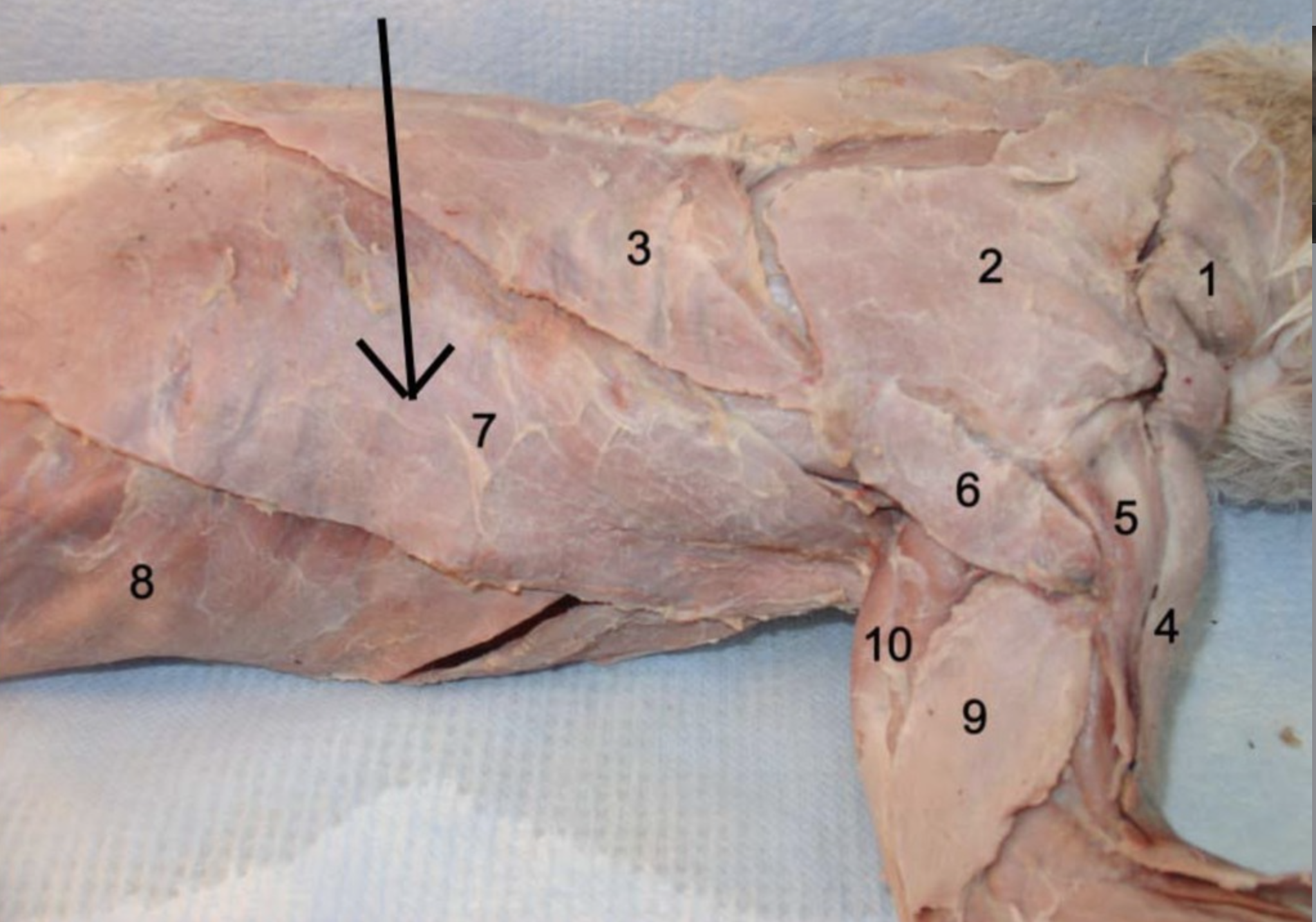
latissiumus dorsi
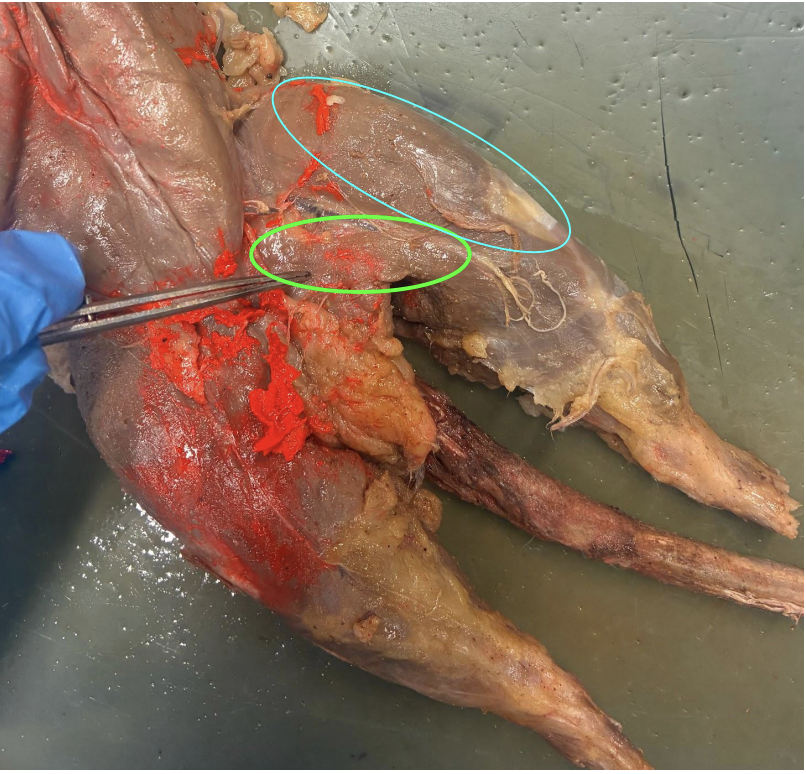
A=green circle B=blue circle
A: gracillus
B: sartorius
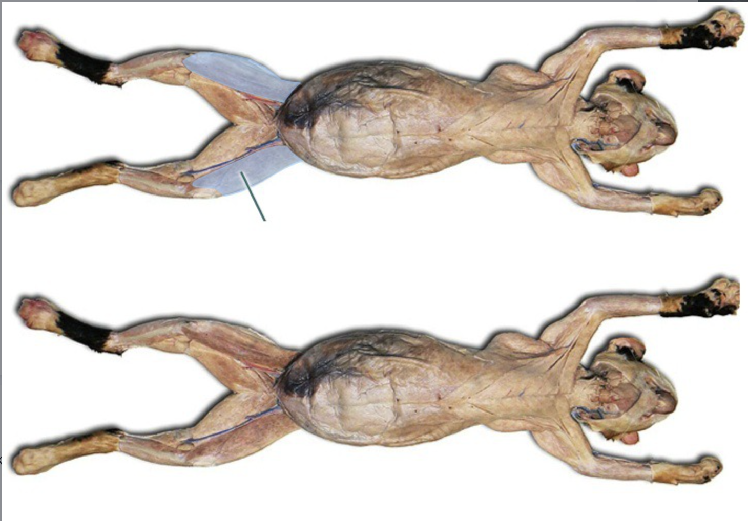
sartorius
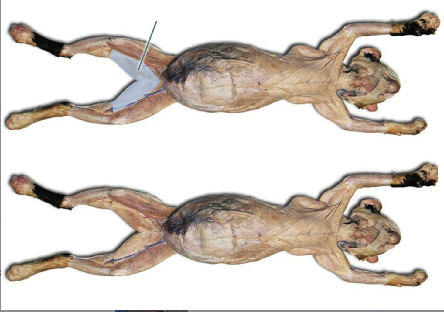
gracillus
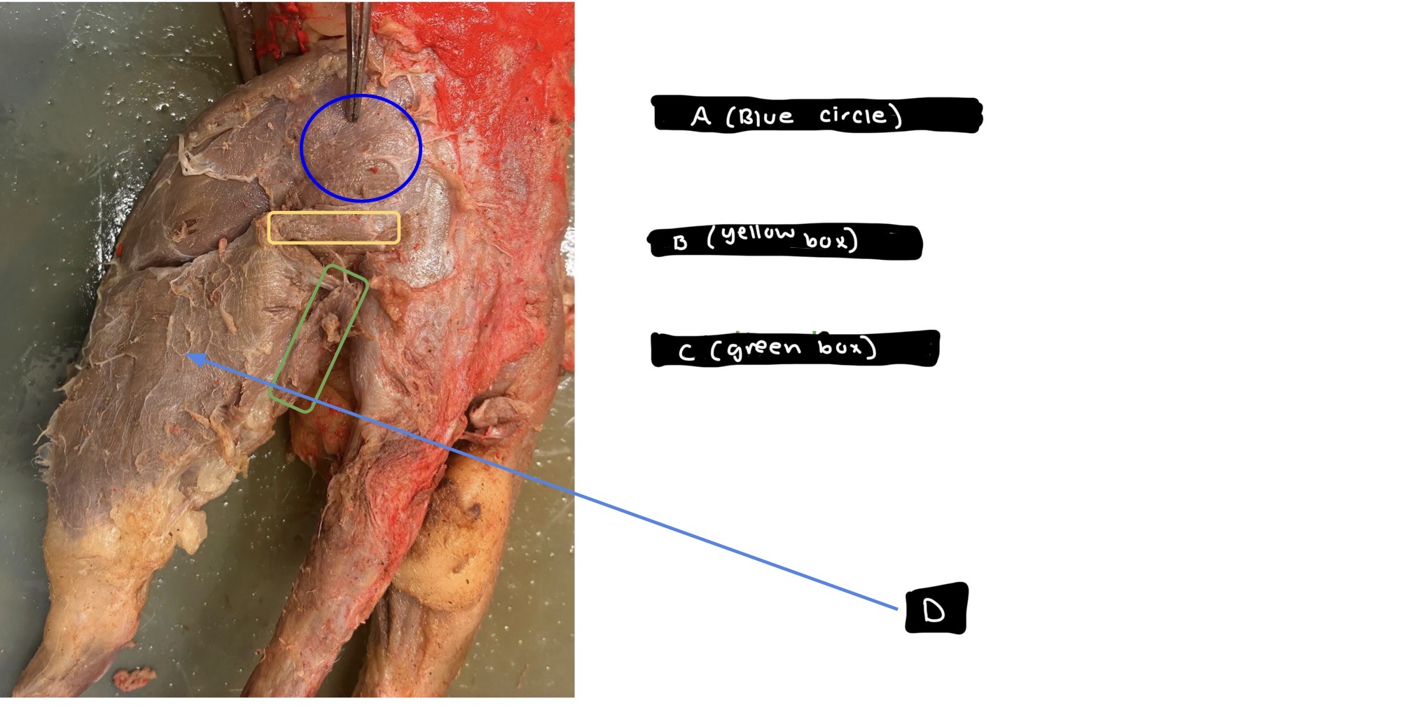
A: gluteus maxiumus
B: caudofemoralis
C: semitendinosus
D:bicep femoralis
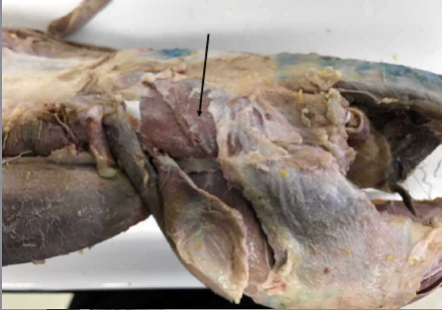
gluteus maximus

caudofemoralis
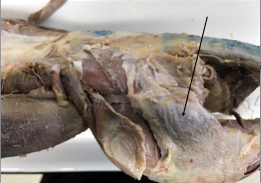
bicep femoralis

gastrocnemius
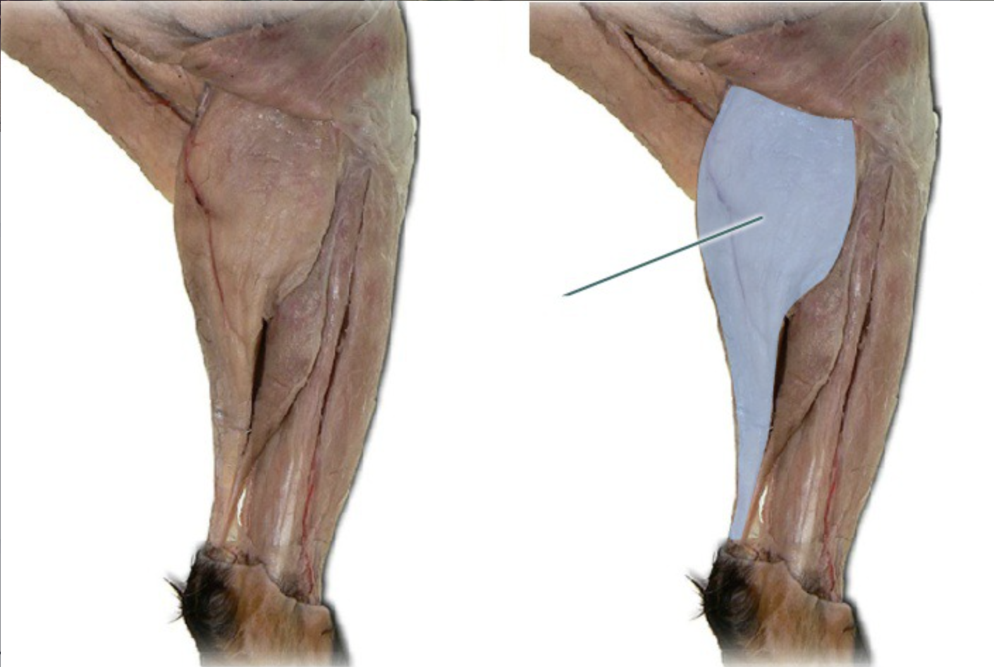
gastrocnemius
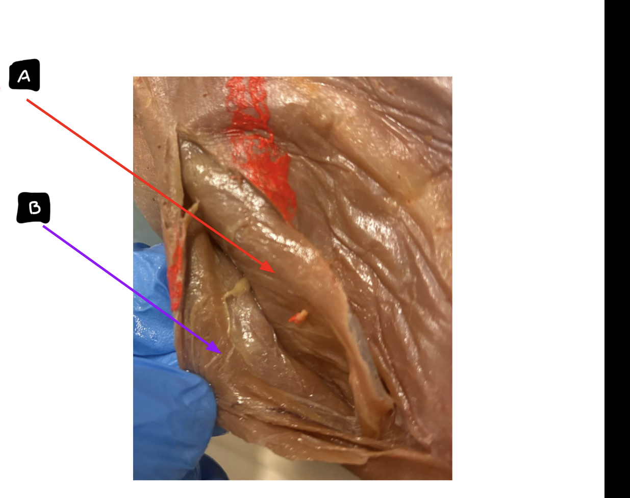
A: transverse abdominus
B: internal abdominal oblique
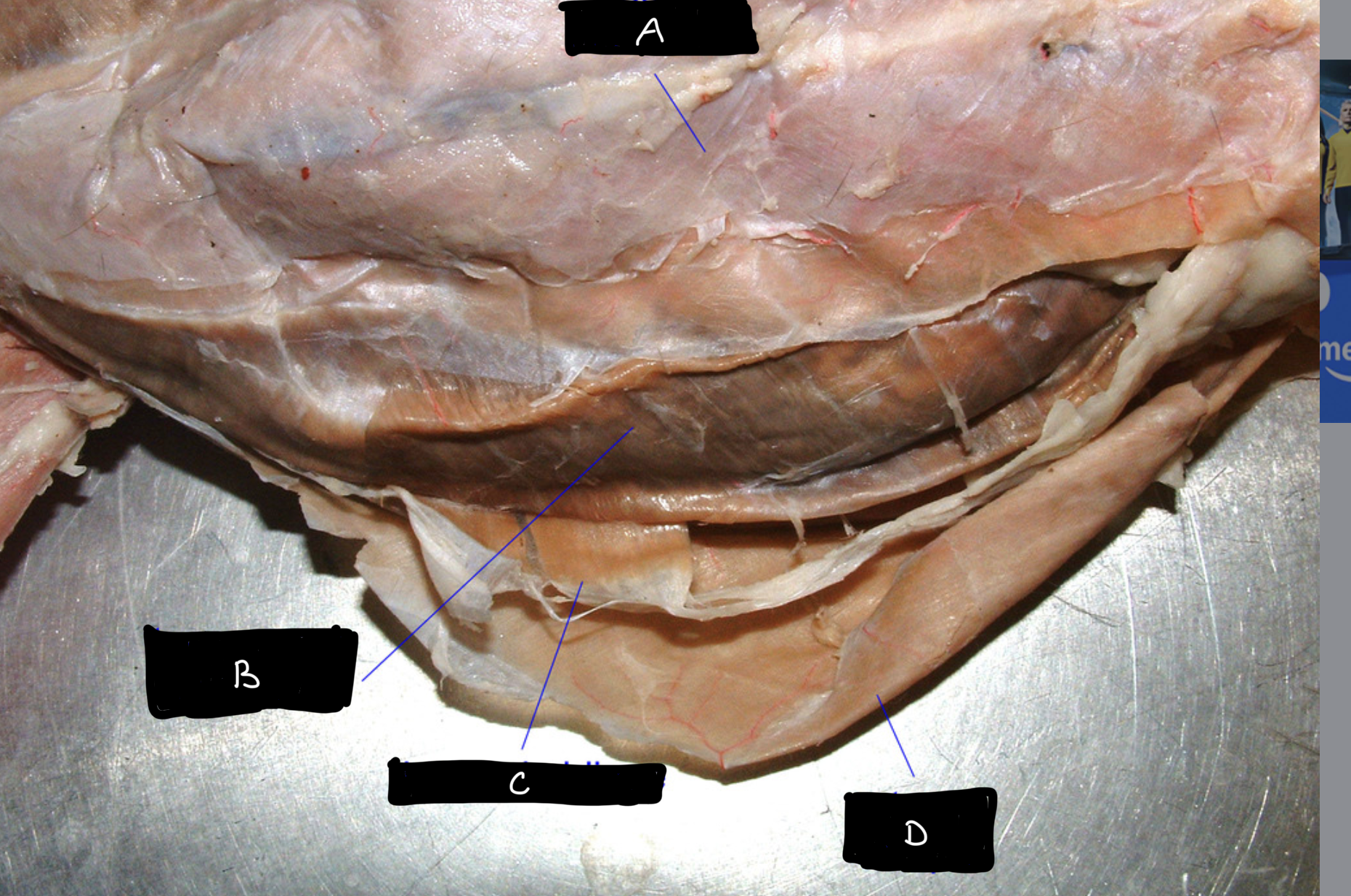
A: rectus abdominus
B: transverse abdominus
C: internal abdominal oblique
D: external abdominal oblique
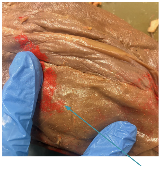
external abdominal oblique

rectus abdominis

A: mylohyoid
B: digastric
C: sternothyroid
D: sternohyoid
E: sternomastoid
F: clavotrapezius
G+M: pectoralis major
H+L: pectoralis minor
I: latissimus dorsi
J: external abdominal oblique
K: masseter
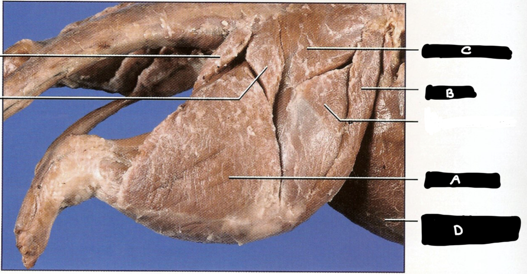
A: biceps femoralis
B: sartorius
C: gluteus maximus
D: external abdominal oblique
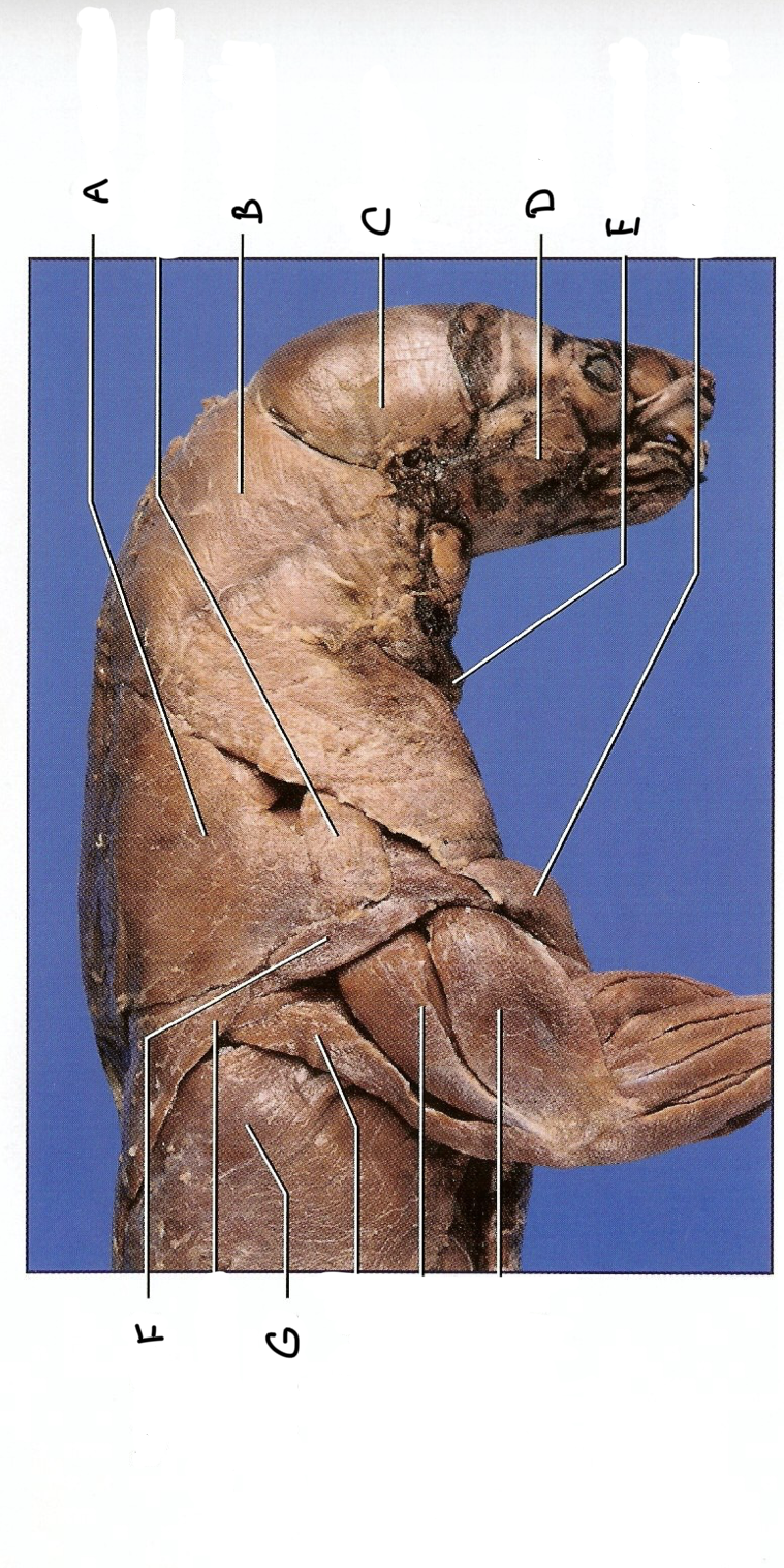
A: acromiotrapezius
B: clavotrapezius
C: temporalis
D: masseter
E: sternomastoid
F: spinodeltoid
G: Latissimus dorsi
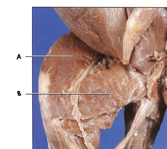
A: sartorius
B: Gracilis