head and neck
 Call Kai
 Learn
Learn Practice Test
 Spaced Repetition
 Match
Match Flashcards
 Knowt Play
1/19

There's no tags or description
Looks like no tags are added yet.
Name | Mastery | Learn | Test | Matching | Spaced | Call with Kai | Chat |
|---|
No analytics yet
Send a link to your students to track their progress
20 Terms
pharyngeal arches appear in which Stage of development?
Fourth and fifth week
Extend of scalp
It extends anteriorly up to the eyebrows (superciliary arches),
posteriorly up to the superior nuchal lines, and laterally on
each side up to superior temporal line
Layers of scalp
Skin.
2. Connective tissue (superficial fascia).
3. Aponeurosis (occipitofrontalis muscle and its
aponeurosis).
4. Loose areolar tissue.
5. Pericranium.
3rd layer of scalp

Surgical layers of scalp
First 3 layers
Black eye
Peri orbital hematoma following head injury
Blood and fluid accumulates in loose area or layer of a scalp and spread anteriorly because frontal muscle has no body attachments
Emissary vein
The vein connecting veins outside cranium with the intercranial dural Venus sinus by passing through foramen in cranium
loose areolar tissue is layer traversed by emery bin
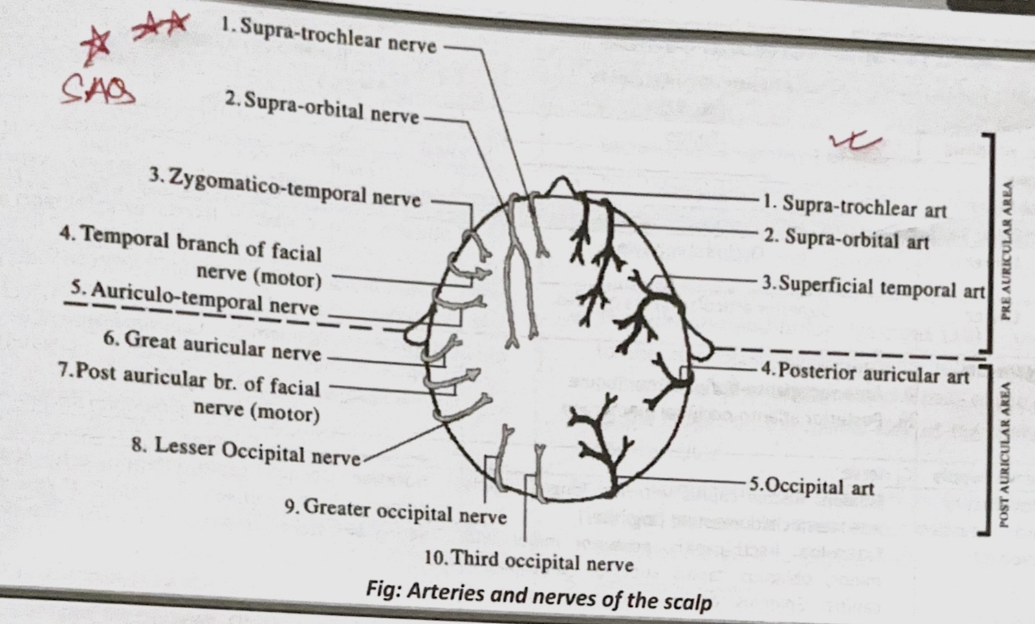
Nerve supply of scalp
Arterial supply of scalp
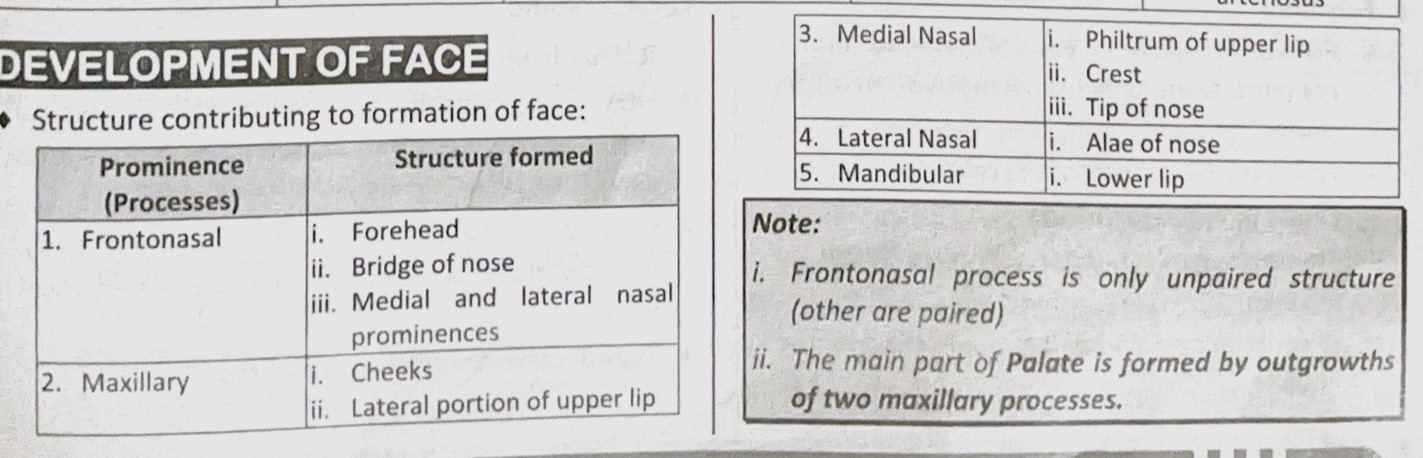
Development of face
Pharyngeal pouches?
Pharyngeal clefts (grooves)?
Pharyngeal pouches: Endodermal outpocketings of the pharyngeal endoderm that develop into various head and neck structures.
Pharyngeal clefts (grooves): Ectodermal invaginations between the pharyngeal arches on the external surface of the embryo.
First pharyngeal arc
Nerve
Artery
Muscle
Skeleton
Nerve: mandibular branch of trigeminal nerve
Artery: maxillary artery
Muscles: mastication (masseter, temporalis, medial and lateral pterygoids), Mylohyoid, tensor Palatine, tensor Tympani, anterior belly of digastric
Skeleton: Maxilla, mandible, malleus, incus, interior ligament of malleus, sphenomandibular ligament, Zygomatic bone, part of temporal bone
Second pharyngeal arc
Nerve
Artery
Muscle
Skeleton
Nerve: facial nerve
Artery: stapedial hyoid artery
Muscle: facial expression (frontalis, Platysma, buccinator, auricularis, orbicularis oris, orbicularis oculi, posterior belly of digastric, stapedius, stylohyoid
Skeleton: stapes, stylohyoid ligament, styloid process, lesser horn, and upper part of the body of hyoid bone
third pharyngeal arc
Nerve
Artery
Muscle
Skeleton
Nerve: glossopharyngeal nerve
Artery: common carotid artery, Internal carotid artery
Muscle: stylopharyngeus
Skeleton: greater horn and lower part of the body of hyoid bone
fourth pharyngeal arc
Nerve
Artery
Muscle
Skeleton
Nerve: superior laryngeal branch of vagus nerve
Artery: proximal part present Subclavian and brachiocephalic artery
Muscle: cricothyroid, constrictors of pharynx, levator Palatine
Skeleton: cartilage of larynx
sixth pharyngeal arc
Nerve
Artery
Muscle
Skeleton
Nerve: recurrent laryngeal branch of vagus nerve
Artery: right and left pulmonary artery and ductus arteriosus
Muscle: intrinsic muscles of larynx
Skeleton: cartilage of larynx
Cartilage of 1st and 2nd arch
Meckel’s cartilage
Reichert’s cartilage
Facial expressions

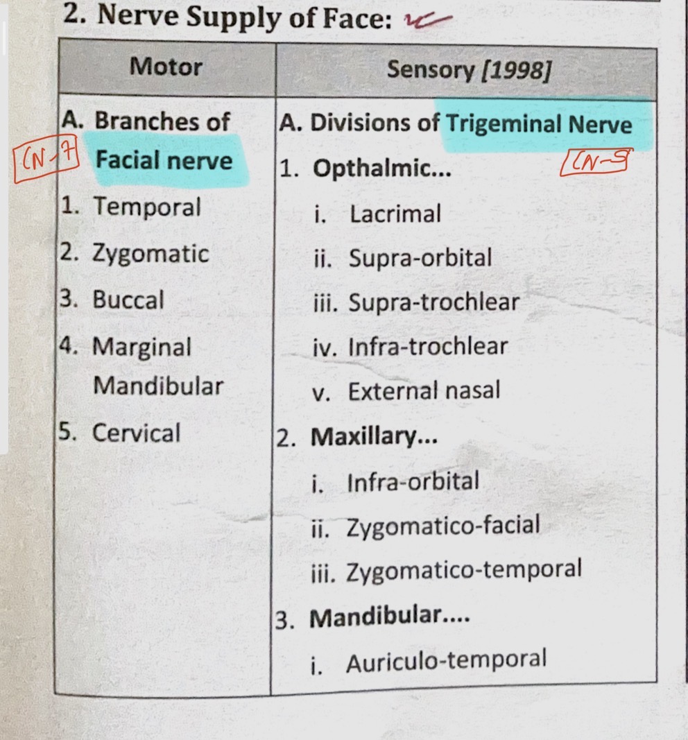
Nerve supply of face
Arterial supply office
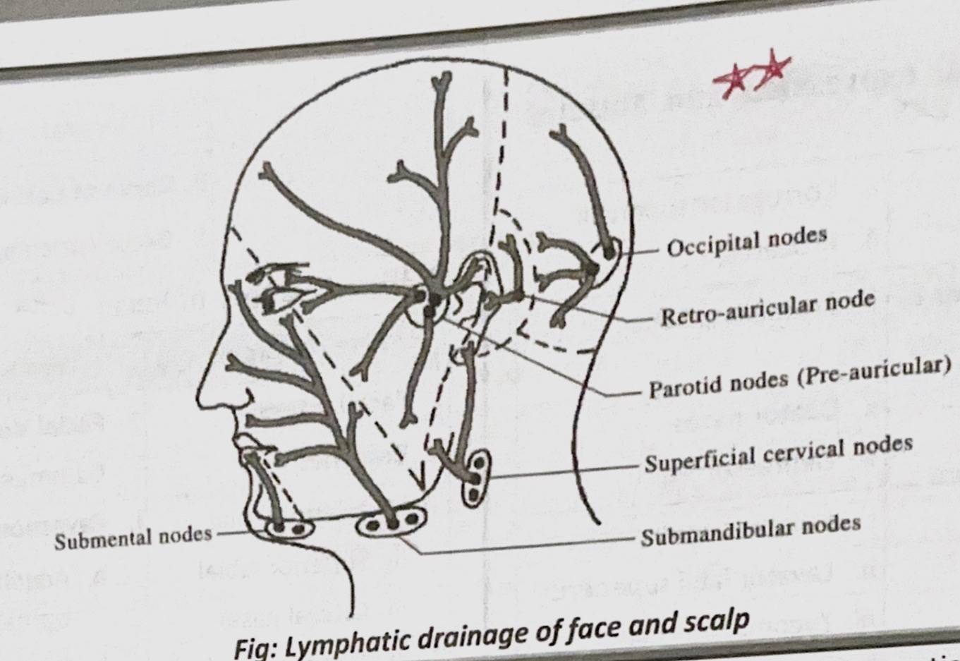
Lymphatic drainage of face