Case 1: Mandy Wallsmith Pt 1 - Transverse Myelitis
 Call Kai
 Learn
Learn Practice Test
 Spaced Repetition
 Match
Match Flashcards
 Knowt Play
1/48

There's no tags or description
Looks like no tags are added yet.
Name | Mastery | Learn | Test | Matching | Spaced | Call with Kai | Chat |
|---|
No analytics yet
Send a link to your students to track their progress
49 Terms
Spinal Cord: Grey Matter
Centre
Surround central canal containing CSF
Contain:
Cell bodies
Dendrites
Proximal axons
Parts:
Ventral horn
Dorsal horn
Lateral horn
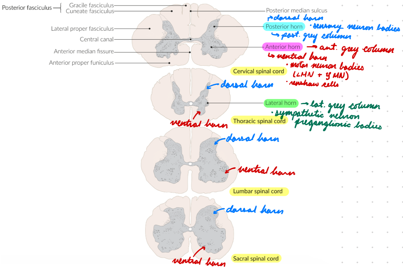
Spinal Cord Grey Matter: Ventral Horn
Anterior grey column
Contain:
Motor neuron cell bodies
Alpha/lower motor neurons (LMN)
Axons → Periphery
Innervate: Skeletal muscle = Contraction
Gamma motor neurons
Innervate: Muscle spindles = Stretch sensitivity
Renshaw Cells: Inhibitory neurons
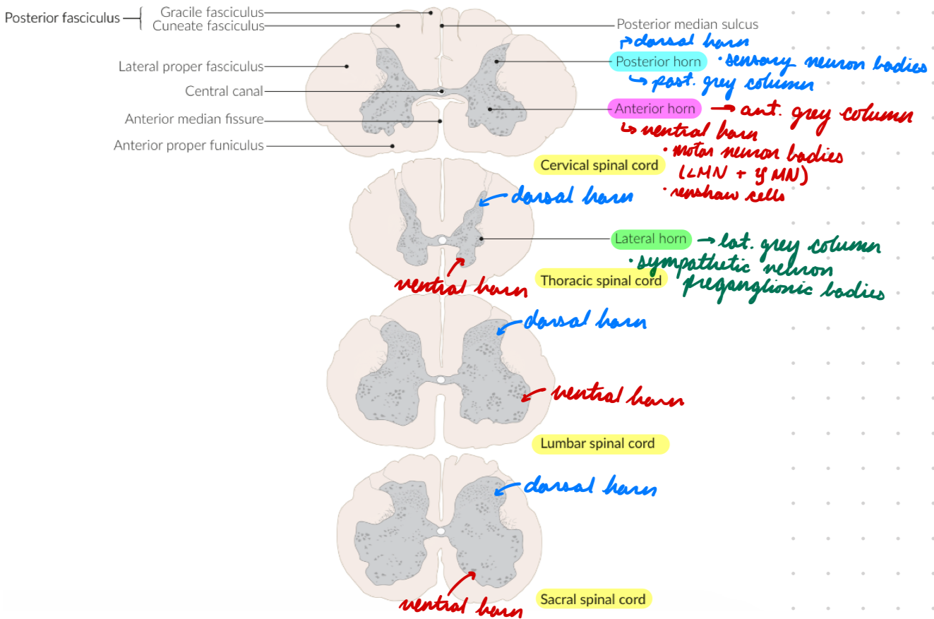
Spinal Cord Grey Matter: Dorsal Horn
Posterior grey column
Contain: Sensory neuron cell bodies
Spinal reflexes
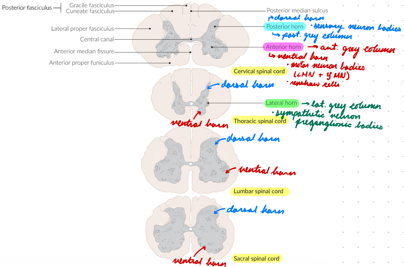
Spinal Cord Grey Matter: Lateral Horn
Lateral grey column
Contain:
Sympathetic neuron preganglionic cell bodies
Clarke Nucleus: Posterior thoracic
Unconscious proprioception
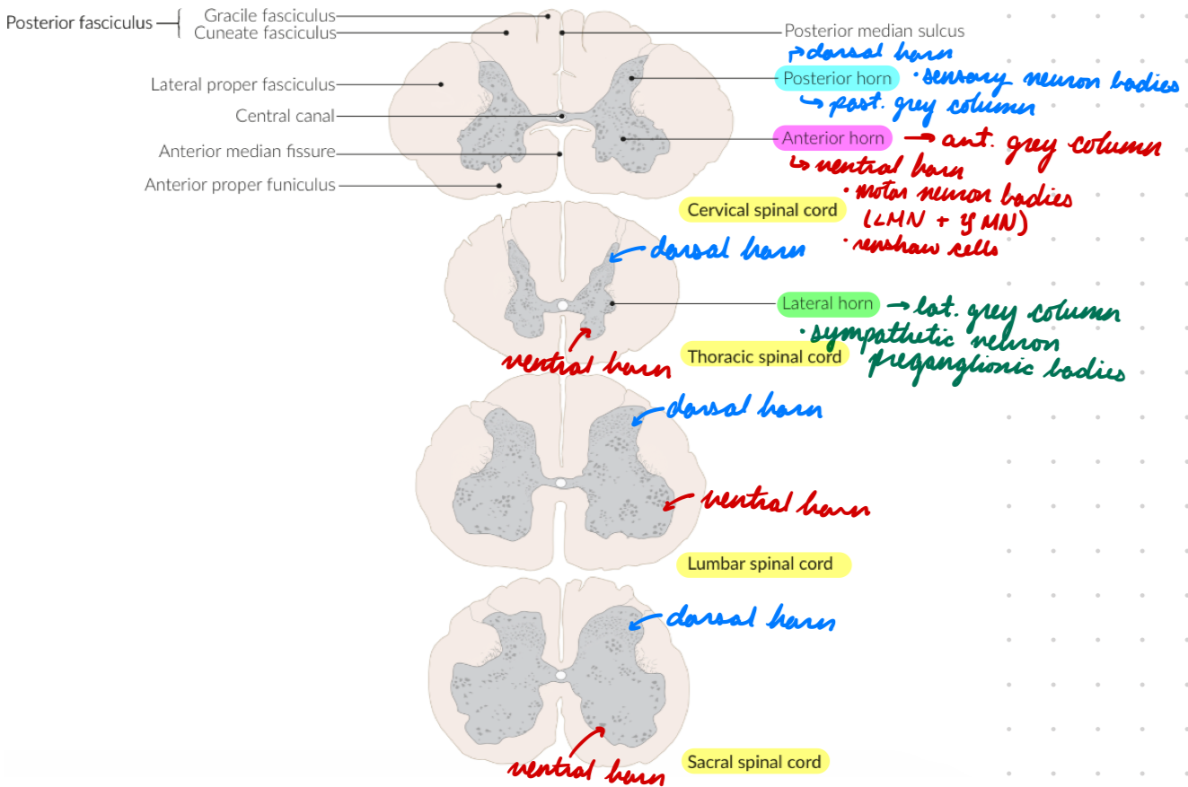
Spinal Cord: White Matter
Surround grey matter
Contain:
Myelinated axons
Nerve fibre bundles
No dendrites
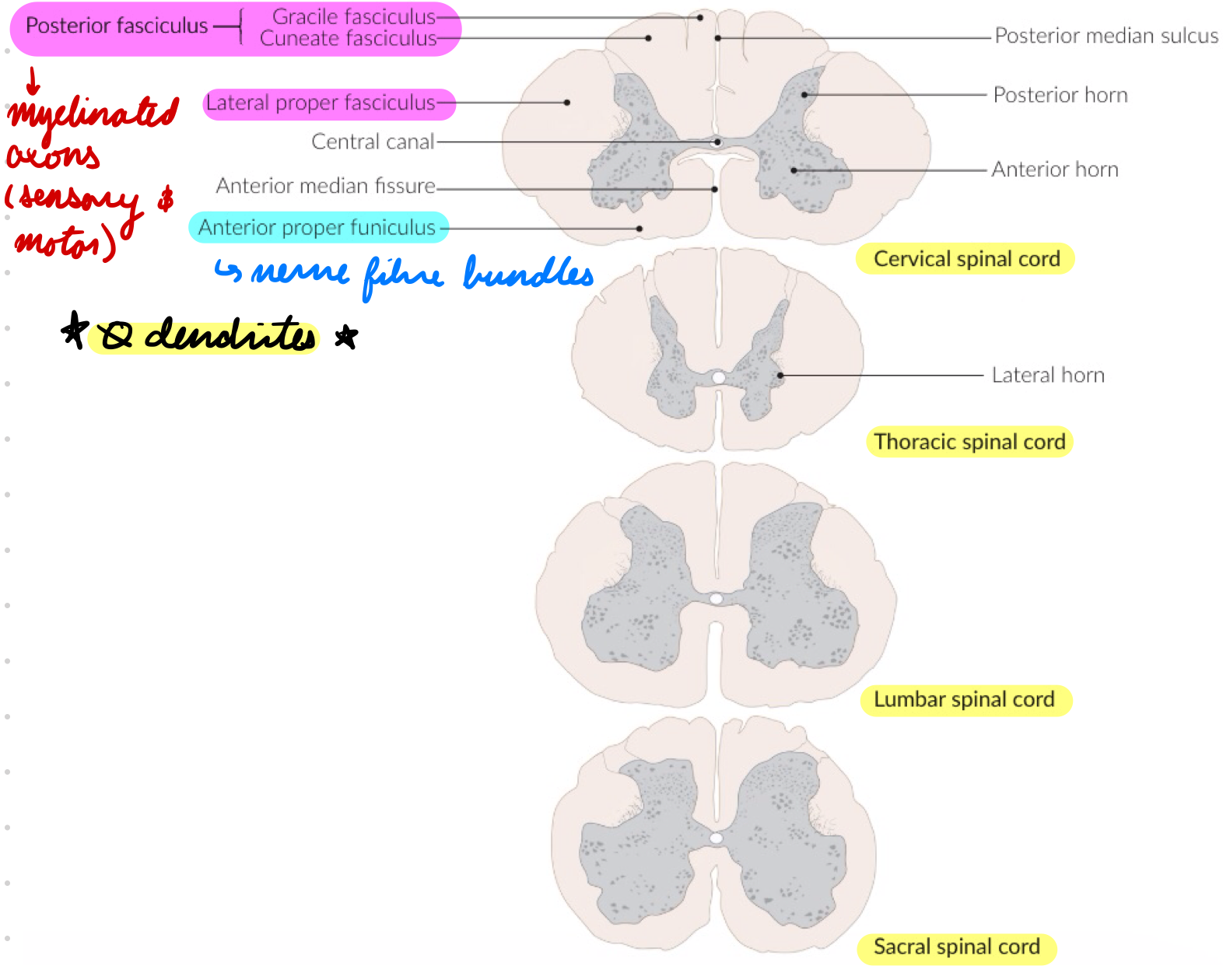
Spinal Cord White Matter: Myelinated Axons
Tracts/fasciculi
Sensory (ascending) + motor (descending) tracts transmit signals between CNS and PNS
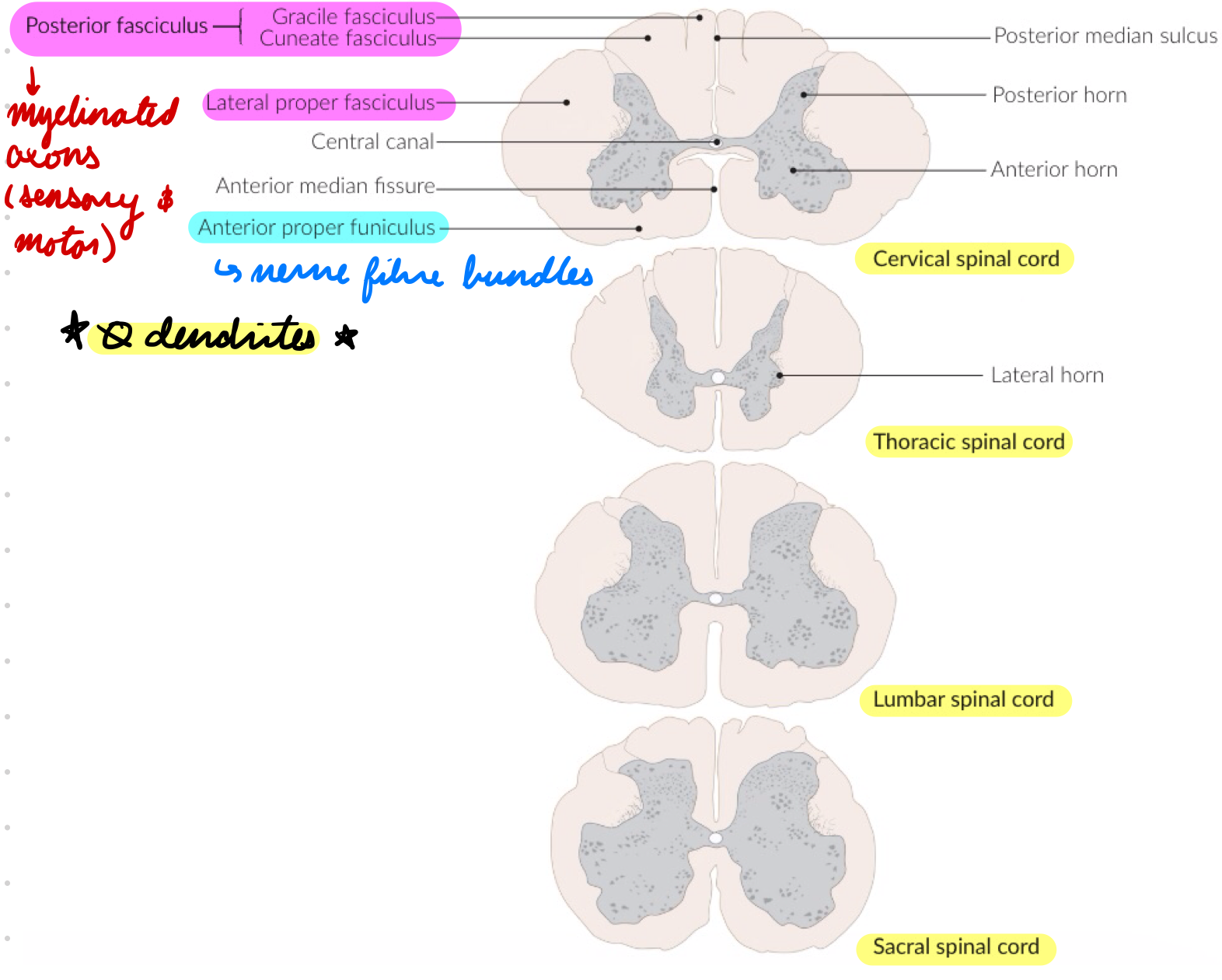
Spinal Cord White Matter: Nerve Fibre Bundles
Funiculi
Anterior
Lateral
Posterior
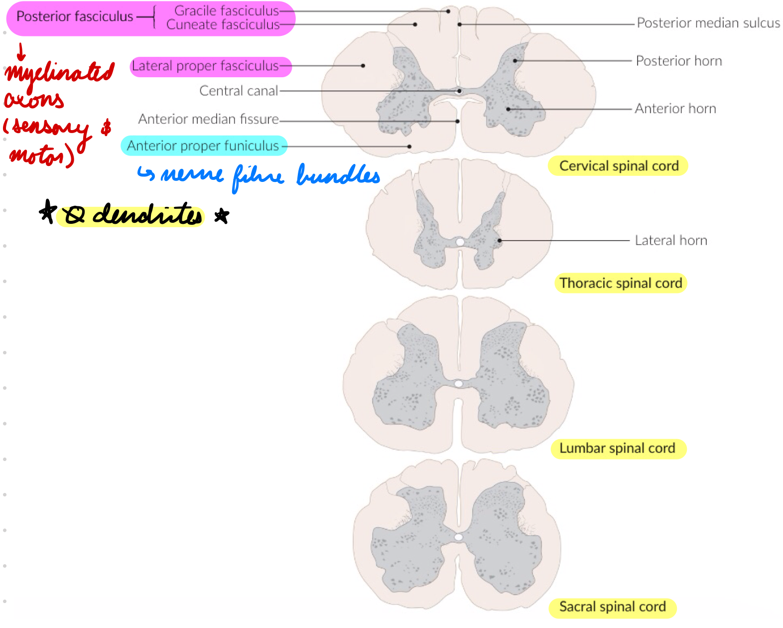
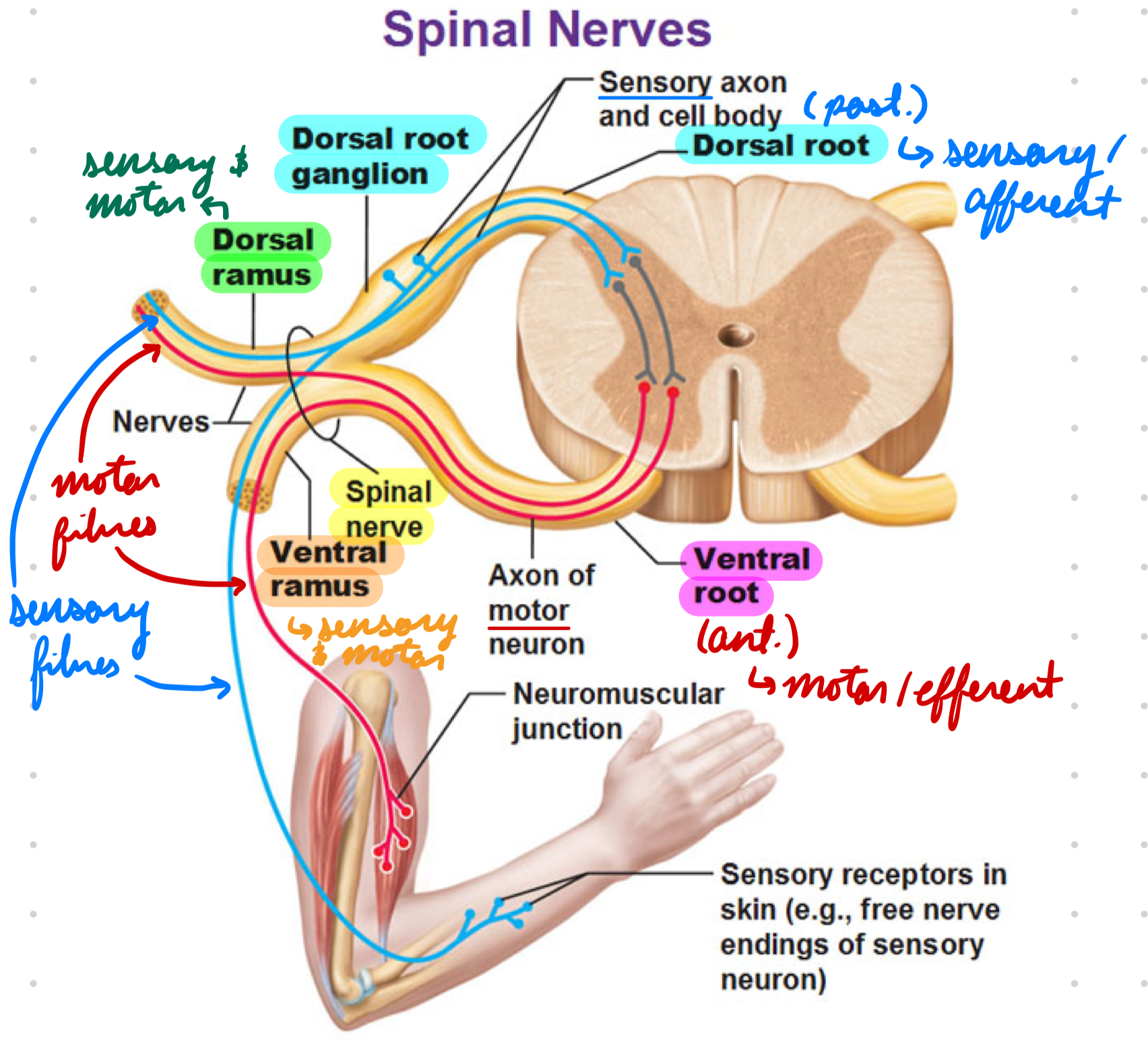
Spinal Nerves
Exit spinal canal → Innervate periphery
Parts:
Roots
Rami
Spinal Nerves: Roots
Nerve exiting intervertebral foramina
Anterior: Motor/efferent fibres
Dorsal: Sensory/afferent fibres
Dorsal Root Ganglia: Outside vertebral column
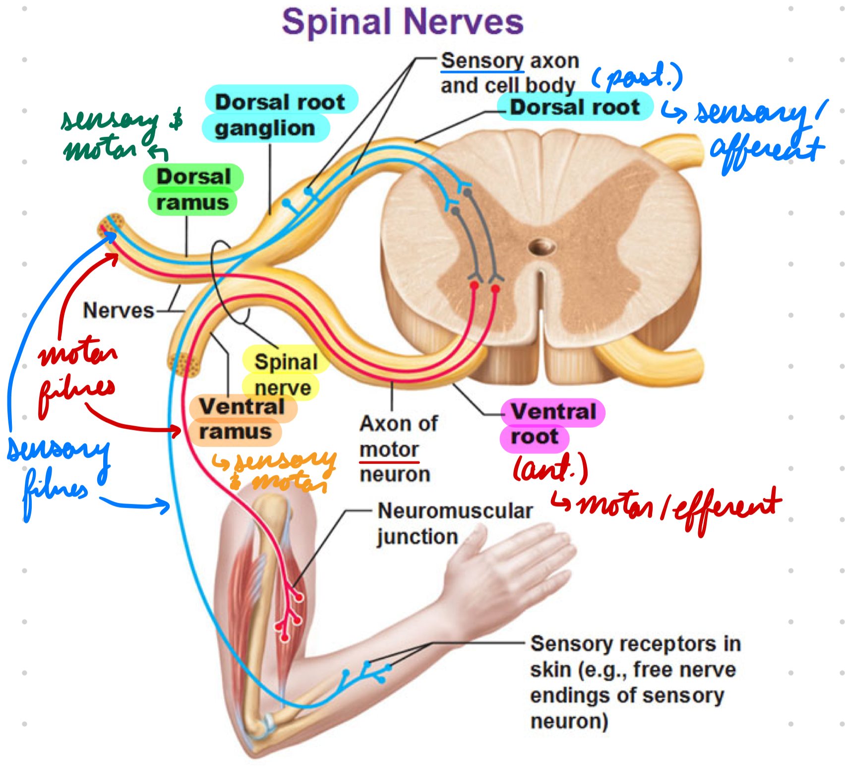
Spinal Nerves: Rami
Spinal nerve sections + sensory and motor fibres
Ventral: Anterior
Dorsal: Posterior
Spinal Cord: Sensory Systems
Ascending tracts
2 major conscious sensation pathways
Dorsal Column-Medial Lemniscus (DCML) System
Anterolateral (Spinothalamic) System
2 major unconscious sensation pathways
Spinocerebellar tract
Spinoolivary tract
Spinal Cord Sensory: DCML System
3 neurons
Transmit:
Proprioception
Epicritic sensation
Fine touch
Vibration
Pressure
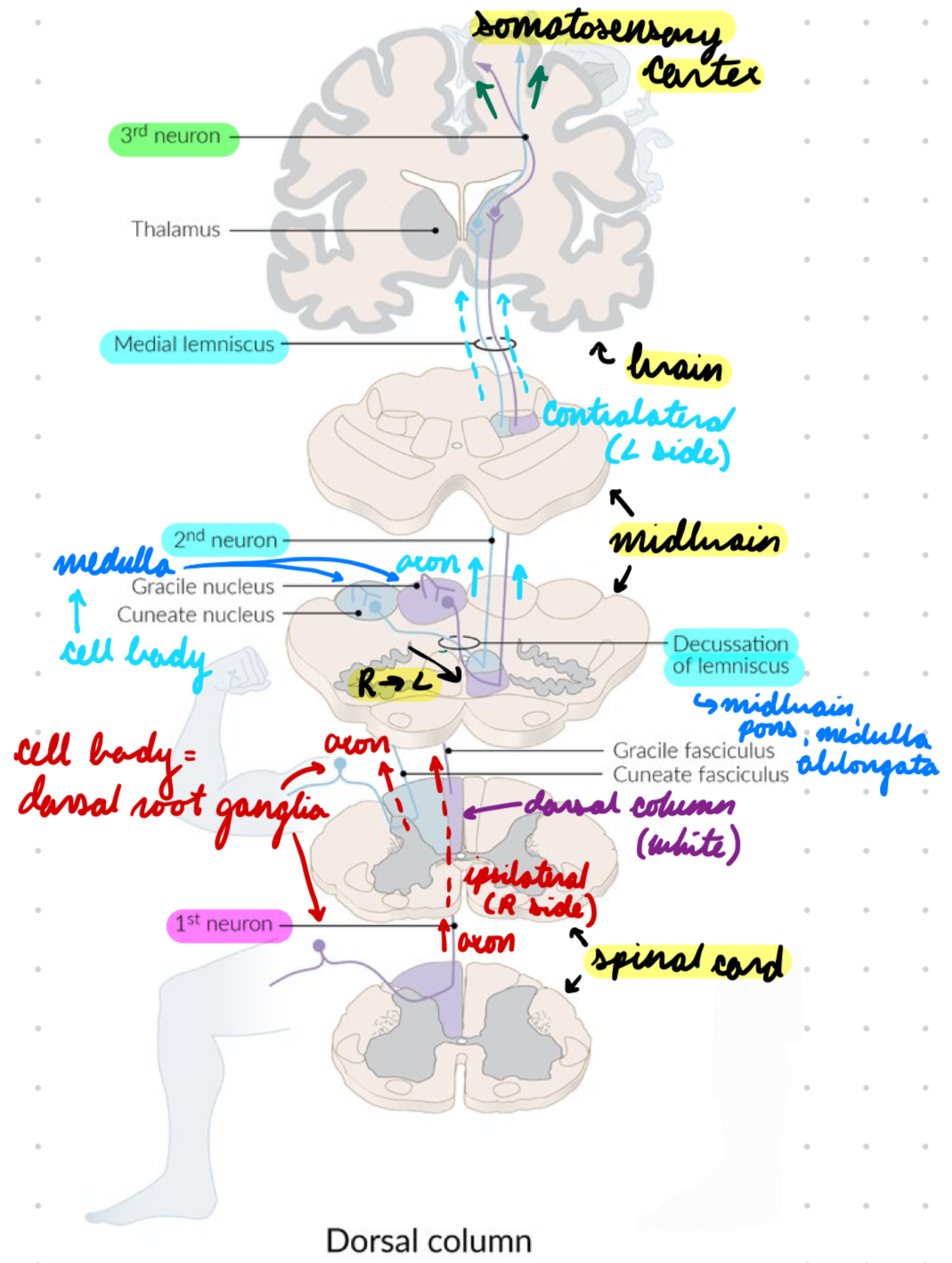
DCML System: 1st Order Neuron
Cell body in dorsal root ganglia
Sensory nerve fibres
Axons → Spinal cord dorsal column (white matter) → Ascend ipsilaterally to medulla oblongata
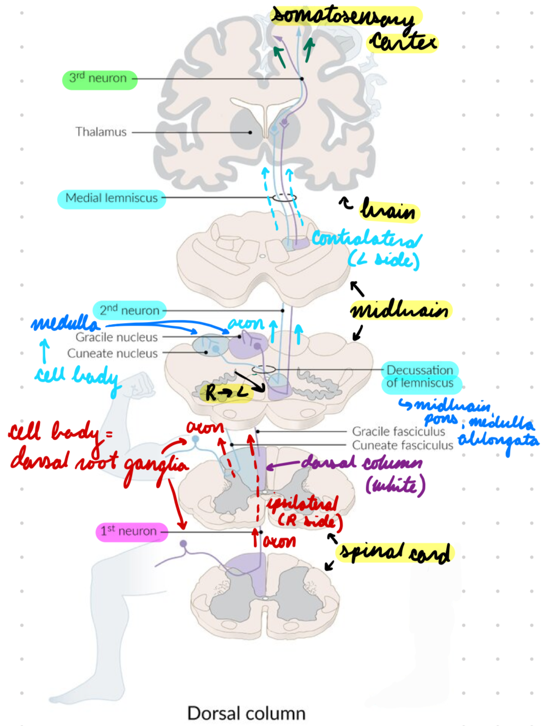
DCML System: 2nd Order Neuron
Cell body in dorsal column nuclei (medulla)
Axons decussate in medulla + ascend contralaterally in medial lemniscus (brainstem)
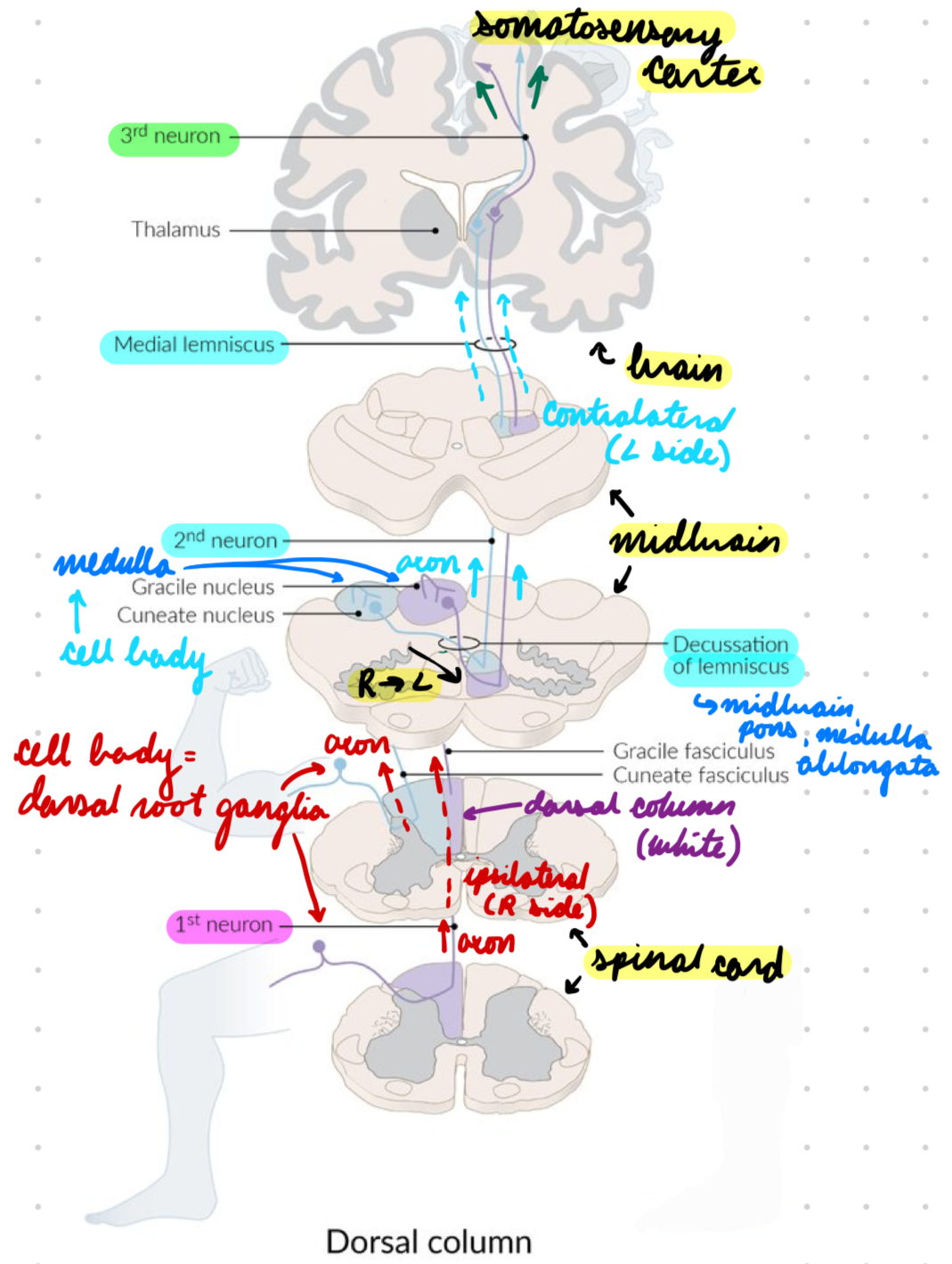
DCML System: 3rd Order Neuron
Somatosensory cortex
Spinal Cord Sensory: Spinothalamic System
3 neurons
Transmit:
Pressure (anterior)
Protopathic sensation
Temp (lateral)
Pain (lateral)
Crude touch (anterior)
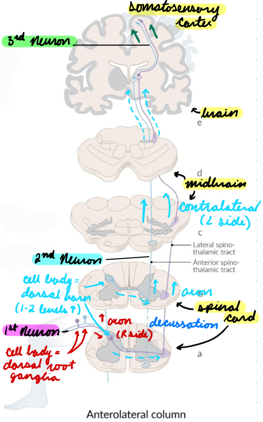
Spinothalamic System: 1st Order Neuron
Cell body in dorsal root ganglia
A-delta and C fibres
Axons → Spinal cord → Synapse in dorsal horn (grey matter) (1-2 spinal levels above)
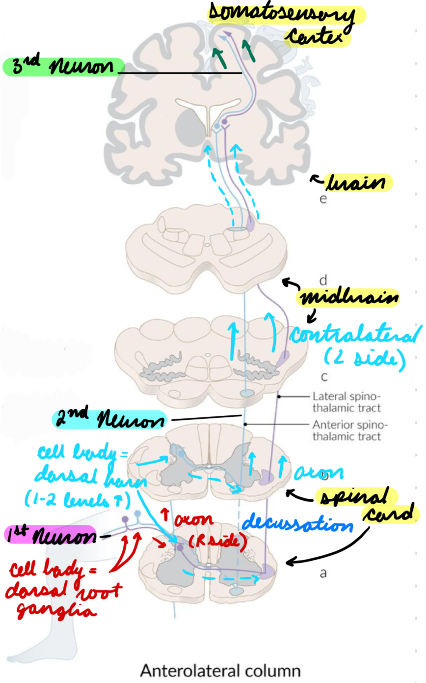
Spinothalamic System: 2nd Order Neuron
Cell body in dorsal horn
Axons decussate in spinal cord + ascend contralaterally
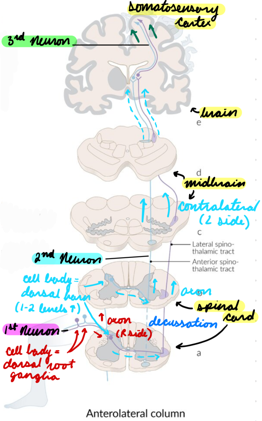
Spinothalamic System: 3rd Order Neuron
Somatosensory cortex
Spinal Cord: Unconscious Sensation
Transmit: Proprioception
Spinal Cord: Motor Systems
Descending tracts
General:
Upper Motor Neuron (UMN): Descending inhibition
Inhibit excess firing from LMNs
Lower Motor Neuron (LMN): Excitatory signals
Constant excitatory neuron firing = Spinal cord reflexes
2 major types
Corticospinal (Pyramidal) Tract
Extrapyramidal Tracts
Spinal Cord Motor: Pyramidal Tract
2 neurons
Control: Voluntary movement of contralateral side
Spinal Cord Motor: Extrapyramidal Tracts
2 neurons
Control: Motor neurons
Involuntary movement (reflexes)
Muscle tone
Facial muscles
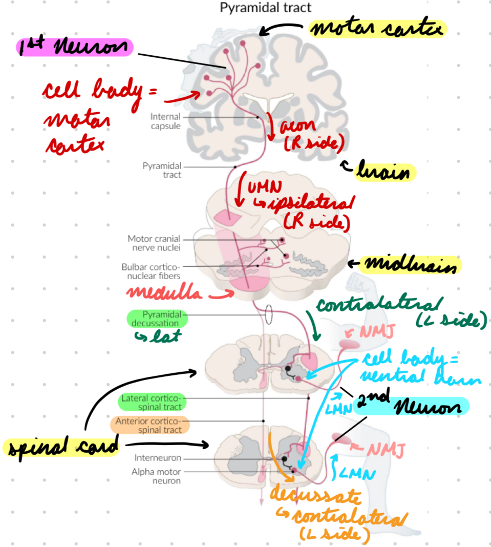
Pyramidal Tract: 1st Order Neuron
Cell body in motor cortex
Types:
Upper motor neuron
Axons descend ipsilaterally
Lateral corticospinal tract
Axons decussate in medulla oblongata → Descend contralaterally in spinal cord
Anterior corticospinal tract
Axons decussate at innervation level
Synapse in ventral horn (grey matter)
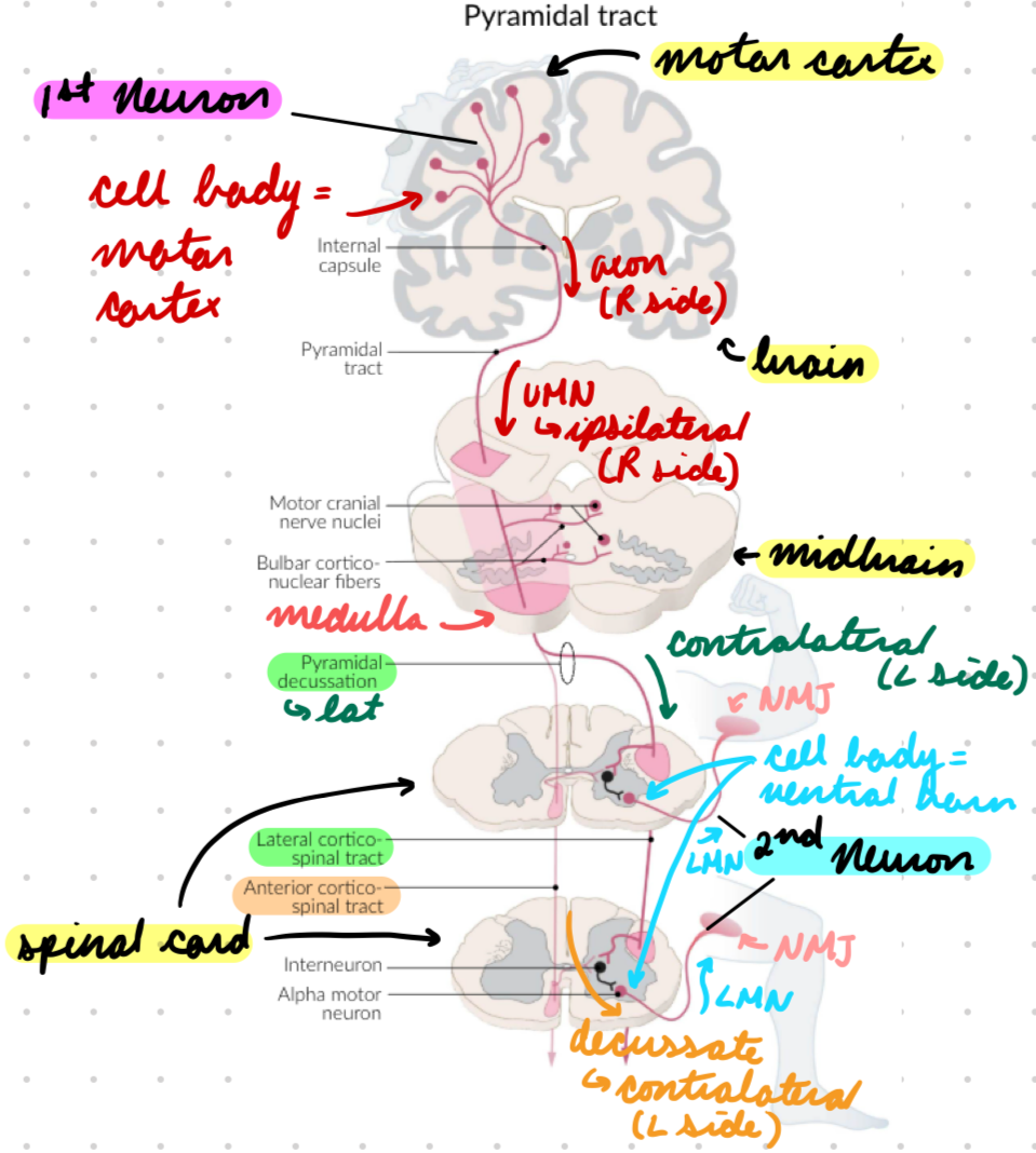
Pyramidal Tract: 2nd Order Neuron
Cell body in ventral horn
LMN → NMJs
Upper + lower limbs
Extrapyramidal Tracts: 1st Order Neuron
Different pathways
Rubrospinal pathway
Proprioception
Reticulospinal tract
Posture + tone
Vestibulospinal tract
Ear
Tectospinal tract
Eye
Synapse in ventral horn (grey matter)
Extrapyramidal Tracts: 2nd Order Neuron
Cell body in ventral horn
LMN → End locations depending on tract
Sensory Neuron Dysfunction: Peripheral
Lesions in peripheral nerves (post-rami)
Affect all senses together (pain, temp, touch, vibration, proprioception) in predictable pattern
Peripheral Sensory Neuron Dysfunction: Types
Polyneuropathy: Stocking-glove distribution
Symmetric sensory loss in toes → Ascend symmetrically to knees → Fingertips
Mononeuropathy: Affect 1 peripheral nerve = 1 nerve territory
Radiculopathy: Dermatome pattern
Sensory Neuron Dysfunction: Central
Lesions in CNS (spinal cord + brain)
No pattern
Central Sensory Neuron Dysfunction: Types
Sensory Level: Sensory loss below spinal cord lesion level
Dissociated: Spinal cord tract lesion = Affect different sensory modalities
Spinothalamic: Pain/temp
DCML: Vibration/proprioception
Bowel/bladder dysfunction
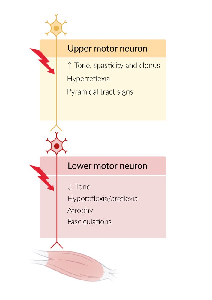
Motor Neuron Dysfunction: UMN
Lesion before ventral horn
Loss of LMN inhibition
Normal muscle
Central paresis: No voluntary movement +
Increased tone, spasticity (uncontrolled movement), clonus
Decreased power
Hyperreflexia
Detrusor muscle hyperreflexia + incoordination
Upgoing Babinski reflex (abnormal)
Big toe up
Toes 2-5 down
Motor Neuron Dysfunction: LMN
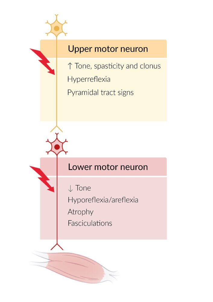
Lesion between ventral horn + muscle
Loss of excitatory firing
Muscle atrophy + fasciculation (twitching)
Peripheral paresis: No voluntary movement +
Decreased tone
Decreased power
Hyporeflexia/areflexia
Overflow incontinence
Bladder overfills = Involuntary urine leakage
Downgoing Babinski reflex (normal)
Toes down
Transverse Myelitis: Description
Acute/subacute inflammatory myelopathy (neurologic disorder from spinal cord injury)
Affect ascending + descending neural pathways
Transverse Myelitis: Epidemiology
Bimodal peak incidence
10-19 years
30-39 years
Transverse Myelitis: Etiology
Idiopathic
Parainfectious
Enterovirus
EBV
CMV
B. burgdorferi (Lyme disease)
CNS demyelination disorders
MS
Paraneoplastic syndromes
Drugs
TNF-a inhibitors
Chemotherapy
Transverse Myelitis: Pathophysiology
Autoantibodies cause… = Neuroinflammation
Lymphocyte + monocyte collection in spinal cord
Spinal cord demyelination
Axon injury
Astroglial + microglial activation
Nonneuronal support cells in CNS
Transverse Myelitis: Clinical Presentation
Onset: Acute/subacute (4 hours to 21 days)
Spinal shock: Loss/depression of sensorimotor functions + reflexes below injury level
UMN dysfunction
Spasticity
Hyperreflexia
Upgoing Babinski sign
Progression: Neurological dysfunction
Motor
Paresis
Paraplegia
Sensory
Numbness
Paresthesias
Autonomic
Bladder/bowel incontinence
Sexual dysfunction
Transverse Myelitis: Investigations
Imaging
Lab tests
CSF analysis
Transverse Myelitis Investigations: Imaging
MRI ± gadolinium contrast
Focal gadolinium-enhancing lesion in spinal cord
T2-weighted sequence (hyperintensity) = Bright CSF (edema) + adipose tissue (white brain matter)
Transverse Myelitis Investigations: Lab Tests
Serology: Determine cause
Viral infections
Syphilis
HIV abs
Inflammatory markers
Increased ESR + CRP
Serum abs
ANA
RF
Antiphospholipids abs
Vit B12 + methylmalonic acid (buildup in B12 deficiency)
TFTs: R/O thyroid dysfunction
Transverse Myelitis Investigations: CSF Analysis
Inflammatory markers
Pleocytosis: Increased cells
Increased IgG
Oligoclonal Bands: IgG subfractions on electrophoresis = Increased IgG
Cultures
Transverse Myelitis: Acute Treatment
First-Line: High-dose IV corticosteroids
Second-Line:
Plasmapheresis
Cyclophsophamide
Supportive care
Transverse Myelitis Acute Treatment: Plasmapheresis
Insufficient corticosteroid response
Filter plasma from whole blood = Remove autoantibodies
Replace with donor plasma or albumin solution
Transverse Myelitis Acute Treatment: Cyclophosphamide
Immunosuppressant
Alkylating DNA = Prevent production
Transverse Myelitis Acute Treatment: Supportive Care
Prevent ulcers + thrombosis
Manage complications
Urinary retention
Gastroparesis
Constipation
Resp failure
Transverse Myelitis: Chronic Treatment
Neurological rehab (physiotherapy)
Treat underlying cause
Transverse Myelitis: Complications
Resp failure
Immobilization complications
Atelectasis
DVT
Pressure ulcer
Dysphagia
GI dysfunction
Osteoporosis
Autoimmune disorders
Orthostatic hypotension
Thermodysregulation
Dysreflexia
MRI Sequences (Results)
T1-Weighted:
Hyperintense: Fat/adipose tissue (white brain matter)
Hypointense:
Water/CSF/edema
Bone
Ligaments
Tendons
Air
T2-Weighted:
Hyperintense: Water/CSF/edema
Hypointense:
Bones
Ligaments
Tendons
Air