8/24: Research Methods
 Call Kai
 Learn
Learn Practice Test
 Spaced Repetition
 Match
Match Flashcards
 Knowt Play
1/27

There's no tags or description
Looks like no tags are added yet.
Name | Mastery | Learn | Test | Matching | Spaced | Call with Kai | Chat |
|---|
No analytics yet
Send a link to your students to track their progress
28 Terms
brain damage
→ the best way to identify and study which parts of the brain are responsible for a certain function
Is this the best research method for neurosci T _ T ???
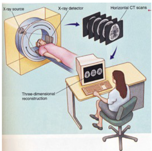
computerized tomography
CT → imaging technique that uses x-ray beams (high energy light/radiation)
Patients lie in a cylindrical x-ray source to get images of their body at all angles, 360°
Following scanning, x-ray detections are computerized and reconstructed into a 3D model of the patient’s body
CT scans are cheap and fast, but not the best for soft tissue (like the brain)
Sometimes contrast agents (fluids like iodine) are administered to improve resolution
magnetic resonance imaging
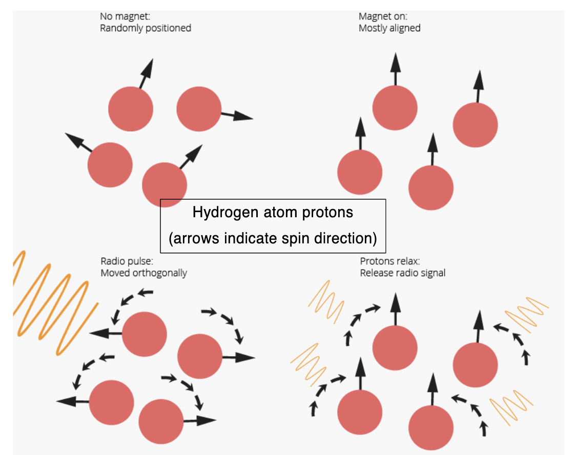
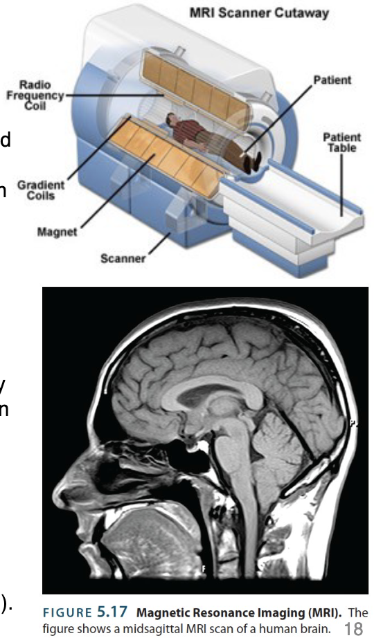
MRI→ imaging technique that uses magnetic fields/radio waves (low energy light)
Patients lie in a cylindrical radio frequency coil (magnet) to get images of their body at all angles, 360°
As magnetism is administered, the hydrogen atom protons in a patient’s body absorb the energy, orient themselves in line with the magnetic fields, and return emissions
Following scanning, radio wave detections are computerized and triangulated to form an estimate of the relative density of hydrogen atoms throughout the scanned
MRI scans have good spatial resolution for imaging tissues (hydrogen atoms are especially prevalent in lipids … myelin is a kind of fat and there’s plenty of that in the brain)
Safer than CT; orientation of an atom is inconsequential to internal biochemistry
diffusion tensor imaging
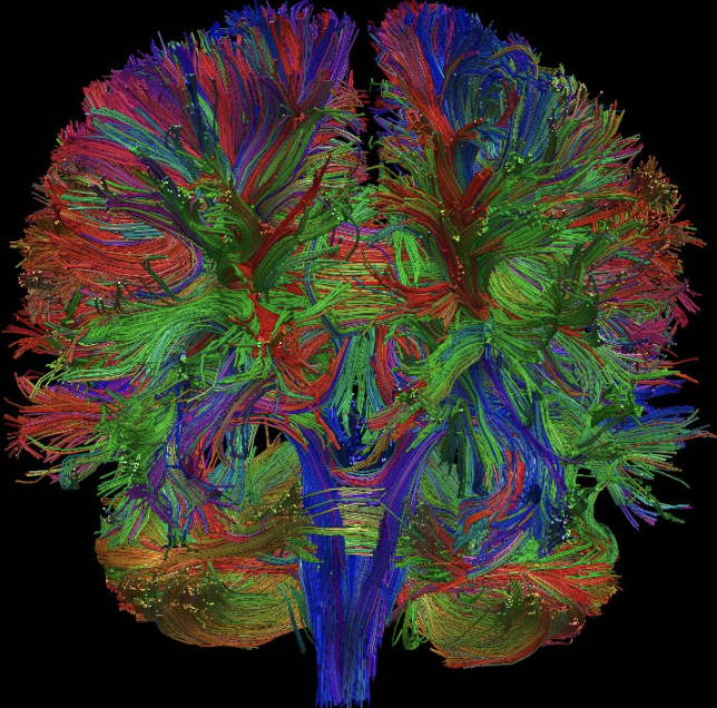
DTI → MRI technique that measures the direction and speed of the diffusion of water molecules in the brain
Alternative to normal MRI (whose magnets are optimized for lipids)
Used to identify axon tracts
Colours indicate the direction of water molecule diffusion
functional magnetic resonance imaging
fMRI → imaging technique that uses a rapid series of MRI scans measuring the movement of magnetic field distortions over time
Same process as MRI, but with measuring blood oxygen levels in a locale
As magnetism is administered, blood oxygenation in a given area changes;
An area’s rapid ↑ in blood flow correlates positively with ↑ neural activity
fMRI scans have good spatial resolution and temporal resolution, providing great structural and functional information
Popular for being non-invasive
fMRI upcoming modification
→ to measure fluctuations in neurotransmitter signaling
Would involve using “enzyme-activated magnetic resonance contrast agents” to distort magnetic fields differently when they bind to neurotransmitters
positron emission tomography
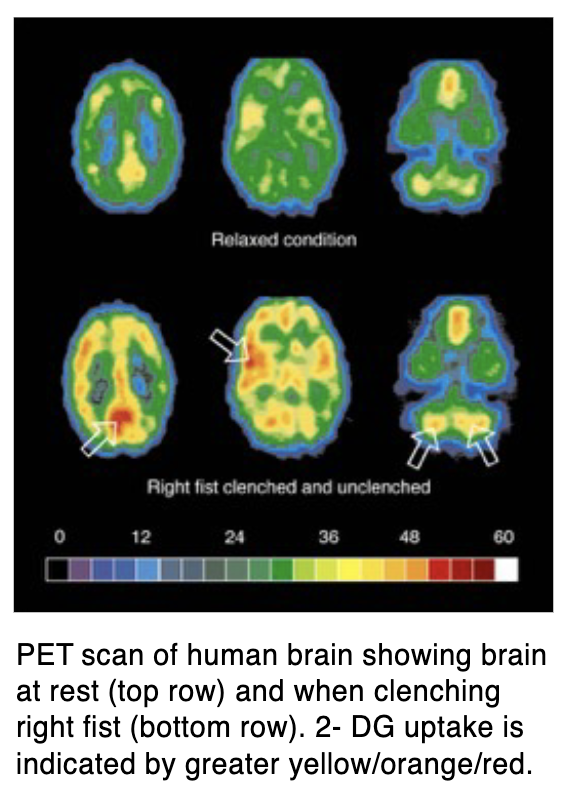
PET → imaging technique that involves injections of radioactive compounds
Radioactive sugar molecules are commonly used to detect changes in energy use
2-DG → similar to glucose (consumed by active cells), however, it’s not metabolized as easily (endures)
Scanning where and how radioactive sugar molecules move over time
PET substances can also measure changes in the expression levels of neurotransmitter receptors across weeks
Main disadvantages of PET scans are operating costs and safety precautions (radioactive molecules decay from the moment they’re produced, so they have to be made on-site and as close to the procedure as possible)
More recently superseded by fMRI
electroencephalogram
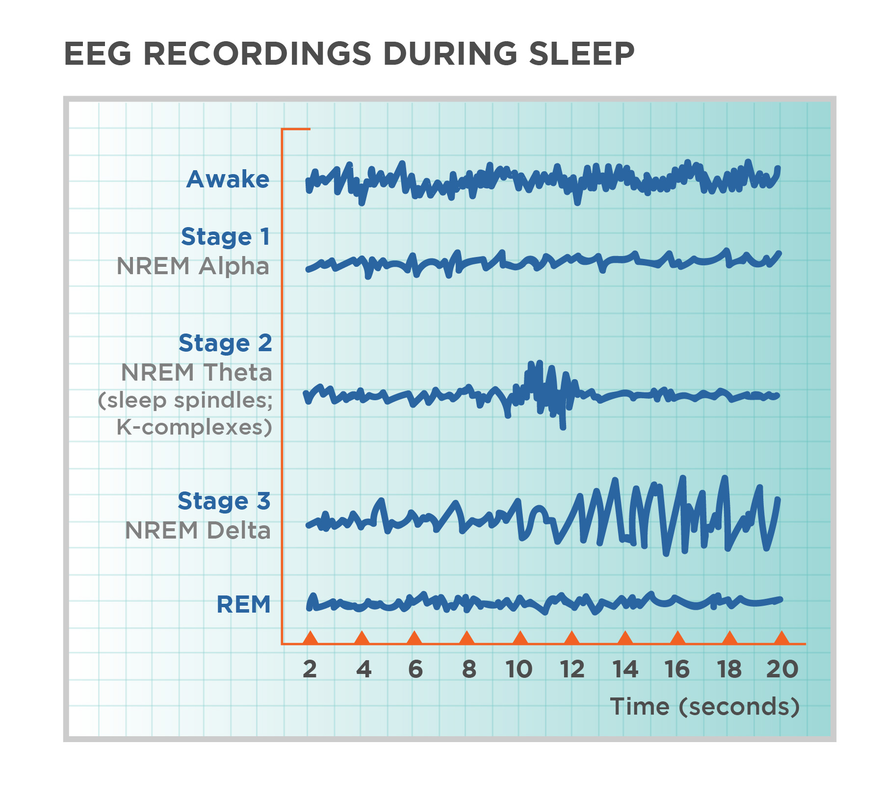
EEG → measures electrical activity in the brain
Macroelectrodes attached to the scalp to record summed population-level activity of millions of neurons
Can be used a diagnostic tool since specific patterns of EEG activity are associated with different states of consciousness (awareness), subconsciousness (sleep stages), and cerebral atrophy (neural loss)
experimental ablation
→ lesion study; involves the procedural removal or destruction of a portion of the brain
Inferential findings: Functions that no longer work after the procedure are probably controlled by the lesioned brain area
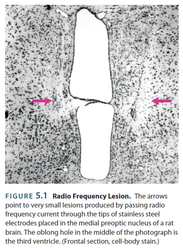
radiofrequency lesion
→ small lesions made by passing an electric current through a metal wire insulated everywhere but the tip (whose heat will burn surrounding cells)
Shape and size of lesion is determined by the duration and intensity of the current
Shortcoming is that passerby axons are burnt in the process
excitotoxic lesion
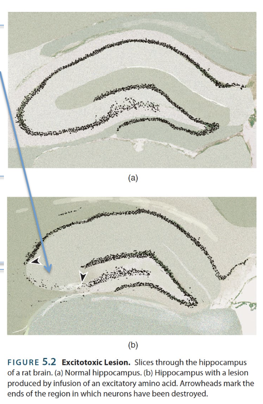
→ small lesions made by injecting a glutamate receptor agonist
Localized drugs cause so much excitation (Ca2+ influx) such that the affected neurons undergo apoptosis
This method spares passerby axons
ex.
Kainic acid
sham lesion
→ not an actual lesion; made by mimicking the procedures for an actual experimental ablation, except the damaging part
reversible lesion
→ temporary “lesions” made by injecting drugs that block or reduce neural activity in a given region
Localized drugs cause so much excitation (Ca2+ influx) such that the affected neurons undergo apoptosis
This method spares passerby axons
ex.
Voltage-gated sodium channel blockers → stops action potentials
GABA receptor agonists → hyperpolarizes cell bodies
microelectrodes
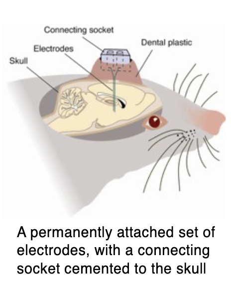
→ neural activity implants made from thin metal wires with a fine tip that record single-unit recordings (the electrical activity of individual neurons; chronic or acute)
Can be used in behaving animals to record action potentials
Wires are connected to a “socket” in the animal’s head so that it can be “plugged” into a recording system at any time
electrical stimulation
→ method of manipulating neural activity that involves passing an electrical current through a wire inserted into the brain
Affects all receptor components in the area (axons of neurons)
Overstimulation (rapid stimulation frequencies) actually produces the same behavioural effects as lesioning the area (deliberate damage)
chemical stimulation
→ method of manipulating neural activity that involves administering drugs into a particular brain region
Usually pushed through a guide cannula (hollow tube)
Anesthetics → can completely shut down neural activity, entirely or locally
Receptor agonists/antagonists are alternatives since they don’t affect passerby axons
optogenetics
→ refers to the use of light to control light-sensitive neurons
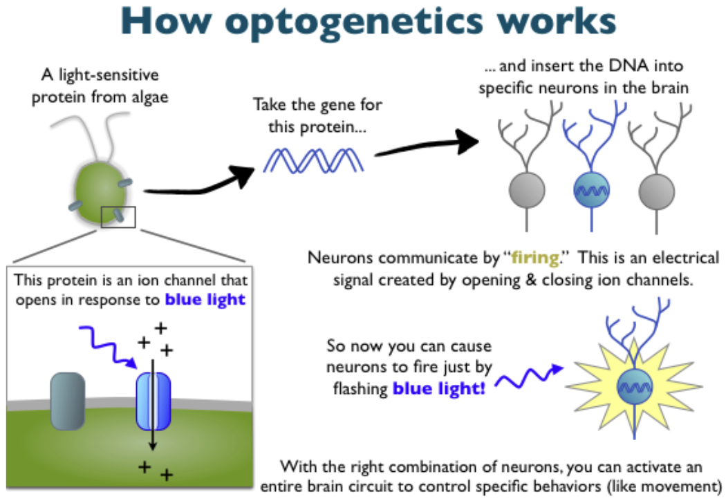
Neurons are made light-sensitive through the introduction of foreign DNA, which provides instructions to make light-sensitive proteins called opsins
Opsins in eye are metabotropic receptors with 30ms~ delay
Similarly, opsins optogenetically manipulated are ion channels that open and close instantly in response to light
First opsins (ex. ChR2) were discovered in bacteria and algae
Excitatory opsins → light delivery generate action potentials
Inhibitory opsins → light delivery prevent action potentials
channelrhodposin-II
ChR2 → excitatory photosensitive ion channel permeable to sodium (Na+) ions
Responds to blue light, depolarizing neurons
This opsin evolved from single-cell organisms
IC++
→ inhibitory photosensitive ion channel permeable to chloride (Cl-) ions
Responds to blue light, hyperpolarizing neurons
This opsin was designed by humans
virus
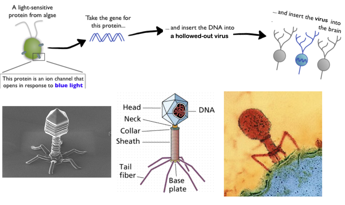
→ small infectious agent that replicates by injecting its DNA into normal cells of a host organism
Replication-deficient → when a virus has its DNA removed
Microbiologists can insert foreign DNA into replication-deficient viruses
This lab-made foreign DNA encodes proteins for a cell to express, such as fluorescent proteins or optogenetic proteins
viral-mediated gene delivery
→ using viruses to deliver expressive DNA into neurons
Modified viruses (replication-deficient viruses filled with foreign DNA) are injected into the brain. Surrounding cellular components become infected.
When viral DNA accesses the nucleus of a cell, that cell will start to transcribe the viral DNA to make its associated proteins.
fluorescent calcium imaging
→ imaging technique that involves implanting a fiber optic cable into the brain to measure neural activity triggered by calcium-sensitive proteins
Green fluorescent protein (GFP) → bioluminescent protein discovered in jellyfish
GCaMP→ modified GFP to bind to calcium (which is involved in action potentials) and to fluoresce much brighter when it does
GOOGLE SAYS: GFP + calmodulin (CaM) + M13, a peptide sequence from myosin light-chain kinase
tracing methods
→ reveal the input and output structures involved in a particular brain region involved with a particular function
Retrograde
Anterograde
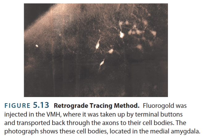
retrograde labeling
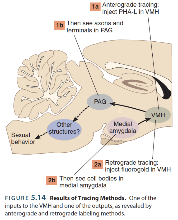
→ determining neural connections by tracing afferent axons; inputs
Asks “what brain areas send their axons here?”
Retrograde tracer:
Taken up by axon terminals and transported back to the cell body
Retrograde tracer → chemicals taken up by axon terminals and transported back to the cell body
ex. Fluorogold
anterograde labeling
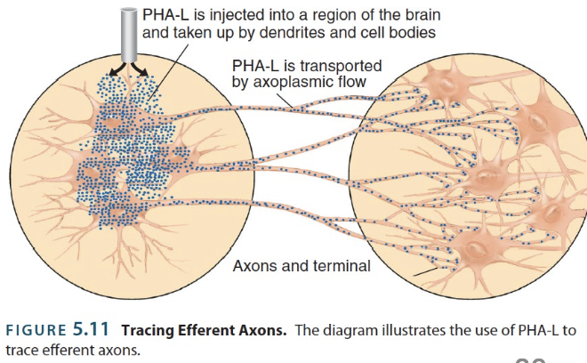
→ determining neural connections by tracing efferent axons; output
Asks “where do these axons go?”
Anterograde tracer → chemicals taken up by cell bodies and transported back to axon terminals
ex. PHA-L
stereotaxic brain surgery
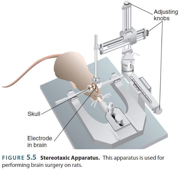
→ surgical intervention that uses a stereotaxic apparatus to administer electrical or chemical stimulation
Injections: drugs, viruses, tracers, etc.
Implantations: cannula, electrodes, fiber optic cables, wires, etc.
Lesions: radiofrequency, excitotoxic, reversible, etc.
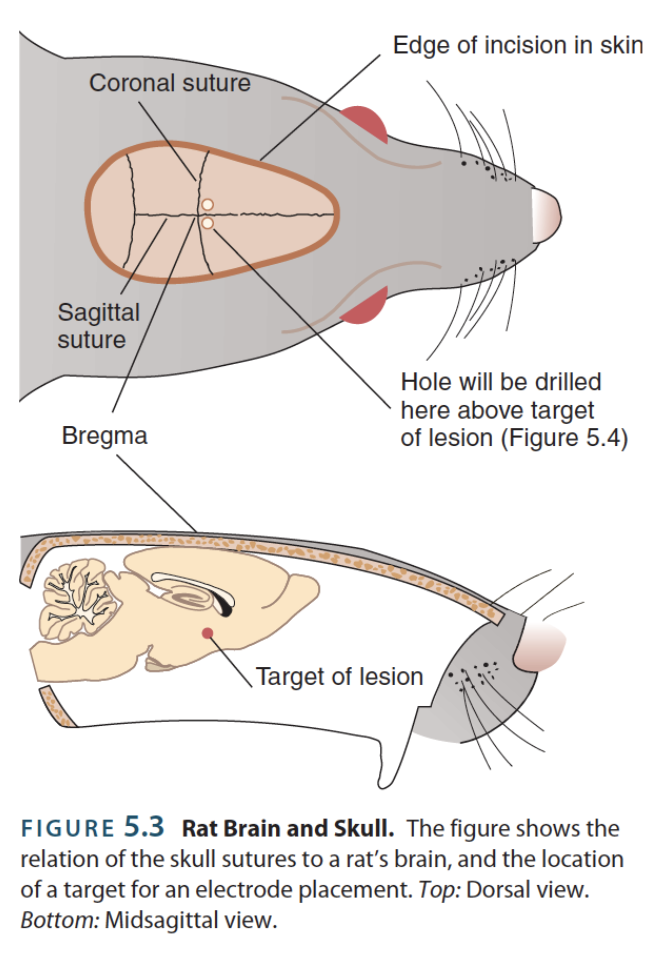
Bregma → junction where pieces of the skull fuse together; often used as a reference point for stereotaxic surgery
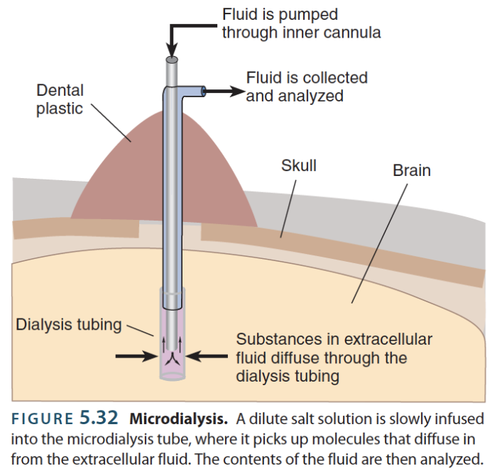
microdialysis
→ old-fashioned approach to measuring fluctuations in neurotransmitters in behaving animals
Dialysis → refers to the use of a semipermeable membrane to either deliver or measure the concentration of molecules in some solution or area
Microdialysis probe → small insertable metal tube that holds dialysis tubing
Sampling rate → frequency of sampling the concentration of molecules over the dialysis membrane
Since equilibration takes time, sampling is usually once every 10 minutes, but it could be more frequent
man-made fluorescent receptors
→ modern approach to measuring fluctuations in neurotransmitters in behaving animals
Executed via viral-mediated gene delivery → method to deliver protein receptors that fluoresce when bound to neurotransmitters