Unit 5: Integumentary System
 Call Kai
 Learn
Learn Practice Test
 Spaced Repetition
 Match
Match Flashcards
 Knowt Play
1/129

There's no tags or description
Looks like no tags are added yet.
Name | Mastery | Learn | Test | Matching | Spaced | Call with Kai | Chat |
|---|
No analytics yet
Send a link to your students to track their progress
130 Terms
integumentary system
the skin and its appendages that make up a complex set of organs that serve several function
layers of skin
epidermis
dermis
epidermis
composed of epithelial cells, the outermost protective shield of the body
dermis
makes up the bulk of the skin; tough, leathery layer composed of mostly dense connective tissue
hypodermis/subcutaneous tissue
lies deep to the skin; superficial to the tough connective tissue wrapping (fascia) of skeletal muscles
4 cell types of the epidermis
keratinocytes
melanocytes
dendritic cells
tactile epithelial cells
keratinocytes
produce keratin, the fibrous protein that helps give the epidermis its protective properties
epidermal growth factor
peptide produced by various cells throughout the body
melanocytes
spider-shaped epithelial cells that synthesize the pigment melanin
melanocytes location
deepest layer of the epidermis
where are melanocytes synthesized
in granules called melanosomes and are then transferred through the cell processes to nearby keratinocytes
dendritic cells
star-shaped cells that arise from bone marrow and migrate to the epidermis
dendritic cells function
ingest foreign substances and are key activators of the immune system
tactile epithelial cells (merkel cells)
present at the epidermal-dermal junction; associated with a sensory nerve ending
tactile epithelial cells function
sensory receptor for touch
thick skin
covers the palms, fingertips, and soles of the feet
5 layers of epidermis (thick skin) *thin skin does not have stratum lucidum
stratum correum
stratum lucidum
stratum granulosum
stratum spinosum
stratum basale
stratum basale
deepest epidermal layer that is actively mitotic
stratum spinosum
several cell layers thick; cells contain thick bundles of intermediate filaments, which consist of a tension-resisting protein, pre-keratin
stratum granulosum
1-5 cell layers in which keratinocyte appearance changes drastically, and the process of keratinization begins
2 types of granules that cells accumulate in keratinization
keratohyaline granules help form keratin in upper layers
lamellar granules
lamellar granules
contain a water-resistant glycolipid that is secreted into the extracellular space; glycolipid slows water loss across the epidermis
how does the epidermis receive its nourishment?
from the capillaries in the dermis
stratum lucidum
few rows of flat, dead keratinocytes
stratum correum
broad zone of 20 to 30 cell layers that accounts for up to ¾ of the epidermal thickness
desomsomes
help to hold the cells together during abrasion and stress
4 cell types of the dermis
fibroblasts
macrophages
mast cells
WBCs
2 layers of the dermis
papillary
reticular
papillary dermis
areolar connective tissue in which fine interlacing collagen and elastic fibers form a loosely woven mat with many small blood vessels
dermal papillae
projections that indent the overlying epidermis
contents of the dermal papillae
capillary loops or free nerve endings (pain receptors) and touch receptors called tactile corpuscles
reticular dermis
dense irregular connective tissue; network of blood vessels that nourish this layer; dermal vascular plexus lies between this layer and the hypodermis; ECM contains thick bundles of interlacing collagen fibers
flexure lines
dermal folds that occur at or near joints, where the dermis is tightly secured to deeper structures
melanin
polymer of AA tyrosine
2 forms of melanin
reddish yellow
brownish black
melanin transport
from the melanocytes to the basal keratinocytes
why is melanin pigment only found in the deeper layers of the epidermis?
lysosome break down the melanosomes
carotene
yellow to orange pigment; stratum correum; can be converted to vit A
hairs or pili
flexible strands produced by hair follices and consist largely of dead, keratinized cells
hair follicles
tubular invaginations of the epidermis from which the hairs grow
advantages of hard keratin (hair, nails) over soft keratin (epidermal cells)
tougher and more durable
individual cells do not flake off
chief regions of hair
root and shaft
3 connective layers of keratinized cells
medulla
cortex
cuticle
medulla
central core; consists of large cells and air spaces; soft keratin; is absent in fine hairs
cortex
bulky layer surrounding the medulla, consists of several layers of flattened cells
cuticle
outermost part formed from a single layer of cells overlapping one
hair pigment synthesis
made by melanocytes at the base of the hair follicle and is transferred to the cortical cells
papilla of a hair follice
protrudes into the hair bulb
peripheral connective tissue (fibrous sheath)
the connective tissue sheath is derived from the dermis; forms the external layer of the follicle wall
glassy membrane
at the junction of the fibrous sheath and the epithelial root sheath. the basement membrane of the follicle epithelium
2 components of the epithelial root sheath
external root sheath: direction continuation of the epidermis
internal root sheath: derived from the matrix cells
components of a the wall of a hair follicle
peripheral connective tissue
glassy membrane
epithelial root sheath
how does hair grow
the cells in the bulb of the follicle divide rapdily
how are hair matrix cells replenished as they divide
by stem cells that migrate down to the bulb from a region closer to the skin surface called the hair bulge
arrector pili muscle attachment
its contraction pulls the upright and dimples the skin surface to produce goosebumps
arrector pili muscle function
contractions force sebum out of hair follicles to the skin surface where it acts as a skin lubricant
vellus hair
body hair of children and adult women
terminal hair
coarser, longer hair of eyebrows, eyelashes, and scalp
androgen
male sex hormones
hair follice growth cycles
an active growth phase is followed by a resting phase in which the hair matrix is inactive and the follicle shrinks somewhat
male pattern baldness
a genetically determined, gender-influenced conditions that change the response of the hair follicles to androgens
drugs used to treat male pattern baldness functions
inhibit the production of androgens
increase blood flow to the skin and hair follicles
structure of nail
proximal root (embedded in skin)
nail plate or body (visible attached portion)
free edge
nail bed
contains the deeper layers of the epidermis; where the nail rests upon
nail matrix
thickened proximal portion of nail bed
nail matrix function
responsible for nail growth
lunule
the region that lives over the thick nail matrix
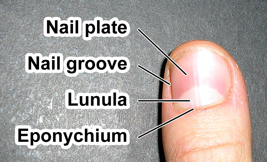
hyponychium
the thickened region beneath the free edge of the nail where dirt and debris tend to accumulate
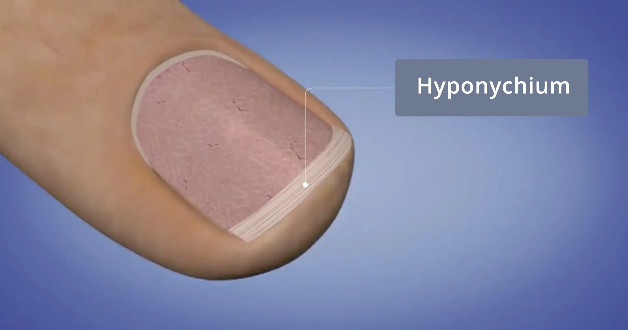
hyponychium function
secures the free edge of the nail plate at the tip of the finger or toe
2 types of sweat glands
eccrine
apocrine
what regulates sweating
autonomic (involuntary) nervous system
eccrine sweat glands structure
simple, coiled, tubular gland; secretory part lies coiled in the dermis, and the duct extends to open in a tunnel-shaped pore at the skin surface
eccrine sweat glands secretion
sweat
sweat
hypotonic filtrate of blood that passes through the secretory cells of the sweat glands and is released by exocytosis
sweat composition
99% water, traces of metabolic wastes (urea, uric acid, ammonia), and dermicidin
dermicidin
microbe-killing peptide
sweat function
prevent the body from overheating
apocrine sweat glands location
axillary and urogenital areas
apocrine sweat gland secretion
sweat, fatty substances, and proteins (bacteria decomposing can lead to BO)
when do apocrine glands begin functioning
at puberty
3 lines of evidence that suggest that apocrine glands are the human equivalent of animal’s sex scent glands
sexual foreplay increases their activity
enlarge and recede with the phases of a women’s menstrural cycle
may act as pheromone
2 types of modified apocrine glands
ceruminous glands
mammary glands
ceruminous glands
modified apocrine glands found in the lining of the external ear canal; secretion mixes with sebum to form earwax
mammary glands
secrete milk
sebaceous glands structure
simple branched alveolar glands
sebaceous glands secretion
sebum
sebum function
softens and lubricates the hair and skin, slows water loss from the skin, prevents hair from becoming brittle, bactericidal action
acne
active inflammation of the sebaceous glands accompanied by pimples, whiteheads, or blackheads
functions of the skin
protection
BT regulation
cutaneous sensation
metabolic functions
blood reservoir
excretion
3 barriers of the skin
chemical
physical
biological
acid mantle
low pH of skin secretions
defensins
skin cell secretion; natural antibiotic
eathelicidins
protective peptides that prevent infection by group A streptococcus bacteria; secreted by wounded skin
substances that penetrate the skin in limited amounts
lipid-soluble substances: O2; CO2; vits A, D, G, K; steroids (estrogens)
oleoresins of certain plants (poison ivy)
organic solvents: acetone
salts of heavy metal (lead and mercury)
drug agents called penetration enhancers that help ferry other drugs into the body
biological barriers
dendritic cells in epidermis
macrophages in dermis
dendritic cells
part of immune system; engulf foreign invaders that have penetrated the epidermis; leave the skin and migrate to the recent lymph node where they display their prey to immune cells—triggers immune response
dermal macrophages
dispose of viruses and bacteria that manage to penetrate the epidermis; initiate immune response
insensible perspiration
routine and unnoticable sweating
sensible perspiration
noticable output of sweating
constriction of dermal blood vessels function
allows warm blood to bypass the skin temporarily and allows skin T to drop to that of the external environment