Unit 1
 Call Kai
 Learn
Learn Practice Test
 Spaced Repetition
 Match
Match Flashcards
 Knowt Play
1/41

There's no tags or description
Looks like no tags are added yet.
Name | Mastery | Learn | Test | Matching | Spaced | Call with Kai | Chat |
|---|
No analytics yet
Send a link to your students to track their progress
42 Terms
Cell Theory
Living organisms are made of cells
Cells are the basic unit of structure and function
All cells are made of other cells
History of Cells
~3900-2500 mya
Prokaryotes appear, chemoautotrophs, use CO2 as carbon source and inorganic materials for energy
1850 mya
Unicellular eukaryotes appear
Evolutionary Tree
Bacteria and archaea are prokaryotes
Archaea and eukaryotes are more similar, meaning they have a more recent common ancestor
Characteristics of Life
Genetic information is stored as DNA
DNA used as a template for copying the genome
Plasma membrane surrounds cell, creates barrier
RNA used as an intermediary *vulnerable
Proteins are used as catalysts for almost all reactions *enzymes, vulnerable
Free energy must be consumed
Eukaryotes vs. Prokaryotes
Prokaryotes
Small
No nucleus
No membrane-bound organelles
Eukaryotes
Large
Nucleus, DNA storage
Membrane-bound organelles
Endosymbiotic Theory
Archaea ate bacteria
Formed symbiotic relationship
Formed mitochondria
Again with photosynthetic cell
Formed chloroplast
Covalent Bonds
Non polar covalent
Equal sharing of e- due to similar electronegativities
Greater potential energy
Ex. CH4
Polar covalent bonds
Unequal sharing of e- due to differing electronegativities
Not as reactive, creates dipole moments
Ex. H2O
Non Covalent Bonds
Weaker than covalent bonds
Individually weak, collectively strong
Ionic
Taken, not shared e-
Little importance in cells
Ex. NaCl
Hydrogen
Requires hydrogen in a polar bond with + charge
Another polar atom with - charge
Hydration shells surround most biological materials
Ex. H2O and H2O
Van der Waals interactions
Weak non polar bonds
Ex. C2 and C2
Hydrophobic Effect
Forming hydration shells is unfavorable, low entropy
Non polar molecules in water associate with each other
Entropy=disorder, higher disorder is energetically favorable
Hydration shells=water spread out into shell over non polar molecules
Form separately or together around molecules
Together is more favorable, takes less water
Molecular Complementarity
Generally protein-protein, protein-DNA, etc. are not covalent
Why use weaker non covalent bonds? They’re reversible
Why do non covalent interactions matter?
Reversible, motor protein “foot”, moves to area of more complementarity
Complementary shape and portions of molecules
Polar: Not always on outside, can interact with each other
Non polar: Can be outside, interact with each other
Redox Reactions
OIL RIG
Oxidation is lost, reduction is gained
Gain or loss of time with electrons
Ex. Methane to methanol , C-OH bond losing time with C-H bonds
Building Blocks
Sugars → Polysaccharides
Nucleotides → Nucleic acids
Amino acids → Proteins
Fatty acids → Fats
Sugars, amino acids, and nucleotides form long chain polymers
Monosaccharides
General form (CH2O)n
Aldose, top of sugar, top of alphabet
Ketose, middle of sugar, middle of alphabet
Carbon number (3=triose, 5=pentose, etc.)
Disaccharides and Polysaccharides
Defined by a and B links
B hydroxyl, OH group sticks up the same as the outside carbon
a hydroxyl, OH group sticks down opposite as outside carbon
Oligosaccharides- short chains
Polysaccharides- Long chains
Branched sugar, whether sugar has a or B hydroxyls
pH
Measuring how basic or acid a substance is using amount of hydrogen ions
Amino acid zwitterions
Low pH, protonated
Neutral, not charged but charges within
High pH, deprotonated

Polymerization
Condensation reaction
Anabolic, builds polymer and H2O is a byproduct
Hydrolysis
Catabolic, use H2O to break apart polymer
Composition of a cell
70% water, 30% chemicals (proteins, small others)
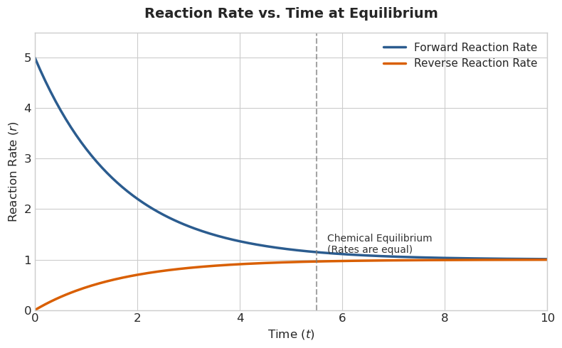
Reaction Rate
Reactants are converted to products
Catalysts lower activation energy and speed up reaction
Intracellular reactions are more complex and usually include many reactants
Energetics
Δ G = Δ H - TΔ S
Δ G, endergonic or exergonic reactions
Endergonic- spontaneous, losing energy
Exergonic, non spontaneous, needs catalyst
Δ H, exothermic or endothermic
Exothermic, releases heat
Endothermic, takes in heat
Δ S, disorder
More disorder is energetically favorable
Energy is needed to lower disorder
Enzyme and Reaction Rates
Some RNA act as catalysts
Catalysts lower activation energy
Enzymes as catalysts
Sucrose binds to sucrase
Converts products, releases products
Separate enzyme needed for reverse reaction due to complementarity

Protein Structure and Function
Primary
Sequence of AA, properties of side chains n → c terminus
Secondary (local folding)
Folding of localized areas, AAs near
Tertiary (overall conformation)
Folding of secondary structures
Quaternary (multimeric structure)
Two or more proteins
Supramolecular (large-scale assembly)
Function (regulation, structure, movement, catalysis, signalling, transport)
Condensation Reaction
Amino acid (carboxyl reacts with amino group, forms peptide bond)
Peptide (N-terminus, amino end and C-terminus, carboxyl end)
Protein
Zwitterion (+ and - charge, charge is neutral, backbone of protein
Anatomy of a Protein
N-terminus: amino (+)
C-terminus: carboxyl (-)
Side chains
Peptide bonds
Amino Acid Rotation
Rotation is constrained around the peptide bond
Phi and psi angles
Acidic AA (-)
Aspartic acid: Asp, D
Glutamic acid: Glu, E
Basic AA (+)
Arginine: Arg, R
Lysine: Lys, K
Histidine: His, H
Uncharged Polar AA
Asparagine: Asn, N
Glutamine: Gln, Q
Serine: Ser, S
Threonine: Thr, T
Tyrosine: Tyr, Y
S, T, Y are most commonly phosphorylated
Non Polar Amino Acids
Alanine: Ala, A
Glycine: Gly, G (smallest)
Valine: Val, V
Leucine: Leu, L
Isoleucine: Ile, I
Proline: Pro, P (cyclic, sharp kinks)
Phenylalanine: Phe, F
Methionine: Met, M
Tryptophan: Trp, W
Cysteine: Cys, C (can form disulphide bonds only when oxidized, usually extracellular)
Secondary Structures
α-helices
Backbone twist
Hydrogen bonds between
β-strands
Can be antiparallel (easy) or parallel (long route, extra AA)
Hydrogen bonds between strands
Zig-zag pattern
β-turns
Sharp turns
Usually by proline
Random coils
Unstructured
Intrinsically Disordered Proteins
Binding
Flexible region wraps around partner molecule to bind
Signalling
Flexible structures allow phosphate groups to easily be added or removed, molecular switch
Tethering
Flexible leash to hold 2 domains together while allowing movement
Diffusion barrier
Protein network forms filters that regulate what passes through
Elastin
Forms network that stretches under force, giving tissues elasticity, held together by disulfide bonds
Secondary to Tertiary Structures
Decreasing entropy as moves to tertiary structure
Intermediate tertiary structures form but are not stable or functional
Native state is the lowest energy but is the most stable
Driving force is side chain interactions: Hydrophobic effect, ionic bonds, disulfide bridges, hydrogen bonding, van der Waals forces
Representations of Protein Structures
Backbone trace (no side chains)
Ribbon diagram (no side chains)
Ball and stick (side chains)
Space filling model (shows specific shape)

Protein Domains and Quaternary Structure
Structurally, usually functionally distinct subunits
Assembly of quaternary structure
Dozens of the same protein lined up
Homo (same protein) or hetero (different protein)
Monomer, dimer, trimer…=# of proteins in a complex
Chaperones and Chaperonins
Chaperones
Helps protein fold correctly
Unfolded protein binds to nonpolar binding site (short segment)
ATP hydrolyses to ADP, “mouth” closes
Protein folds correctly, binding domain gets a new ATP
Protein releases
Chaperonins
Double barrel shape
Incorrect or incompletely folded protein enters cage
GroES cap and add ATP
Shake up protein, hydrolyse ATP to ADP
Barrel lengthenes
Add new ATP and release protein
Post-Translational Modifications
Phosphorylation: Kinase adds phosphate to protein, phosphatase pulls it off
Phosphate is from ATP
Can turn protein “on” or “off”
G-proteins
GAPs: Accelerate GTP hydrolysis
GEFs: Facilitate GDP to GTP exchange, inactive
Co-Factor Binding
Vitamins derivatives as coenzymes
Vitamins need to be converted to coenzymes for enzyme catalytic activity
Hem around protein: Hemoglobin
Cofactor binds to enable visual signalling
Proteolytic Activation
Rapidly activate proteins through cleavage
Single polypeptide chain, folding by disulfide bonds
Central peptide is removed
Active insulin released
Polyubuqitylation
Ubiquitin attaches to target protein to regulate its function
Monoubiquitylation- Histone regulation
Multiubiquitylation- Endocytosis
Polyubiquitylation- Lys48 (proteasomal degradation) or Lys63 (DNA repair)
Protein Interactions
Surface-string
Helix-helix
Surface-surface
Proteins need to find their binding partner and form a strong bond
Protein-Ligand Interactions
Forms non covalent bonds
Binding site formation via folding
Unfolded: AA side chains are far apart
Protein folds to native state
Binding site is for a specific ligand
Ligand=non enzyme, substrate=enzyme
Rates of Binding
Kon=rate constant for formation of complex
Koff=rate constant for dissociation of complex
R (receptor) + L (ligand) →← RL (ligand receptor complex)
Equil, Kon=Koff
Koff=[R][L]/[RL]=Kd
The more stable and tight RL complex, smaller Kd
10-100nM is very tight
Kd is binding affinity
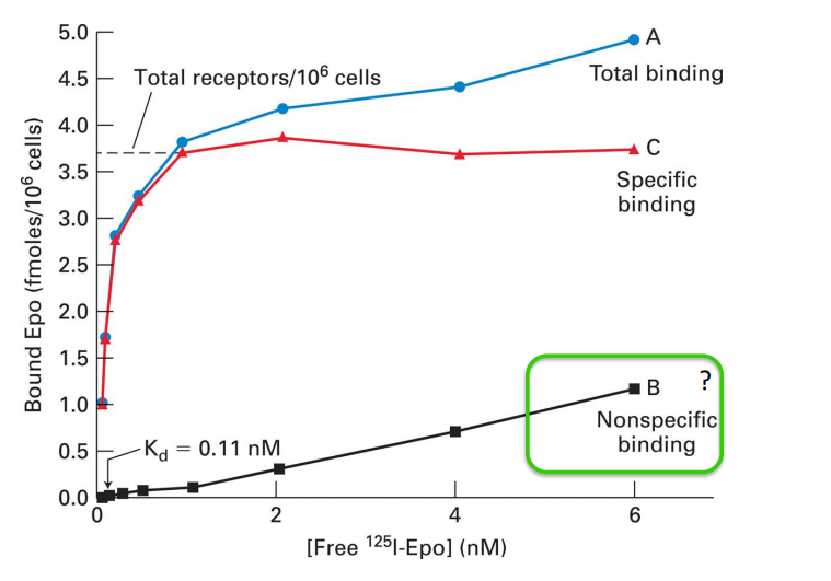
Binding Curves
To find Kd, y-axis is highest point on line. Then divide in half and see the Kd reflected on the x-axis