McGraw Hill ch. 16 Senses
1/52
There's no tags or description
Looks like no tags are added yet.
Name | Mastery | Learn | Test | Matching | Spaced |
|---|
No study sessions yet.
53 Terms
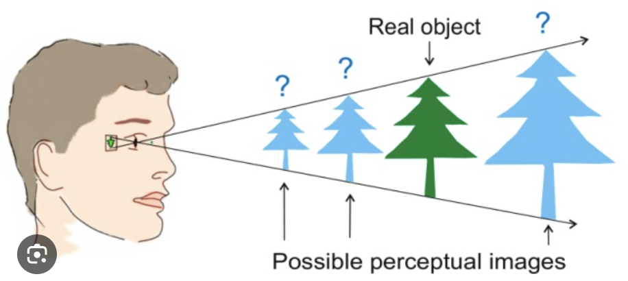
Describe the general function of sensory receptors as transducers. (What is "transduction"?)
respond to stimulus give info of internal/external environment giving info to brain
transduction: converts sensory signal to electrical signal processed in specialized area in brain
Describe the general structure of a sensory receptor, and explain the significance of a receptive field.
convey signals to CNS by sensory neurons
receptive field: distribution area of the endings of a sensory neuron
(area a receptor gathers information)
Define a sensation
a stimulus we are constantly aware of
consciousness signal must reach cerebral cortex
Explain the various characteristics of a stimulus that sensory receptors provide to the CNS.
modality: sensory receptor relaying sensory input along designated sensory neurons to specific regions of the CNS
location: sensory information is relayed either from different regions of a sensory receptor or from different locations
intensity: change in number of nerve signals that are arriving along a designated nerve
duration: decrease in sensitivity to a continuous stimulus is called adaptation

Distinguish between tonic and phasic receptors
tonic receptors: limited adaptation respond continuously head position receptors in inner ear all pain receptors
phasic receptors: undergo adaptation adapt rapidly only respond to a new stimuli pressure receptors
Identify and describe the three criteria used to classify receptors
distribution: general sense receptors
visceral and somatic sensory and special senses
modality: chemoreceptors, thermoreceptors, photoreceptors, mechanoreceptors, nocioceptors
taste smell temperature vision distortion pain
origin: exteroceptors, interoceptors, proprioceptors
cutaneous, in smooth muscle, found in joints
Classify the various types of sensory receptors based upon each of the three criteria.
somatic sensory: skin joints touch receptor
visceral sensory: blood vessels stretch receptor in stomach wall
special senses: head 5 senses
exteroceptors: external environment skin
interoceptors: detect stimuli in skin 5 senses
proprioceptors: detect stimuli in joints muscle spindles
chemoreceptors: detect chemicals taste receptors
thermoreceptors: temp change in skin hypothalamus
photoreceptors: change in light intensity color in retina
mechanoreceptors: deformation of plasma membrane skin
nociceptors: detect painful stimuli pain receptors

Compare and contrast unencapsulated and encapsulated tactile receptors.
unencapsulated tactile receptors: dendrite endings sensory neuron no protective covering
FREE NERVE ENDING/ ROOT HAIR PLEXUS/ TACTILE DISC
encapsulated tactile receptors: neuron ending wrapped in ct covered by glial cells and neurolemmocytes
END BULB/ LAMELLATED CORPUSCLES/ BULBOUS CORPUSCLES
Define referred pain, and explain its significance in diagnosis
inaccurate localization of sensory signals
region of pain isnt for where the medical problem is
heart attack pain in arm for men
back pain for liver failure
Briefly discuss how pain perception is moderated in 3 ways
stop pain message from being formed lidocaine numb mouth
block pathway of pain message nerve block procedure
alter CNS processing like narcotics
Which "sense" information does NOT pass through the thalamus?
smelling olfactory tracts
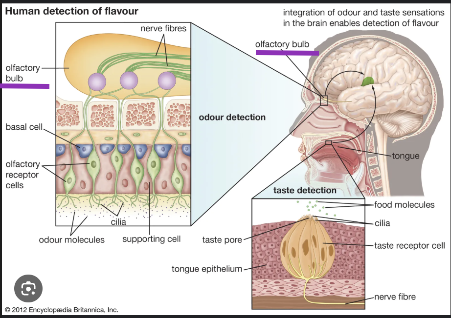
Name the components of the olfactory receptors, and discuss their mode of action.
Olfactory Epithelium- epithelium tissue inside nasal cavity olfactory, supporting and basal cells responsible for detecting odors
Olfactory Tract-bundle of axons connecting to mitral and tuffted cells of olfactory bulb to target regions of brain (amygdala, piriform & entorhinal cortex)
Olfactory bulb-structure brain receives neural input from nasal cavity sense of smell.
Olfactory nerve- bipolar neurons congregate to form olfactory nerve receptors in the mucosa of the nasal cavity relays info to the brain
Describe the path of a nervous impulse from your olfactory receptors to the cerebrum - which part? Is there just one path/one final destination?
olfactory never project through skull cribriform plate enter olfactory bulb
1)olfactory sensations start odorant binds to protein stimulate receptor cell
2) g-protein activate leading to depolarization of ion channels
3)action potential triggered on axon
4)secondary neuron conducts signals to CNS area
5)olfactory receptors adapt quickly bc synaptic inhibition in olfactory bulb

Describe the structure and function of papillae of the tongue 4 types
Fungiform: mushroom shaped, present at tip. Innervated by facial nerve.
Filliform: thin, long papillae "V" shaped and most numerous. However, not involved with gustation
Foliate: ridges & grooves towards posterior roof of mouth innervated by facial nerve and glossopharyngeal nerve
Circumvallate: only about 10-14 on most people. Present at the back of tongue arranged in a circular row. Innervated by glossopharyngeal nerve
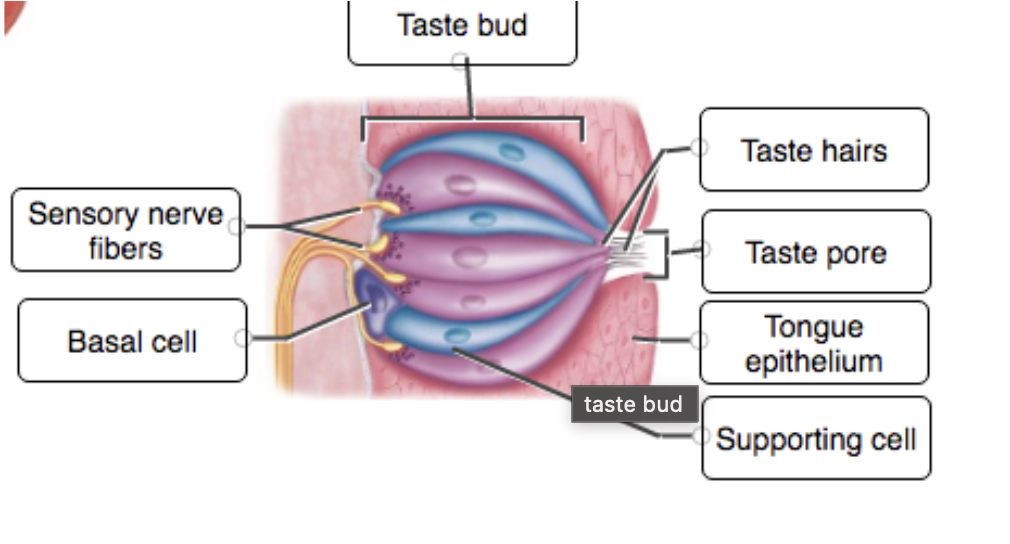
Discuss the structure and location of gustatory receptors
receptors that detect various specific tastes, like sweet, salty, bitter, sour, and umami spread across the tongue
Describe the five types of tastes, and explain the association of smell with taste.
Sweet: Produced by organic compounds sugar or artificial sweeteners
Salt: Produced by metal ions Na+ & K+
Sour: Associated w/ acids vinegar
Bitter: Produced by alkaloids unsweetened chocolate
Umami: Taste related to amino acids producing meaty flavor
80% OF TASTE IS SMELL
Describe the path of a nervous impulse from your gustatory receptors to the cerebrum - which part? Which nerves carry this information?
Sensory neuron connect to gustatory cells in tongue project to medulla
sensory medulla neurons project to thalamus
tertiary thalamic neurons project to primary gustatory cortex in insula
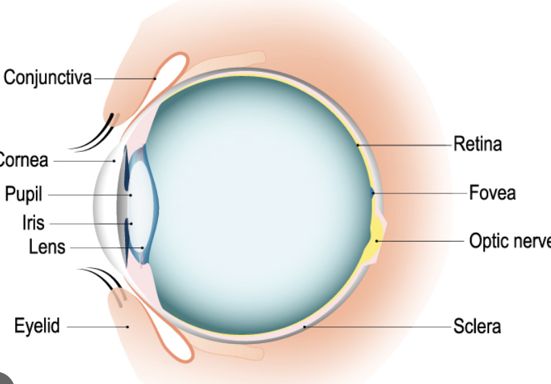
Describe the accessory structures of the eye, and list their functions
Eyebrow, Eyelash, Eyelids: are to protect the eye from any potential irritant
Conjuctiva: thin-transparent membrane that covers the sclera. Helps lubricate the eye by producing mucous and tears
Which way do tears flow across your eye? What do tears do?
lacrimal gland above eye in bone produces tears
lacrimal apparatus collects/drains fluids
secretes through ducts in superolateral orbit
tears moisten eye and cleans, reduces eyelid friction
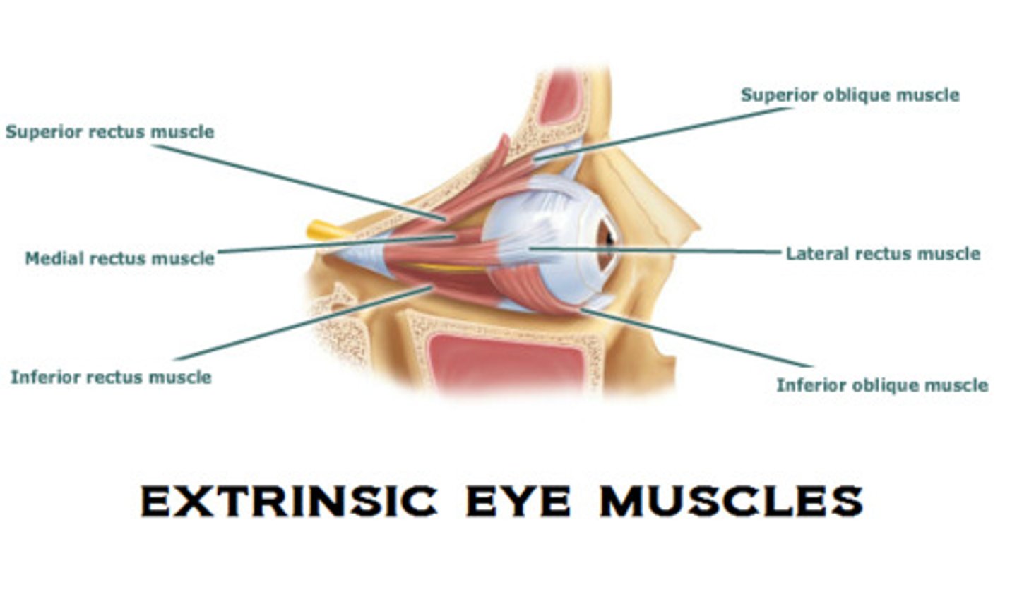
Describe the extrinsic eye muscles
superior inferior medial lateral rectus move eye up down laterally medially
superior inferior obliques move eye to or away from nose
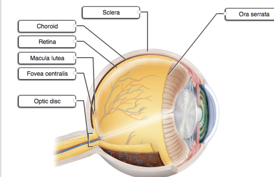
Describe the structures of the eye
Retina-recieve light and convert the light into neural signals that are sent to the brain for visual recognition
Ciliary Body- focuses
Ciliary muscles-smooth muscle connected to lens
Lens-changes shape to focus on objects at various distances
Sclera-white of eye
Suspensory ligaments- controls lens
Iris- controls pupil diameter sphincter constrict pupil dilator dilate pupil
Describe refraction of light
when light passes b/w media of different densities like air and cornea through curved surfaces such as the lens
Discuss how light is focused on the retina
Light passes through the front of the eye (cornea) to the lens
The cornea and the lens help to focus the light rays onto the back of the eye (retina)
The cells in the retina absorb and convert the light to electrochemical impulses which are transferred along the optic nerve and then to the brain
Explain what a "blind spot" is
the optic disc is called the blind spot bc no photoreceptors are there
Explain how light gets from outside your eye to your photoreceptors...listing all the structures and fluids it passes through on the way
light passes through cornea
through aqueous humor
through the pupil hole
through the lens
through the vitreous humor
through the ganglion cells
through bipolar cells
hits rods and cones photoreceptors
Describe the location and function of the lens. What is myopia, hyperopia, emmetropia, and presbyopia?
emmetropia- normal vision
presbyopia-age related change in vision need convex lenses
hyperopia- farsighted
myopia- nearsighted
Why will everyone need glasses at some point in their lives?
presbyopia age related change in vision lens get stiffer less stable becomes more spherical get convex lenses
Define phototransduction
converting light to electrical signals by photoreceptors of rods and cones
Compare and contrast the two general types of photoreceptors, including their photopigments
rods long narrow in dim light
cones see color activated in high intensity light
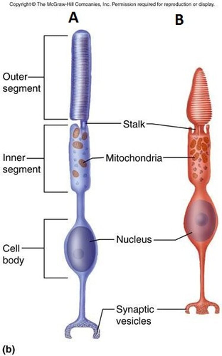
Where is the highest concentration of cones located?
concentrated in fovea centralis
Why can you see better at night out of the corner of your eye than looking directly at something?
because your lens does not have to focus and change shape
in dim light your eyes do not need to adjust as much as in bright light
rods are present at the extremes of the eye
What is color blindness?
resessive condition in males absence in one type of cone cell for red and green most common
Explain the bleaching reaction and how it relates to dark adaptation and light adaptation
light causes reconfiguration to trans-retinal which dissociates from opsin (bleaching retraction)
dark adaptation bleached rods make rhodopsin
light adaptation processing from low to bright light rods work better in dim light take time to adjust
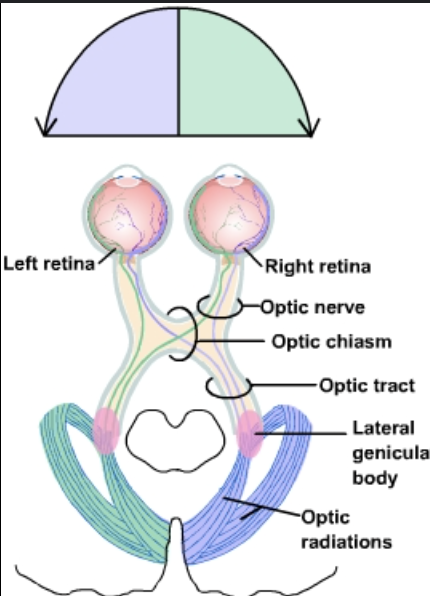
Describe the visual pathway from the photoreceptors to the brain
from rods and cones
goes to bipolar cells
then ganglion cells
goes to optic nerve
then optic chiasm
then optic tract
through thalamus
optic radiations occurs
primary visual center in occipital lobe gets the signal
Explain how stereoscopic vision provides depth perception
Stereopsis is depth that arises as a consequence of having two eyes have overlapping visual fields
What happens to the number of action potentials generated in light versus dark? Why?
Rod cells are hyperpolarized in the light and depolarized in the dark
During hyperpolarization, an action potential cannot be generated
so in the dark more action potentials are generated
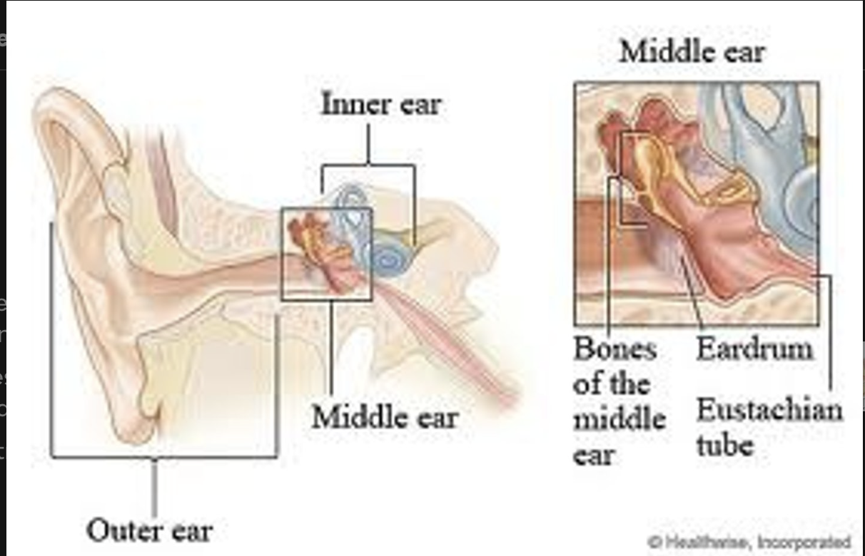
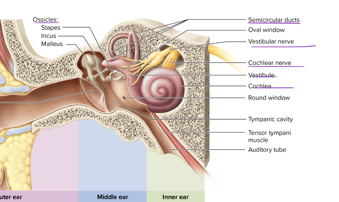
Describe the structures of the outer, middle, and inner ear
external ear has auricle and external acoustic meatus
middle ear has air filled tympanic cavity and auditory tube and ossicles malleus incus stapes
inner ear has cochlea, vestibulae and semicircular vestibule-respond to change in position of head Semicircular ducts-maintain balance, detects motion Vestibulochochlear nerve
Round Window-"sound exit"

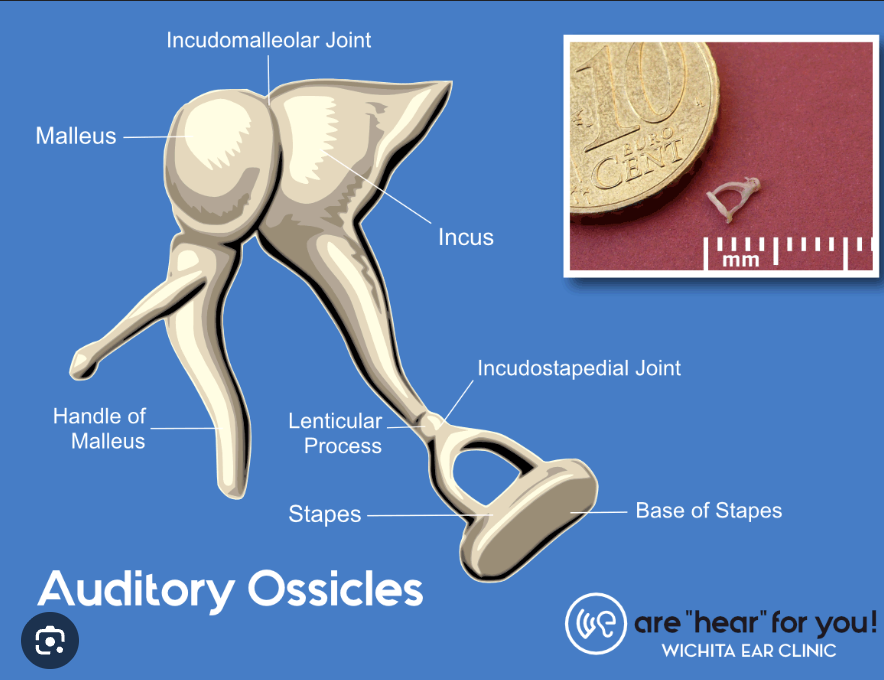
Name the auditory ossicles, and explain how they function in hearing
malleus incus stapes all transmit sound waves to inner ear to amplify sound
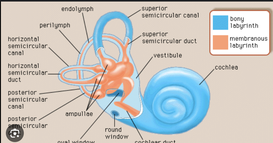
compare and contrast the bony labyrinth and the membranous labyrinth
bone labyrinth: maze in temporal bone has perilymph interstitial fluid fills most of space
membranous labyrinth: tubes has endolymph intracellular fluid with lots of potassium
Explain the components of the cochlea and how they function in the sense of hearing
cochlea receives sound in the form of vibrations, which cause the stereocilia to move in spinal sensory part
The stereocilia then convert these vibrations into nerve impulses which are taken up to the brain to be interpreted

Explain how moving air (which is what sound waves are) gets converted to a nervous signal. And how that nervous signal gets to the processing area in your brain
soundwaves collected by auricle travels down external acoustic meatus
vibrate tympanic membrane moving ossicles and make them vibrate
stapes vibrates moving oval window moving endolymph
endolymph moves basilar membrane moving into ear
hair cells in basilar membrane pushed against tectorial membrane
hair cells bend making depolarization release neurotransmitter
makes action potential in cochlear nerve
goes through vestibulocochlear nerve
moving to medulla oblongata
signal from pons can go to midbrain
nerve signal is now passed to thalamus
relayed to primary auditory cortex to be interpreted
now sound is perceived in temporal lobe of cerebrum
Distinguish between frequency (pitch) and intensity (loudness) of sound.
pitch is frequency of the vibrating object
frequency is rate of vibration
intensity is depends on wave amplitude
Describe the structures of the inner ear involved in equilibrium
vestibule and semicircular ducts-angular acceleration
responsible for balance and equilibrium
utricle and saccule

Explain how the utricle and saccule detect static equilibrium and linear movements of the head, and explain how the semicircular ducts function to detect rotational movements of the head
utricle and saccule detect static equilibrium and linear acceleration from the head bc hair cells have otoliths and when you tilt your head they move with macula knowing up and down
semicircular ducts detect angular acceleration and motion of the head info sent to brain to keep balance
Summarize the nerve pathways involved in equilibrium
signals from macula or ampulla conveyed by 8th CN
axons terminate in vestibular nuclei- medulla control reflexive eye movements and balance
cerebellum-coordinates balance and muscle tone
these send signals to thalamus
thalamus relays info to cerebral cortex of awareness
Clinical view: phantom pain
sensation associated with removed body part bc stimulation still happens to the sensory neurons
Clinical view: detached retina
happens when nearsighted
from outer pigmented and inner neural layers separate
see floaters in affected eye

Clinical View: Macular Degeneration
physical deterioration of macula lutea
cause of blindness associated w/ diabetes ocular infection eye trauma

Clinical View: Cataracts
small opacities in lens result from aging
risk factor of diabetes smoking uv light sunglasses
cant focus on close objects can be removed
Clinical view: glaucoma
increased intraocular pressure
Aqueous humor drainage being blocked is cause
makes reduced field of vision dim vision give halos around light
Clinical view: functional visual impairments
astigmatism: unequal focusing and unequal curvatures in one or more refractive surfaces
rest mentioned already on other card
Clinical view: otitis media
infection in middle ear in children common bc of short horizontal auditory tubes
cause from respiratory infection can spread from pharynx
Clinical view: deafness
any hearing loss
conductive interference of wave in external/middle ear
sensorineural malfunction in inner ear