MODULE 15B - [Anatomy 3.0] CEREBRUM AND DIENCEPHALON
1/209
There's no tags or description
Looks like no tags are added yet.
Name | Mastery | Learn | Test | Matching | Spaced |
|---|
No study sessions yet.
210 Terms
Telencephalon (or the cerebral hemisphere)
Diencephalon (or the core)
Enumerate the two major regions of the Cerebrum
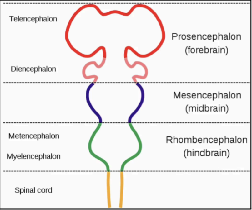
forebrain (prosencephalon)
Cerebrum is formed from the _____, which will give rise to telencephalon (the cerebral hemisphere) and diencephalon (the core)
mesencephalon and rhombencephalon
The _____ and _____ will become the brainstem
Brainstem
this structure will connect the structures to the spinal cord
CEREBRUM
Largest part of the central nervous system (CNS)
Has two basic poles
Anterior (Frontal) Pole
Posterior (Occipital) Pole
It is contained within the cranial cavity, covered by the three layers of meninges, and bathed with cerebrospinal fluid (CSF), which acts as a cushion to the brain.
Anterior (frontal) pole
A more rounded pole of the cerebrum
Posterior (occipital) pole
A more pointed pole of the cerebrum
pressurized
In certain cavities, CSF is _____ so when you move your head, the brain won’t move
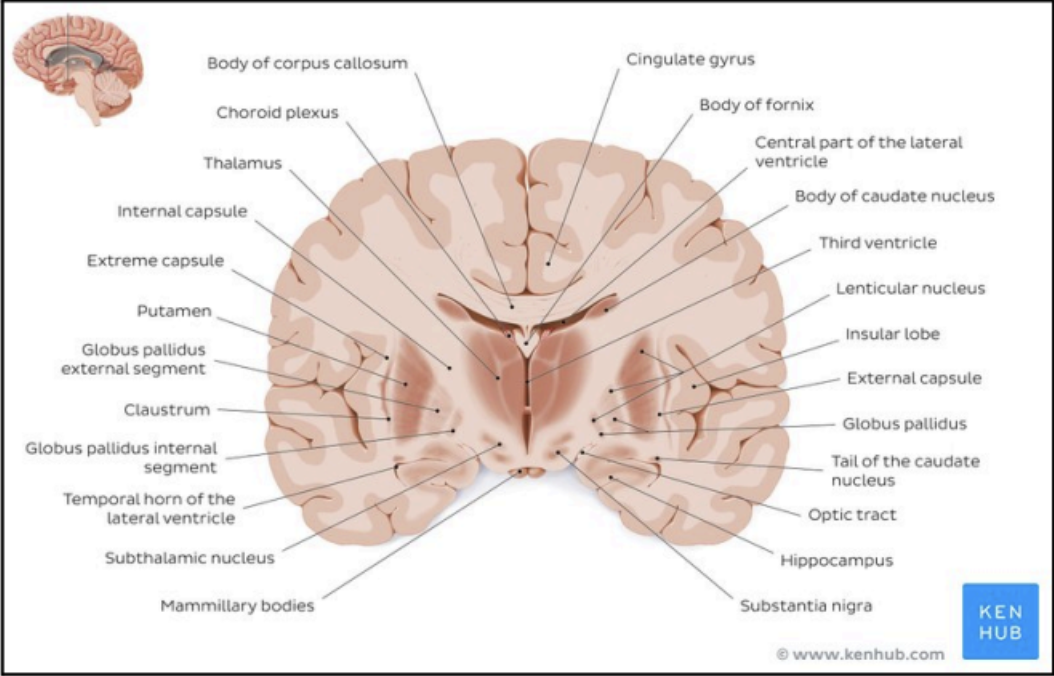
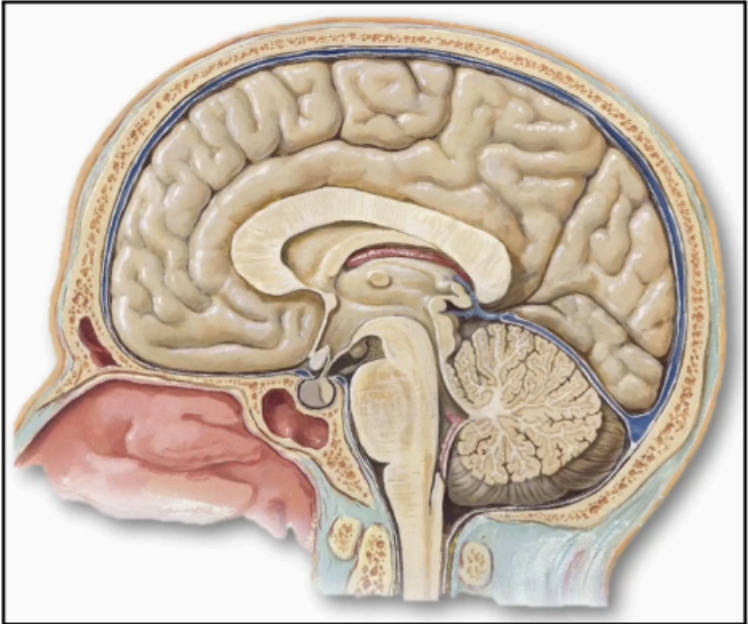
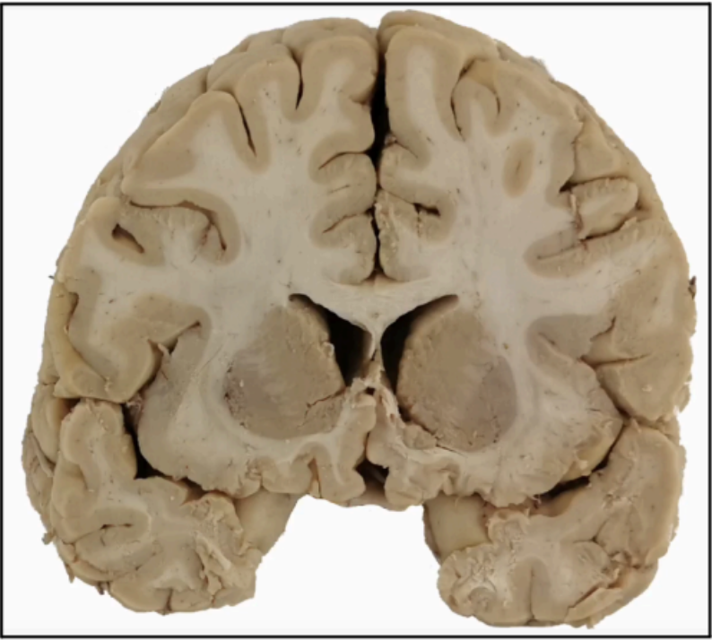
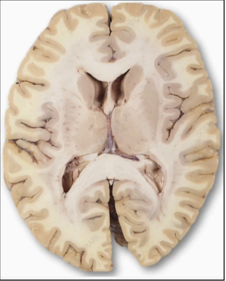
Parts of the brain on a coronal section cut
Parts of the brain on a coronal section cut
mushroom-like
what is the consistency of the brain
GRAY MATTER
Located on the periphery
Composed of cell bodies
Axons from these cell bodies project inwards, forming the central white matter.
Cerebral Cortex
Another name for Gray Matter
Subcortical Gray Matter
Gray matter aggregates within the core of the cerebrum or white matter.
subcortical nuclei
central core of the cerebrum that has aggregates of gray matter which are formed by clusters of cell bodies called
Diencephalon
Parts of the limbic system
Basal nuclei
Enumerate the Groups of Subcortical Nuclei
Both the nucleus and the ganglion are cell bodies
Both have similar functions
The difference is location in the nervous system
Nucleus: CNS (Nucleus = CeNtral NS)
Ganglia: PNS
What is the Similarities and Difference of nucleus and ganglia
WHITE MATTER
Found underneath the cortex
Represents the bundles of axons travelling to and from the cerebral cortex and other areas of the CNS
These axons are covered by a myelin sheath, which makes the area appear white
Schwann cells (in PNS) and Oligodendrocytes (in CNS)
What produces the Myelin sheath
Commissural Fibers
White matter tracts that link or connect the two cerebral hemispheres (left and right)
It will traverse the opposite cerebral hemispheres and interconnect the two
Examples:
Corpus callosum
Anterior commissure
Posterior commissure
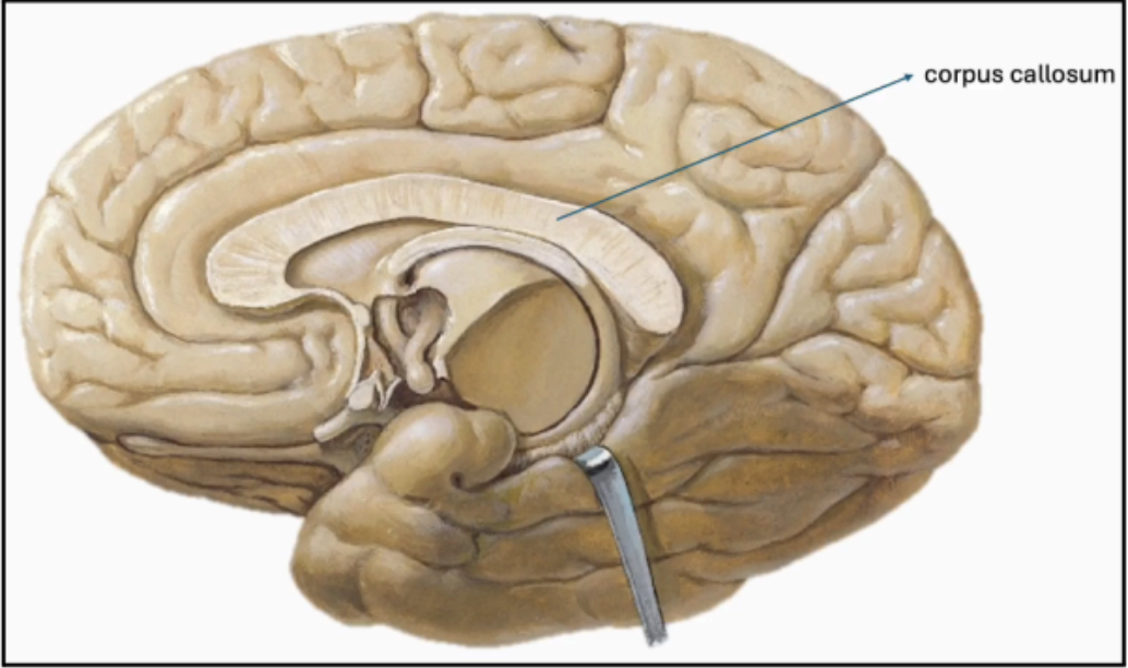
Corpus Callosum
Largest one of the commissural Fibers
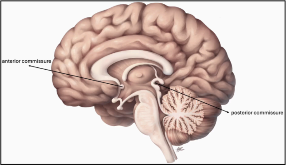
Anterior Commissure
Commissural Fibers of the Limbic System

Posterior Commissure
Commissural Fibers of the Visual System
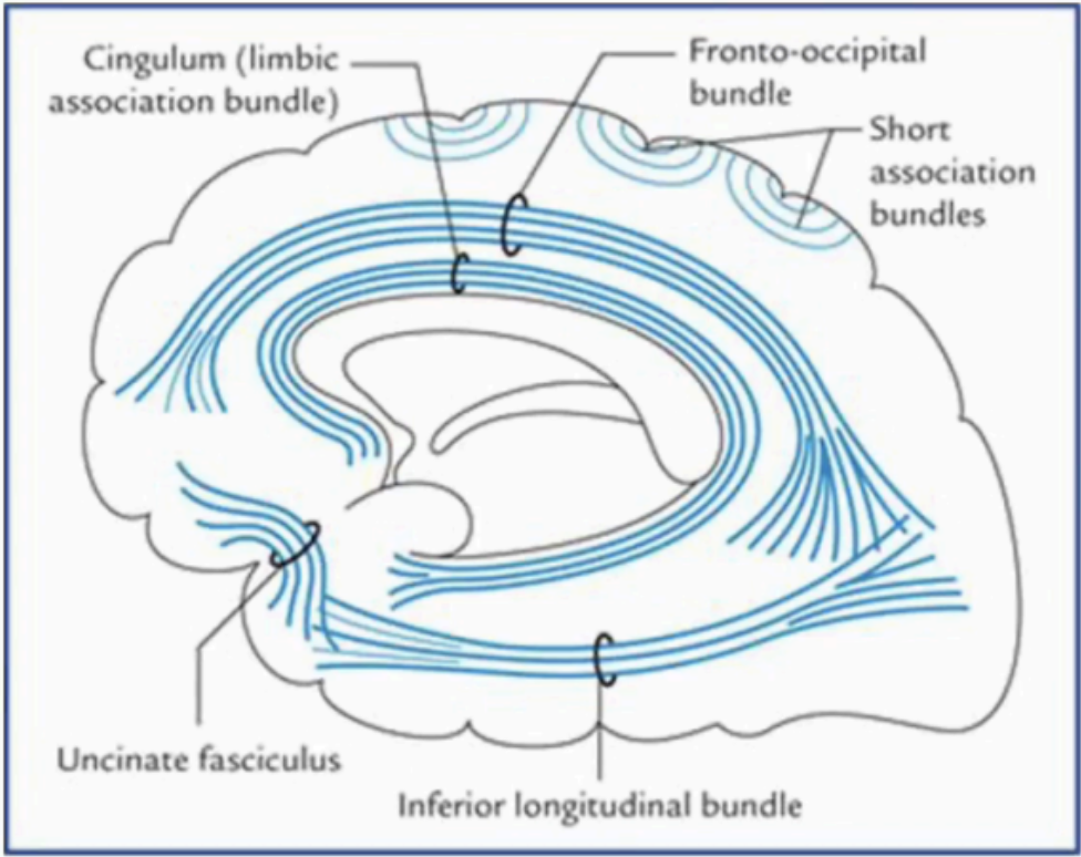
Association Fibers
Fascicles of bundles of white matter connecting one region or lobe on one side of the cerebrum
DO NOT cross the cerebral hemispheres, unlike commissural fibers, and are confined in a particular hemisphere
Short
Long
Enumerate the different types of Association Fibers
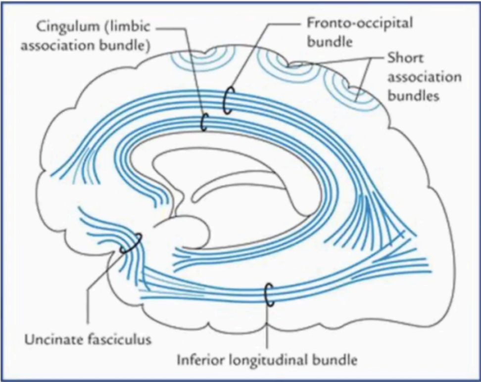
Short Association Fibers
Connecting adjacent regions (or gyrus)
Examples: Uncinate Fibers

Long Association Fibers
Connecting lobes of the cerebral hemisphere
Examples: Fronto-occipital bundle and Inferior Longitudinal Bundle
Fronto-occipital bundle
Long association fibers
Connects the frontal and occipital lobes
Cingulum
Both long and short association fibers
Interconnects the frontal, parietal, and medial temporal lobes, while also linking subcortical nuclei to the cingulate gyrus
Also called the Limbic association bundle
Uncinate Fasciculus
Short Association Fibers
Connects the frontal and temporal lobes
Inferior Longitudinal Bundle
Long Association fiber
Connects the occipital and temporal lobes
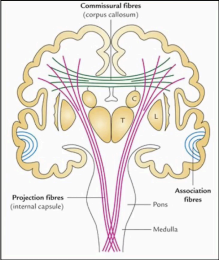
Projection Fibers
White matter tracts or bundles that move out of the cerebrum and connect to other parts of the CNS (i.e., brainstem, spinal cord, cerebellum)
DO NOT cross the opposite cerebral hemisphere
It can go both ways:
From the cerebrum to other parts of the CNS
Other parts of the CNS to the cerebrum
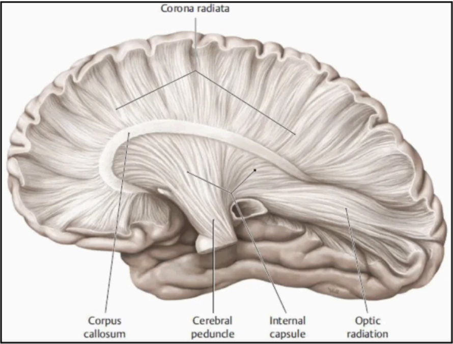
Corona Radiata
The most common type of projection fibers
English translation: “Rays of the Crown”
White fibers that are immediately formed just below the cerebral cortex
The first few fibers that will come out of the cerebral cortex
It is a very fibrous initial segment seen when the specimen is opened
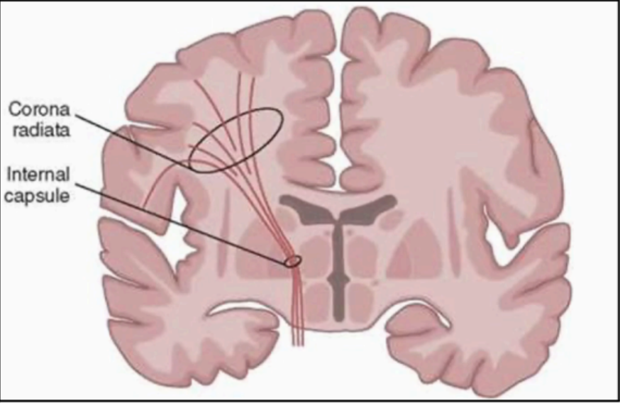
Internal capsule
As the fibers of the Corona Radiata reach the central core of the cerebrum, they organize to form a more solidified bundle of white matter called _____
More compact than the corona radiata.
Embedded (blends) within the subcortical gray matter
Boomerang in shape
Shape of the Internal Capsule (Horizontal View)
Anterior limb
Genu
Posterior limb
Enumerate the three basic parts of the Internal Capsule
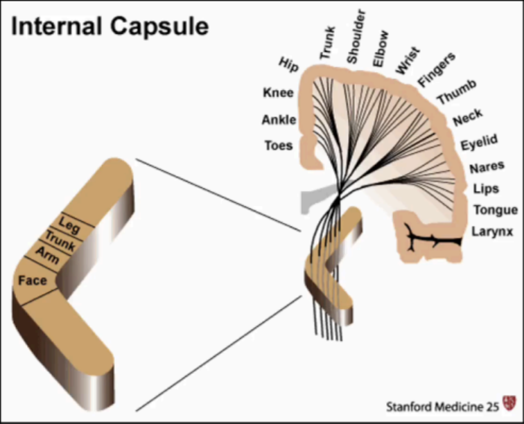
posterior limb
Motor neurons that will supply the arm, trunk, and leg are organized in the _____

genu
Motor neurons that will supply the face are organized in the ______
Left Cerebral Hemispheres
Controls sensation and movement of the RIGHT side of the body
Responsible for:
Analysis and calculations
Time and sequencing
Recognition of words, letters, and numbers
Logic
Right Cerebral Hemispheres
Controls sensation and movement of the LEFT side of the body
Responsible for:
Creativity and spatial ability
Context/perception
Recognition of faces, places, and objects
Artistic expression and music appreciation
It is usually the non-dominant hemisphere
dominant hemisphere
Responsible for speech, language, and comprehension
The left
Which cerebral hemisphere is usually dominant hemisphere
Cerebral dominance
determined by handedness (most of the time)
Right-handed
What handedness is people with a dominant Left hemisphere
Left-handed
What handedness is people with a dominant Right hemisphere
Left brain
Responsible for interpreting words
Right brain
Responsible for interpreting colors
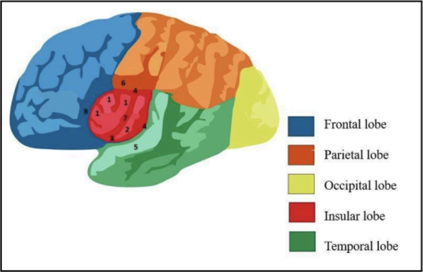
Frontal Lobe
Parietal Lobe
Occipital Lobe
Temporal Lobe
Insular Lobe (or Insula)
Enumerate the five general cerebral lobes most of which derive its name from the cranial bone that lie on top of them
Insular Lobe (or Insula)
A hidden lobe that can only be seen when opening the lateral Sylvian fissure
Gyrus
bumps at the surface of the cerebrum
Sulcus/Fissure
crevices on the brain’s surface
LONGITUDINAL/SAGITTAL FISSURE
Groove that separates the two cerebral hemispheres

CENTRAL SULCUS (SULCUS OF ROLANDO)
The line that separates the frontal lobe and parietal lobe
Has two gyri surrounding it
Precentral Gyrus
Located anterior to the central sulcus
Part of the frontal lobe
Postcentral Gyrus
Located behind the central sulcus
Part of the parietal lobe
LATERAL FISSURE (OR SYLVIAN FISSURE)
Prominent fissure that separates the frontoparietal lobe and temporal lobe
PARIETO-OCCIPITAL FISSURE
A small, posterior fissure that separates the parietal and occipital lobes
It is not that obvious on the lateral surface of the cerebrum
This is more evident on the medial surface of the posterior cerebrum
CALCARINE SULCUS
Separates the occipital lobe and the temporal lobe
Running perpendicular to the parieto-occipital fissure
Appreciated on the medial side of the cerebrum
Brodmann Area (BA)
Useful in determining the functionality of the lobe
A numbering system in neuroanatomy wherein areas of the cerebrum are designated with numbers
These numbers are based on the function of these specific areas
Korbinian Brodmann
Brodmann Area (BA) is coined by
Receptors
This structure’s role to capture stimuli and to convert it to the electric impulse for the brain to interpret
Vision
Receptors for _____ convert light to electrical impulse
Hearing
Receptors for _____ convert sound waves to an electrical impulse
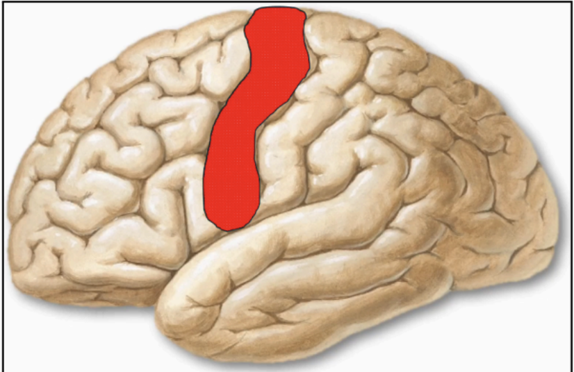
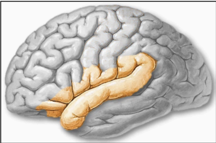
PRIMARY MOTOR CORTEX (PRECENTRAL GYRUS) - BA4 (Red)
The gyrus anterior to the central sulcus is the precentral gyrus which means it is on the territory of the frontal lobe
Periphery of the cerebral hemisphere
Contained within the precentral gyrus
Responsible for the voluntary movement or contraction of all the skeletal muscles on the contralateral side of the body
paresis or paralysis
paresis - weakness on opposite side/contralateral side
Paralysis - cutoff nerves supplying to the muscle
Damage on BA 4 causes
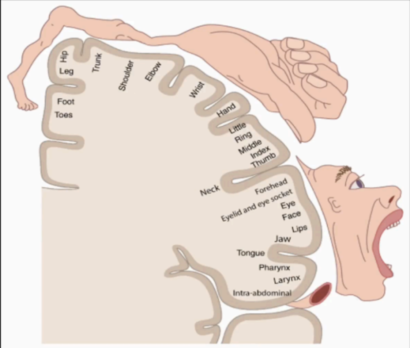
Motor homunculus
homonculus of the BA 4
somatotopic (or topographic) arrangement
In the cerebral cortex, the body is represented in a particular region of the cortex. This representation is referred to as _____
lower limbs
In the Somatotopic Arrangement of the motor homonculus, the _____ are represented on the medial surface of the cerebrum
trunk and upper limbs
In the Somatotopic Arrangement of the motor homonculus, the ____ and the ____ are represented on the upper lateral surface of the cerebrum
face (head and neck)
In the Somatotopic Arrangement of the motor homonculus, the _____ is represented on the lower lateral surface of the cerebrum
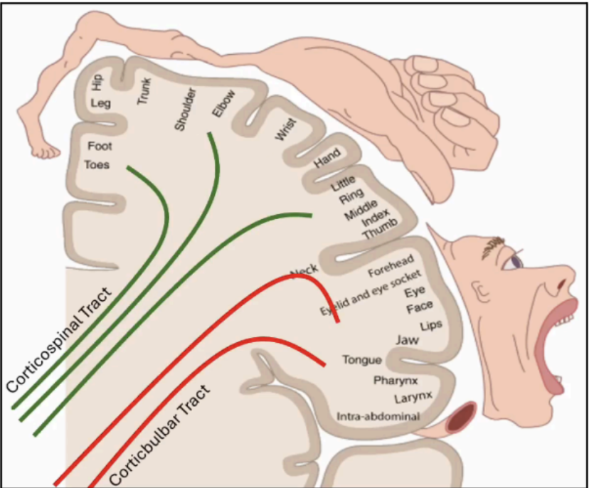
Corticospinal tract
From cortex to opposite side of the spinal cord
Supplies the lower limbs, trunk, and upper limbs
Internal Capsule: Posterior limb
As they elongate, the tracts cross to the other side after crossing the brain stem.
The name of the tract = origin and final destination of the tract.

Corticobulbar Tract
From cortex to brainstem
Supplies the head and neck (bulbar means stem)
Internal Capsule: Genu
As they elongate, the tracts cross to the other side after crossing the brain stem.
The name of the tract = origin and final destination of the tract.
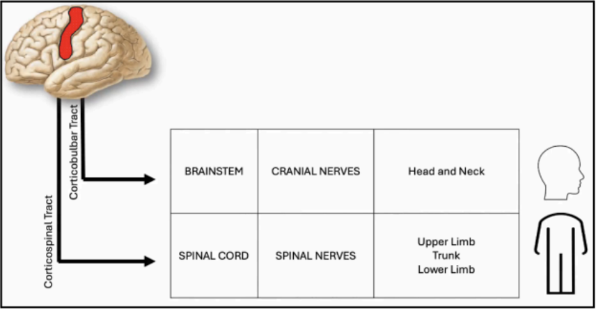
spinal cord and forms the spinal nerve
The Corticospinal Tract will continue until it reaches the _____ and forms the ____, which in turn will supply the upper limb, trunk, and lower limb

brainstem and give rise to the motor cranial nerves
The Corticobulbar Tract will terminate in the _____ and give rise to the _____, which in turn will supply the head and neck musculature
2/3
The Anterior Cerebral Artery (ACA) and Middle Cerebral Artery (MCA) supply the anterior ____ of the brain:
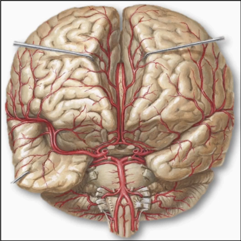
Anterior Cerebral Artery (ACA)
Mainly supplies the medial side of the cerebrum, including the medial side of the frontal lobe
Courses along the longitudinal fissure
On top of the corpus callosum and follows its shape

Middle Cerebral Artery (MCA)
Supplies the lateral side of the cerebrum
Courses inferiorly and exits at the lateral sylvian fissure
Direct continuation of the internal carotid artery
Damaged ACA
manifestation is greater in lower limb than in upper limb
As this artery supplies the medial side and the lower limb is located on the medial side of the homunculus as well
Damaged MCA
Manifestation is greater in the upper limb than in the lower limb
As this artery supplies the lateral side, and the upper limb is located on the lateral side of the homunculus as well
Muscles of the head and neck will also be
affected
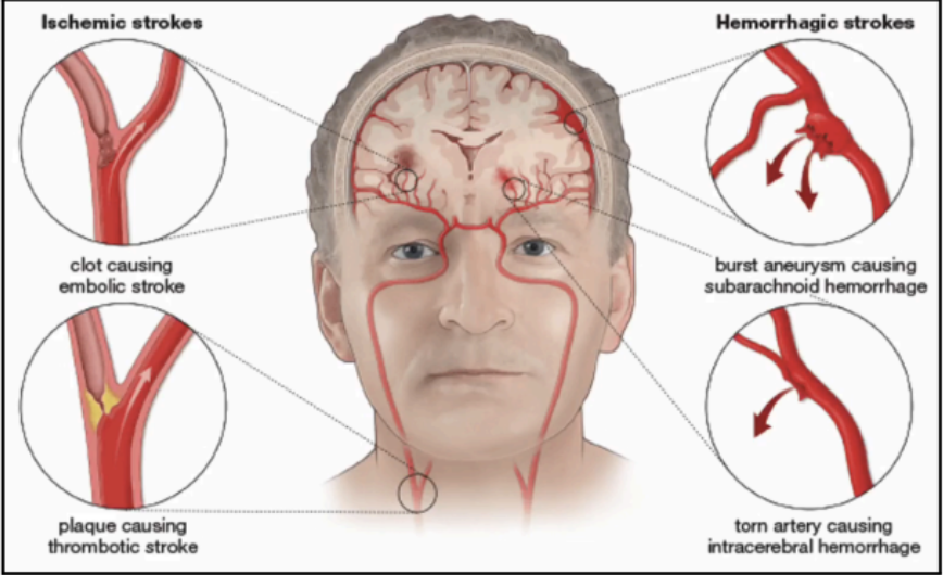
Cerebrovascular Disease (CVD)
Insult to cerebral arteries
Can present in two ways:
Ruptured blood vessel (hemorrhagic)
Obstructed blood vessel (ischemic)
Generic: Stroke
Appropriate: Cerebrovascular disease CVD
Give both the generic and appropriate term
A vascular event that can happen in any part of the body
Blood supply is affected
Hemorrhagic Stroke
Rupture of a blood vessel
Happens in cases of:
Severely elevated blood pressure
(e.g., 230/90)
Cerebral aneurysms or spontaneous rupture secondary to a defect in the arterial wall
The patient may be asymptomatic, and then suddenly fall
Cranial cavity space is minimal, such that if blood
clot forms, the brain will be compressed
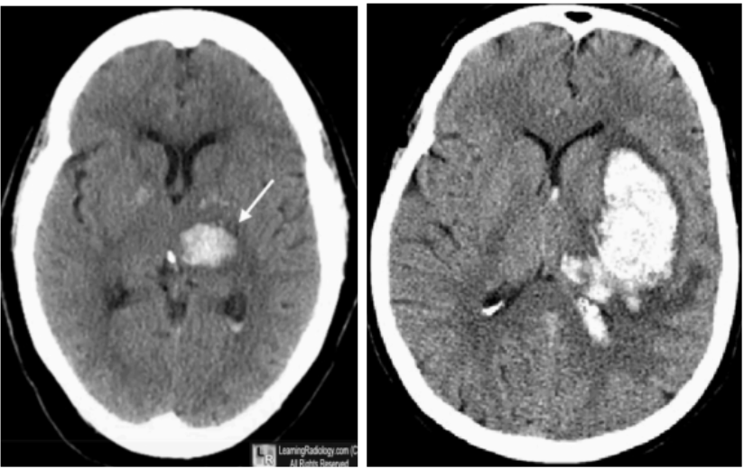
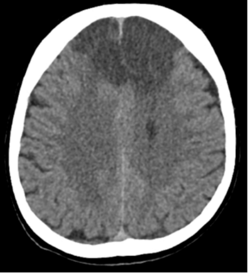
Intracerebral Hemorrhage
Reference point is the bone as it is hyperdense
If there is white in an unusual place (e.g. inside the
white matter), it typically indicates accumulation of
blood clot in the brain
Since the cavity of the skull is very limited, the blood
accumulating inside pushes the other brain
structures to the opposite side
Hence, the midline structures are now
deviated
Cerebral aneurysms or spontaneous rupture
An abnormal swelling or bulge in the wall of a blood vessel, such as an artery
Ischemic Stroke
Obstruction of blood vessel
Happens in cases of impeded blood flow leading to
decrease the blood supply to the area being supplied by the blocked blood vessel, secondary to either:
Atherosclerosis
Cerebral embolism
Causes the distal portion of that artery to become
ischemic
leads to a decrease in oxygen, causing tissue to die
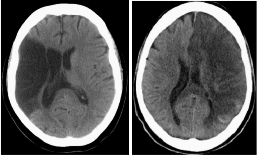
Parts of the brain scan appear black, as it is
hypodense due to poor or no blood supply
Analyzing the CT scan will give the idea that a major artery was blocked
The scan on the right is a massive ischemic stroke affecting the entire half of the cerebrum
The bigger the lesion, the more neurological manifestations will be demonstrated by the patient
In some instances, the shape of the hypodensity will already give us a clue on which artery was blocked
Atherosclerosis
Plaque formation inside the blood vessel
Cerebral embolism
A wandering object or substance that eventually can block the arterial blood vessels
These objects can be a dislodged clot from other areas (such as deep vein thrombosis), air, or a fat globule
Myocardial infarction
Obstruction of vessel supplying the heart
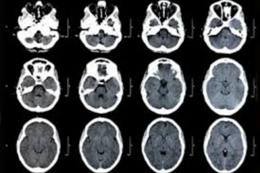
CT scan
One of the most widely used (common) diagnostic imaging modalities to confirm the diagnosis of a hemorrhagic or ischemic stroke
A series of slices is made on the head (with a
specific millimeter thickness) to give a full picture of the brain
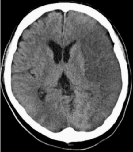
Appearance: Gray and homogenous
Term Used: Normodense
CT Scan interpretation of the Brain parenchyma
Appearance: White
Term Used: Hyperdense
CT Scan interpretation of Bone (skull), blood, or any Ca 2+ - containing substances
Appearance: Black
Term Used: Hypodense
CT Scan interpretation of Ventricular Cavity (contains CSF, which is a fluid)
Appearance: Semi-white
CT Scan interpretation of pineal gland
Appearance: black, because the CSF bathes that location of the brain.
Term Used: hypodense
CT Scan interpretation of the periphery
Non-deviated medial longitudinal fissure
A normal CT scan will always appear as a _____
As deviation may entail compression of one hemisphere to another
Thrombolytics may be given
Don’t give to patients with hemorrhagic type
Management if Ischemic
Stop the medication which could increase bleeding
Management if Hemorrhagic, clot prone
Middle Cerebral Artery (MCA) Obstruction
If this artery is blocked, you expect the patient to have weakness and loss of sensation on the opposite side
side
Especially the upper limbs, head, and neck
If it is the dominant hemisphere, expect a
speech problem
Anterior cerebral artery (ACA) Obstruction
If this artery is blocked, expect difficulty in walking due to a lower limb problem.
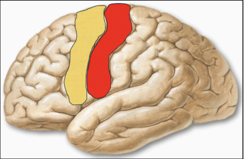
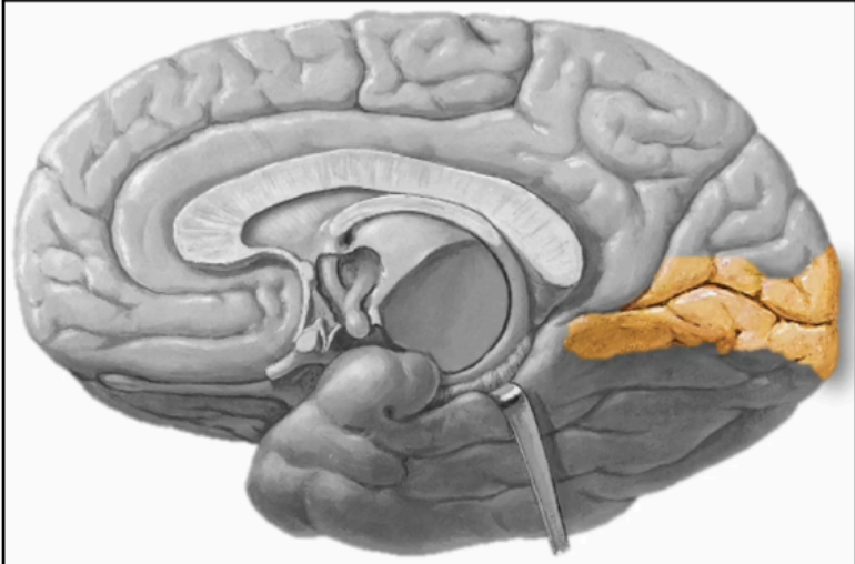
MOTOR ASSOCIATION CORTEX - BA 6 (Yellow)
Located anterior to the precentral gyrus
Adjacent to Brodmann Area 4
Responsible for the planning, sequencing, and
execution of movement
The blueprint of the motor system
Refines the impulse that is needed for BA4
This BA will plan which muscles to contract and relax, and transfer the output to BA4 for final execution.
This BA has connections with other parts of the CNS, such as the cerebellum and the basal ganglia
These structures are the ones responsible for refining of the planned movement
Basal ganglia/nuclei
initiate and stop movements
Output from this structure will be brought back to
BA6
Cerebellum
dictates the coordination (position of the limb, amount of contraction needed)
Output from this structure will be brought back to
BA6
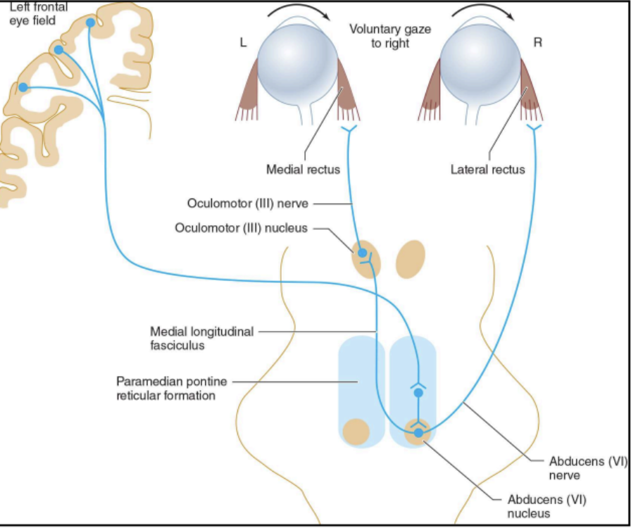
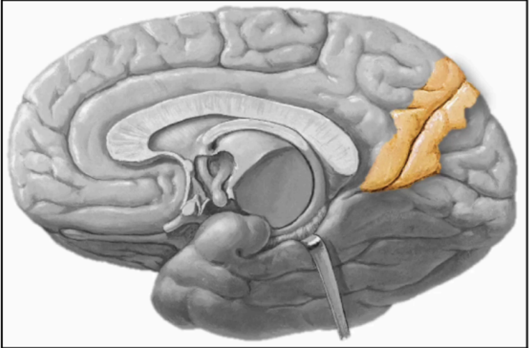
FRONTAL EYE FIELD - BA 8
Located anterior to the motor association cortex
Adjacent to Brodmann Area 6
Responsible for movement involving eyeballs
Responsible for horizontal eye movement or gaze
Movement of the left eye while retaining the right is impossible.
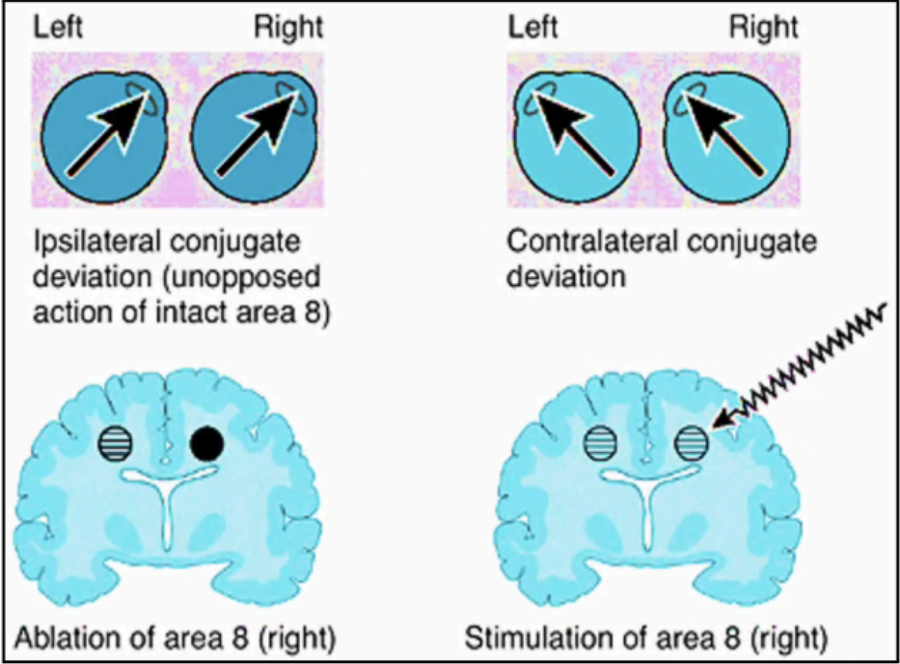
nuclei of the brainstem
From BA8, the fibers will be sent to the _____ responsible for controlling the muscles of the eye