Femur and Hip Anatomy Terms - Medical Study Set
 Call Kai
 Learn
Learn Practice Test
 Spaced Repetition
 Match
Match Flashcards
 Knowt Play
1/88

There's no tags or description
Looks like no tags are added yet.
Name | Mastery | Learn | Test | Matching | Spaced | Call with Kai | Chat |
|---|
No analytics yet
Send a link to your students to track their progress
89 Terms
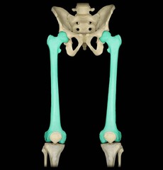
Femur
Largest & Strongest bone in the body, Entire weight is transferred through this bone & associated joints
Lateral epicondyle of femur

Lateral condyle of femur
Rough prominences on the outermost portion of the condyles For attachment of the Medial & lateral collateral ligaments

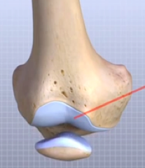
patellar surface (Intercondylar sulcus) (Trochlear groove)
smooth, shallow, triangular depression on the anterior distal femur

patella
kneecap

Medial condyle of femur
Rough prominences on the outermost portion of the condyles For attachment of the Medial & lateral collateral ligaments

medial epicondyle of femur

Adductor tubercle
Slightly raised are on the posterolateral aspect, located on medial condyle

Intercondylar fossa (notch)
deep depression located between the condyles and beneath the popliteal surface
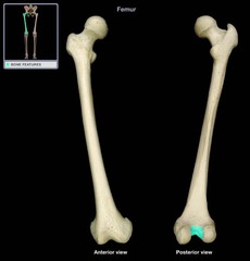
popliteal surface
Triangular surface above notch, behind knee on femur

Patellofemoral joint
between patella and patellar surface of femur
Body or shaft of femur

linea aspera
origin of vastus medialis

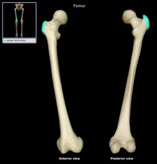
Greater Trochanter
Located superiorly & Laterally to femoral shaft, Palpable bony landmark
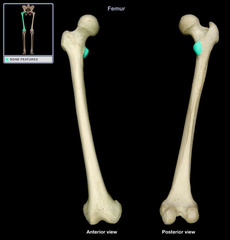
Lesser Trochanter
Smaller, Projects medially & posteriorly from junction of neck and shaft
Intertrochanteric crest
region formed posteriorly between the greater and lesser trochanters

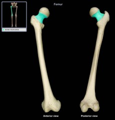
Neck of femur
Strong pyramid process, connects head with body
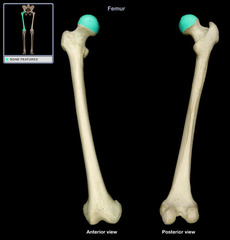
Head of femur
Rounded & smooth for articulation with hip bone, Depression near center
Fovea capitis
Ligament of the head of femur or ligament capitis femoris

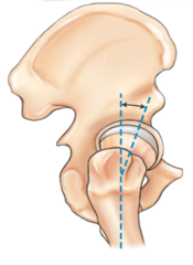
15-20°
What is the angle between the head of the femur and the neck in relation to the body of the femur in a natural position?
Femur must be rotated internally
How must the Femur be rotated to place the femoral neck in true AP?

Anatomical position, feet straight up and down
How is the femur rotated?
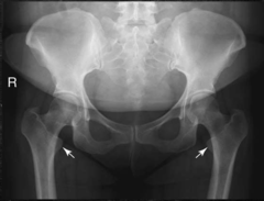
Femur externally rotated, lesser trochanter fully visible
How is the femur rotated?

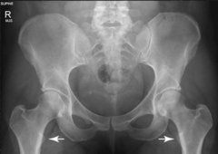
15-20° Internal Rotation, lesser trochanter barely visible (true AP)
How is the femur rotated?
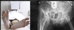
Affected femur placed mediolaterally, unaffected side placed in anatomical position
How would the feet lie to determine a hip fracture?
Find the ASIS (S1-2), go 1-2in medially, then move 3-4in down to neck of femur.
How do you find the the neck of the femur?
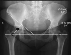
Chondrosarcoma
Malignant tumor of the cartilage, Usually occurs in the pelvis & long bones of men older than 45years, May be removed surgically
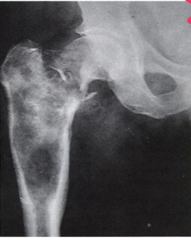
Legg-Calve-Perthes Disease
The most common type of aseptic or ischemic necrosis, Lesions typically involve one hip (head and neck of femur), Occurs predominantly in 5-10 year old boys, Limp is first clinical sign
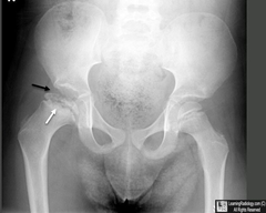
Intercondylar fossa (notch)
11?

Lateral condyle of femur
9?
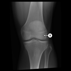
Lateral epicondyle of femur
8?
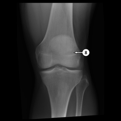
Medial condyle of femur
7?

Medial epicondyle of femur
6?

Adductor tubercle
5?

patella
2?
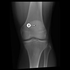
Body or shaft of femur
1?
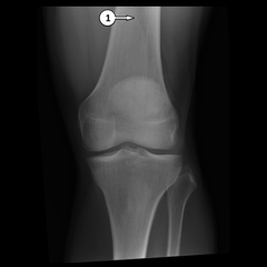
Body or shaft of femur
F?
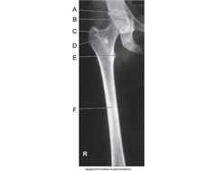
Lesser Trochanter
E?
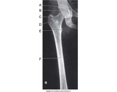
Greater Trochanter
C?
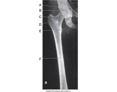
Neck of femur
D?
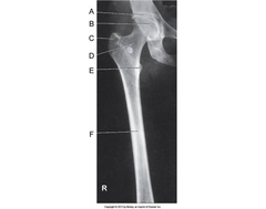
Head of femur
B?

Acetabulum
A?

radiopaque objects, clothing removed placed in gown , long hair, oxygen tubing, and pacemaker leads to avoid reduced detail/scatter
During patient preparation, what must be removed?

1. Limit the number of exposures
2. Collimation
3. shielding (led apron)
4. Position correctly
What 4 things help protect you and the patient against radiation?
BEFORE the examination
When must the anatomical marker be placed?
kVp: 75-80
mAs: 8-12mAs typically
Grid: yes
SID: 40in
What technical factors are needed for a good Femur (kVp, grid, mA, SID)?
AP
Lateral
Routine projections of the Femur?
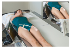
What is the evaluation criteria and procedure for an AP Femur X-ray?
kVp: 75-80
Grid: yes
CR: Perpendicular to body of femur for lower femur and neck for upper femur
Patient position: Patient Supine, leg fully extended,
Lower Femur: Internally rotate affected leg 5° for lower femur (body)
Upper Femur: rotate affected leg 15-20° internally for upper femur (neck)
Breathing instructions: suspend respiration during exposure for upper femur
Evaluation criteria: Mid-distal femur, including knee joint, for detection and evaluation of fractures or bone lesions, proximal portion includes neck and hip, fine trabecular markings
What is the evaluation criteria and procedure for an Lateral Femur X-ray?
kVp: 75-80
Grid: yes
CR: Perpendicular to body of femur for lower femur and neck for upper femur
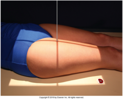
Patient position: Patient Supine, leg placed mediolaterally, Lateral recumbent, flex knee 45° for proximal, 20-30 flex for distal, place cushion under ankle/foot area for good lateral
Breathing instructions: suspend respiration during exposure
Evaluation criteria: Mid-distal femur, including knee joint, for detection and evaluation of fractures or bone lesions, proximal portion includes neck and hip, fine trabecular markings

ILIAC CREST: L4-L5
ASIS: S1-S2
GREATER TROCHANTER: Pubic symphysis
What are the bony landmarks of the pelvis?
Pelvis (os coxae) (Innominate bones)
Serves as the base of the trunk, Forms connection between vertebral column & lower limbs, Consists of 4 bones

Sacrum
5 fused vertebrae

Lumbosacral joint
pertaining to the joint between L5 and the sacrum

Sacroiliac joint
the joint between the sacrum and the ilium
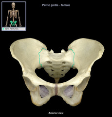
Coccyx
tailbone

Ilium
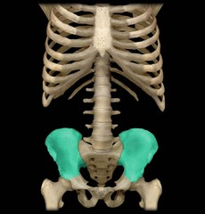
Body of Illium

Ala or wing of Illium
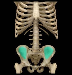
Iliac crests
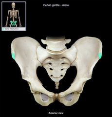
Portion of innominate bones that define the most superior, bilateral aspect of the pelvic cavity. They are palpable external landmarks that aid in evaluating the pelvis.

Anterior Superior Iliac Spine (ASIS)
The attachment for the inguinal ligament. Creates a noticeable bump on the two coxa.
Posterior Superior Iliac Spine (PSIS)
Posterior projection of the iliac crest and serves as an attachment for the posterior sacroiliac ligaments

Anterior Inferior Iliac Spine (AIIS)

Posterior Inferior Iliac Spine (PIIS)

Ischium
the curved bone forming the base of each half of the pelvis.

body of ischium
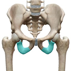
Ramus of ischium
joins the inferior ramus of the pubis anteriorly

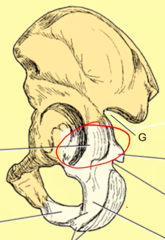
Upper body of ischium
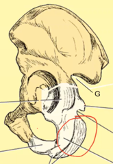
Lower body of ischium
ischial spine
located superior to the ischial tuberosity and projects medially into the pelvic cavity

Greater sciatic notch
allows blood vessels and the large sciatic nerve to pass from the pelvis posteriorly into the thigh
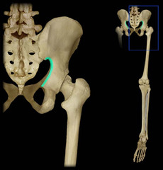
Lesser sciatic notch
inferior to ischial spine
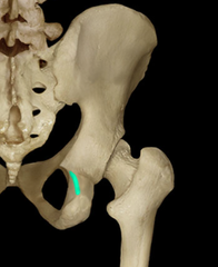
Ischial tuberosities
strongest part of hip bone, what we sit on

Pubis (Pubic bone)
most anterior portion of the hip bone

Superior ramus
Extends anteriorly & medially from body of pubis

Body of pubis
adjacent to the pubic symphysis

Symphysis pubis
a cartilaginous joint that is the point of fusion for two pubic bones
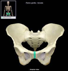
Inferior ramus
Pass down & posterior from symphysis pubis to meet the ramus of ischium
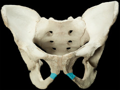
Obturator foramen
opening in hip bone formed by the pubic and ischial rami
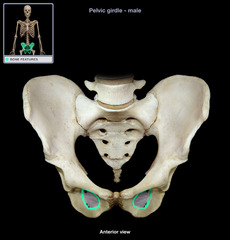
Acetabulum
hip socket, accepts head of femur to form hip joint

ilium
ischium
pubis
What 3 bones make up the acetabulum?
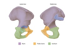
DDH (Developmental Dysplasia of the Hip)
Older term congenital dislocation of the hip (CDH), Hip dislocations that are caused by conditions present at birth, More common in girls or breech babies, US commonly used in newborns, May need frequent radiographs later on

Proximal Femur (Hip) Fractures
More common in older adults/geriatrics with osteoporosis or avascular necrosis, Both lead to weakening or collapse of weight bearing joints, fractures occur with minimum trauma
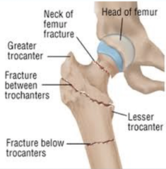
kVp: 70-80 (increase 4-6 for PA)
mAs: Depends
Grid: yes
SID: Varies
What technical factors are needed for a good Hip? (kVp, grid, mA, SID)?
AP Hip (Unilateral)
Lateral (Danelius-Miller Method/Unilateral frog leg)
What are the routine projections of the Hip?
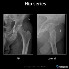
What is the evaluation criteria and procedure for an AP Hip Unilateral X-ray?
SID: 40in
kVp: 70-90
Grid: Yes
CR: perpendicular to femoral neck
Patient position: supine, leg of affected hip internally rotated 15-20°
Breathing instructions: suspend respiration during exposure
Evaluation criteria: No rotation (ASIS equal distance to table), pubic symphysis in middle, proximal 1/3 of femur should be visualized along with acetabulum and adjacent parts (pubis ischium, illium)

What is the evaluation criteria and procedure for a Axiolateral (Trauma hip) (inferosuperior) Danelius-Miller Method X-ray?
SID: 40in
kVp: 70-90
Grid: Yes
CR: perpendicular to femoral neck and IR (IR and CR at 45° in the crotch)
Patient position: Patient supine, Elevate pelvis 1 to 2 inches if possible, Flex & elevate unaffected leg, internally rotate affected like 15 unless possible hip fracture
Breathing instructions: suspend respiration during exposure
Evaluation criteria: Entire femoral head and neck, trochanter and acetabulum as well as any orthopedic device
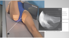
What is the evaluation criteria and procedure for a Unilateral Frog Leg (modified cleaves method) X-ray?
SID: 40in
kVp: 70-90
Grid: Yes
CR: directed at midfemoral neck
Patient position: erect or supine abduct femur 45° from vertical, flex leg and swing out laterally
Breathing instructions: suspend respiration during exposure
Evaluation criteria: Lateral views of acetabulum and femoral head and neck, 1/3 of femur visible, 45° abduction will demonstrate proper femoral neck in profile superimposed by greater trochanter, 20-30° abduction will prevent superimposition of greater trochanter on neck

Modified Axiolateral Clements-Nakayama Method (nontrauma)
What is the special projection of the Hip?

What is the evaluation criteria and procedure for a Modified Axiolateral Projection (Clements-Nakayama Method)
SID: 40in
kVp: 70-90
Grid: Yes
CR: Angle CR 15-20 down into femoral neck (posteriorly) (angle IR 15° from vertical so 75°)
Patient position: supine, leg in natural position
Evaluation criteria: Lateral oblique views of acetabulum, femoral head and neck, trochanteric area visible, minimal superimposition of of great trochanter