Musculoskeletal system
 Call Kai
 Learn
Learn Practice Test
 Spaced Repetition
 Match
Match Flashcards
 Knowt Play
1/86

There's no tags or description
Looks like no tags are added yet.
Name | Mastery | Learn | Test | Matching | Spaced | Call with Kai | Chat |
|---|
No analytics yet
Send a link to your students to track their progress
87 Terms
What are the 2 portal of entry of the musculoskeletal system?
Direct
Hematogenous
What are the 6 defense mechanisms & barrier systems of the musculoskeletal system?
Skin, subcutis & fascia
Vasculature
Immunologic responses
Adequate tissue antioxidant concentrations
Physiologic adaptations
Regenerative capacity of myofibers
This musculoskeletal defense mechanism forms structural barriers to protect against external injury
Skin, Subcutis, Fascia
What are the 3 other causes of muscle dysfunction?
Physiologic
Genetic
Nutritional / Toxicosis
Deficiency in these vitamins are muscle dysfunction under nutritional cause
Selenium & Vitamin E
Success of muscle regeneration depends on these 2:
Presence of an intact basal lamina
Availability of viable satellite cells
T or F: Myofiber necrosis often undergo segmental necrosis.
True
This widespread tissue or cell death of the entire myofibers only happens under severe stress, like:
Extreme pressure to the entire muscle causing crush injury
Widespread ischemia caused by pressure
Thromboembolism of a large artery
Global necrosis
This is a common trigger of necrosis in all cells.
Increased intracellular Calcium
This condition must be considered non-specific response, indicative only of myofiber necrosis.
Myofiber mineralization
These are the stem cells responsible for skeletal muscle regeneration.
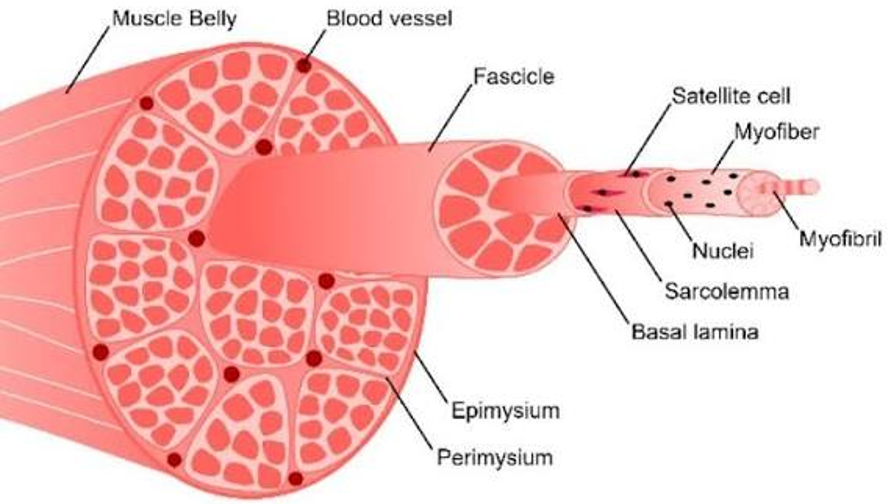
Satellite cells
This part of the muscle fiber is essential for a successful muscle regeneration; otherwise, healing occurs by fibrosis (scar formation).
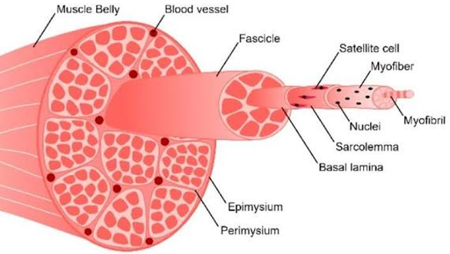
Intact basal lamina
These cells are also known as the muscle stem cells.
Satellite cells
When muscle tissue is injured, the specific type of cell death that occurs is called _________ necrosis.
Coagulative necrosis
Repair of an injured muscle begins when satellite cells move into the damaged area & become _________.
Myoblasts (immature muscle cells)
Myoblasts join together to form a ________ w/c is a new immature muscle fiber.
Myotube
This is the cytoplasm of a muscle fiber
Sarcoplasm
What are the 3 causes of muscle atrophy?
P A D
Physiologic muscle atrophy
Atrophy by Endocrine disease
Denervation atrophy
This is an alteration in myofiber size wherein it’s either a reduction in the volume of the muscle as a whole, or a reduction in the diameter of a myofiber.
Atrophy
A cause of muscle atrophy wherein it is a response to the lack of use, cachexia, or aging is known as __________.
Physiologic muscle atrophy
What type of muscle atrophy occurs as a result of pituitary dysfunction or tumors in aging horses, and is most commonly associated with hypothyroidism and hypercortisolism in domestic animals?
Atrophy by Endocrine Disease
A type of muscle atrophy common in veterinary medicine, exemplified by equine laryngeal hemiplegia (roaring) where affected muscle fibers become progressively smaller in diameter due to loss of nerve supply, is known as __________ atrophy.
Denervation atrophy
An alteration in myofiber size wherein myofibers increase in size by adding myofilaments.
Hypertrophy
What are the 2 causes of muscle hypertrophy?
P C
Physiologic hypertrophy
Compensatory hypertrophy
This type of muscle hypertrophy occurs naturally with exercise.
Physiologic hypertrophy
This type of muscle hypertrophy results from pathologic conditions that either reduce functional myofibers, increasing the load on remaining fibers, or disrupt normal cellular processes.
Compensatory hypertrophy
This term refers to the necrosis of skeletal muscles.
Rhabdomyolysis
This is the invariable feature of rhabdomyolysis, & clinically, myoglobin could be demonstrated in the urine.
Myoglobinuria
The elevated result of this serum level is one of the most important diagnostic criteria of rhabdomyolysis.
Creatine phosphokinase
What are the 3 types of muscle diseases?
D I N
Degenerative
Inflammatory myopathies
Neoplasms
What are the 5 muscle diseases under degenerative type of muscle disease?
Disturbance of Circulation
Nutritional deficiency
Toxic myopathy
Exertional myopathy
Trauma
A medical condition where a restriction or blockage in blood vessels reduces blood and oxygen supply to a part of the body.
Ischemia
A severe, life-threatening medical emergency where tissues or organs are completely deprived of oxygen.
Anoxia
What is the primary factor in assessing the impact of ischemia on muscles?
The varying sensitivity of the different cell types that make up the muscle tissue.
What is myofiber’s sensitivity to anoxia?
Most sensitive
What is Satellite cell’s sensitivity to anoxia?
Less sensitive
What is Fibroblast’s sensitivity to anoxia?
Least sensitive
What occurs first when blood supply to a muscle area is obstructed?
Myofiber necrosis
These cells die as a result of myofiber necrosis.
Satellite cells
What are the 2 factors that influence the extent of skeletal muscle infarcts?
Size of the obstructed vessel
Duration of the blockage
This occurs in downer cows due to increased intramuscular pressure during prolonged recumbency. This leads to localized muscle pallor from myofiber necrosis, resulting from reduced blood flow due to artery compression.
Ischemic necrosis
What are the 4 causes of muscle ischemia?
V E S O
Vasculitis / Vasculopathy
External pressure on a muscle
Swelling of a muscle in a non-expandable compartment
Occlusion of a major blood vessel
A type of degenerative muscle disease that is most common in livestock, including cattle, horses, sheep, & goats. Commonly called selenium / vit E deficiency, but selenium deficiency is the primary cause.
Nutritional myopathy
An essential antioxidant required for glutathione peroxidase, an antioxidant enzyme that protects myofibers from oxidative injury.
Selenium
What muscle types are highly susceptible to nutritional myopathy due to their high oxygen demand & continuous activity?
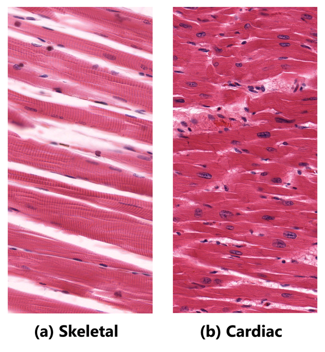
Skeletal muscle
Cardiac muscle
Which life stage in production animals is most commonly affected by selenium-deficiency disorders (such as White Muscle Disease) due to their strict reliance on trace mineral stores acquired during late gestation?
Neonates
In nutritional myopathy, the affected muscle is pale as a result of necrosis, thus, the common name is __________.
WMD - White Muscle Disease
A type of degenerative muscle disease that is common in livestock due to the ingestion of myotoxins from toxic plants, hay, or contaminated feed.
Toxic myopathy
What are the 3 clinical signs of toxic myopathy?
M R M
Muscle weakness
Recumbency
Markedly increased serum muscle enzymes
What is the primary histopathological hallmark of necrotizing toxic myopathy?
Multifocal skeletal muscle necrosis
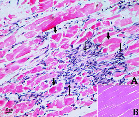
What primary criteria or evidence are used to confirm a diagnosis of toxic myopathy in livestock?
identifying toxin in:
Feed
Pasture
Stomach contents
These drugs can cause calcium overload, leading to skeletal & cardiac muscle necrosis.
ionophore antibiotics
These animals are extremely sensitive to ionophore toxicity.
Horses
These animals are more tolerant to ionophore toxicity, but can be poisoned by excessive doses.
Ruminants
What are the 4 examples of ionophore antibiotics?
L M M N
Lasalocid
Maduramicin
Monensin
Narasin
A type of degenerative muscle disease that is caused by intense or excessive muscle activity, resulting in myofiber necrosis (rhabdomyolysis).
Exertional myopathies
Another widely used and accurate term for exertional myopathy in wild animals is _________.
Capture myopathy
This type of degenerative muscle disease is more common in animals with underlying disorders such as:
Selenium deficiency
Muscular dystrophy
Electrolyte depletion
Glycogen storage disease
Exertional myopathy
Exertional myopathy in horses is also known as ___________ or _____________.
Exertional rhabdomyolysis
Tying-up syndrome
2 examples of working dogs where exercise-associated myopathy is reported.
Greyhound
Sled dogs
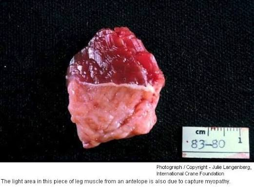
In this type of myopathy, affected muscles may appear pale pink, or diffusely red-tinged, potentially resembling autolysis. Multifocal pale zones can also occur.
Capture myopathy
This type of degenerative muscle disease is caused by physical injury.
Trauma
Trauma, as a degenerative type of muscle disease, can result in ____________ which is common in dogs, cats, & horses.
muscle rupture in the diaphragm
Healing occurs mainly by ________ , leading to scar tissue & reduced muscle function.
Fibrosis
Aside from muscle rupture, trauma may also lead to 2 other conditions:
Myositis ossificans
Musculoaponeurotic fibromatosis
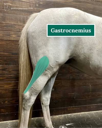
What particular muscle in horses is being damaged to its origin during exercise or while struggling to rise?
Gastrocnemius muscle
Which specific muscle group in cattle is most commonly prone to fiber tearing (split) when an animal slips and falls on a slick surface?
Adductor muscle of the hindlimb
What specific post-traumatic condition is characterized by the development of fibrosis and osseous metaplasia within affected skeletal muscle?
Myositis ossificans
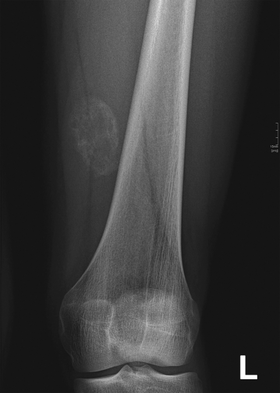
A specific post-traumatic condition, seen primarily in horses, characterized by progressive fibrous tissue proliferation that leads to muscle atrophy is known as __________.
Musculoaponeurotic fibromatosis
This refers to the inflammation of the muscle, may or may not be evident, depending on the presence or absence of fluid exudates.
Myositis
The four patterns of myositis are classified based on the distinct anatomical locations where inflammatory cells infiltrate muscle tissue. Identify these four distinct histological patterns of inflammatory cell infiltration:
Perivascular infiltration
Perimysial infiltration
Endomysial infiltration
Granulomatous infiltration
What pattern of myositis is characterized by the accumulation of inflammatory cells specifically around the blood vessels within muscle tissue?
Perivascular infiltration
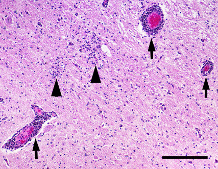
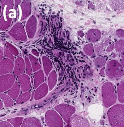
Infiltration of muscle perimysium or connective tissue surrounding individual muscle fascicles.
Perimysial infiltration
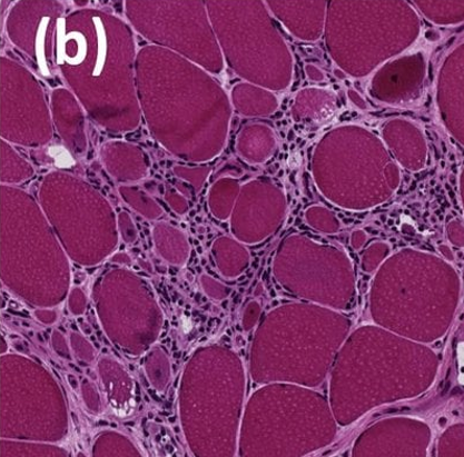
Infiltration of tissues surrounding individual muscle fibers (macrophage is typically involved)
Endomysial infiltration
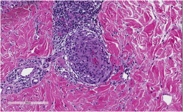
What pattern of myositis is characterized by the formation of granuloma in response to foreign bodies, parasite encystment, fungal infections, or bacterial pathogens?
Granulomatous infiltration
A condition seen in downer cows during prolonged recumbency
Ischemic necrosis
Disease characterized by extremely pale skeletal & cardiac muscles caused by Vitamin E or selenium deficiency
White Muscle Disease
This condition affects the cricoarytenoid muscles due to a loss of nerve supply. Example: Horse’s “"Roaring sounds” during exercise.
Denervation atrophy
This condition is indicated by increased result of serum Creatine Phosphokinase (CPK) or Creatine Kinase (CK).
Rhabdomyolysis
Dark red, hemorrhagic necrosis along with gas production in tissues caused by C. chauvoei
Blackleg
Muscle pigment in urine due to sever muscle damage
Myoglobinuria
A condition characterized by pale, wet, cooked-looking muscle due to transport stress.
Porcine Stress Syndrome
Malignant hyperthermia
Specialized lipid-soluble compounds produced by microorganisms that transport ions across cell membranes to disrupt cellular ion gradients.
Ionophore antibiotics
An example of an ionophore antibiotic that causes Ca overload in cells leading to skeletal & cardiac muscle necrosis
Monensin
A benign tumor made of striated muscles (heart & lungs)
Rhabdomyoma