A&P Nervous System Labs
1/151
There's no tags or description
Looks like no tags are added yet.
Name | Mastery | Learn | Test | Matching | Spaced |
|---|
No study sessions yet.
152 Terms
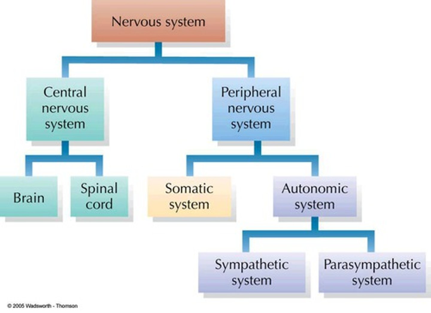
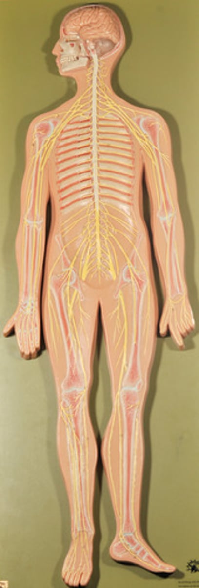
Draw the organization of the Nervous System
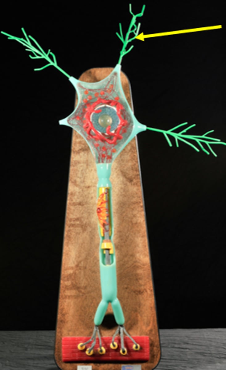
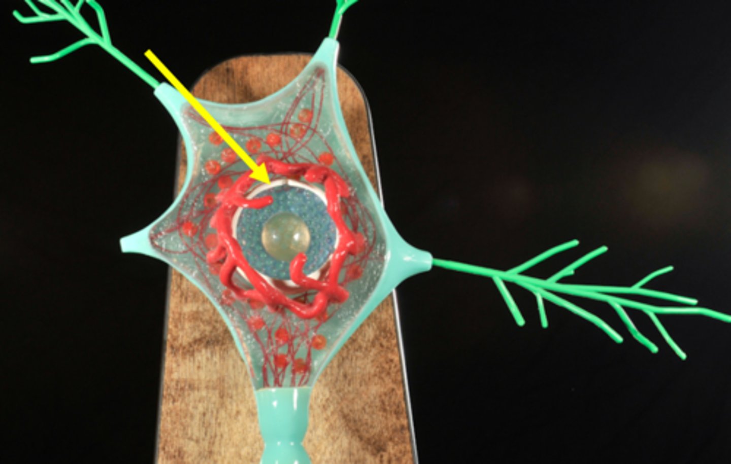
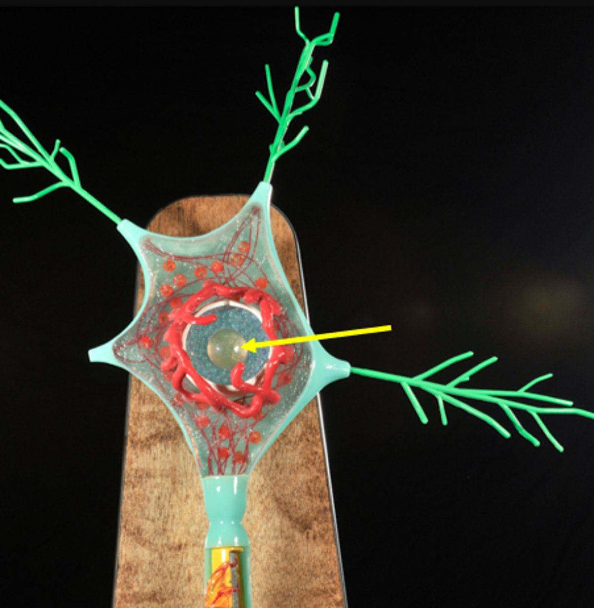
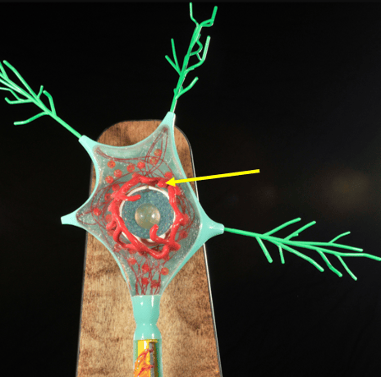
Identify all structures on the Motor Neuron
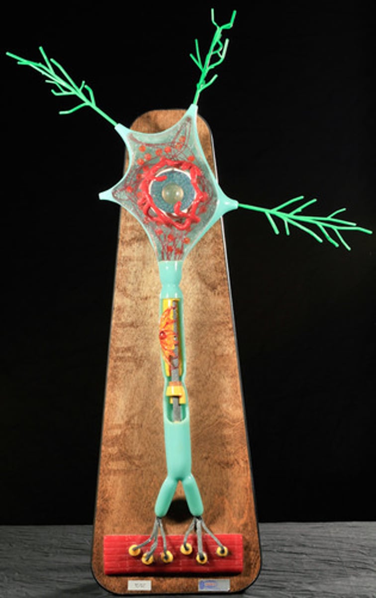
Dendrites
Cell body of neuron
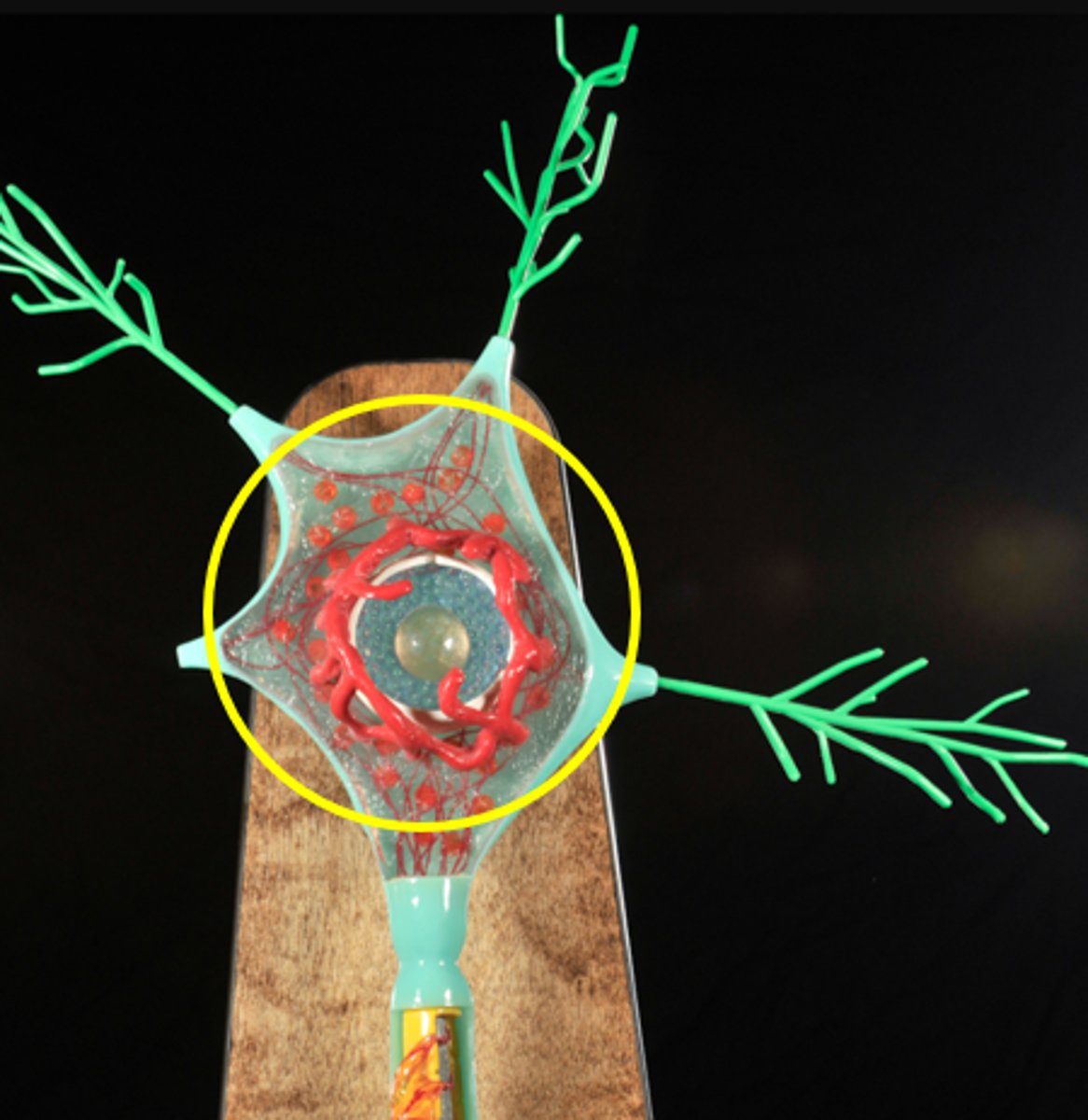
Nucleus of Neuron
Nucleolus of neuron
Chromatophilic substance (Nissl bodies)
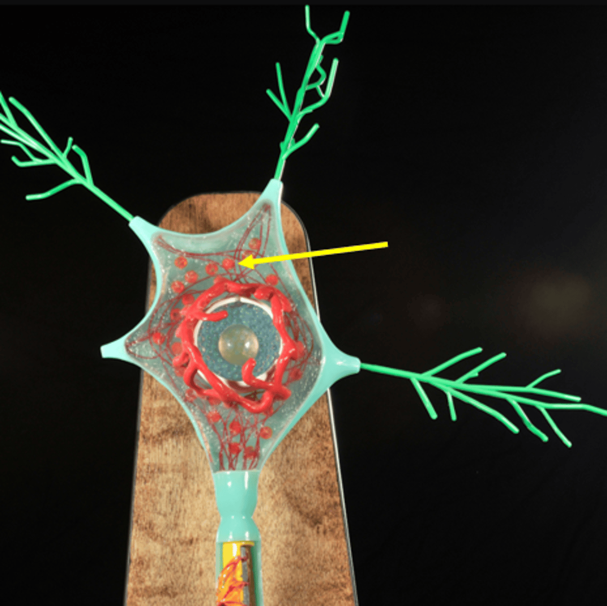
Neurofibrils
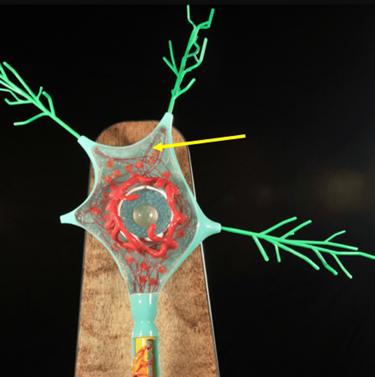
Golgi apparatus of neuron
Axon hillock
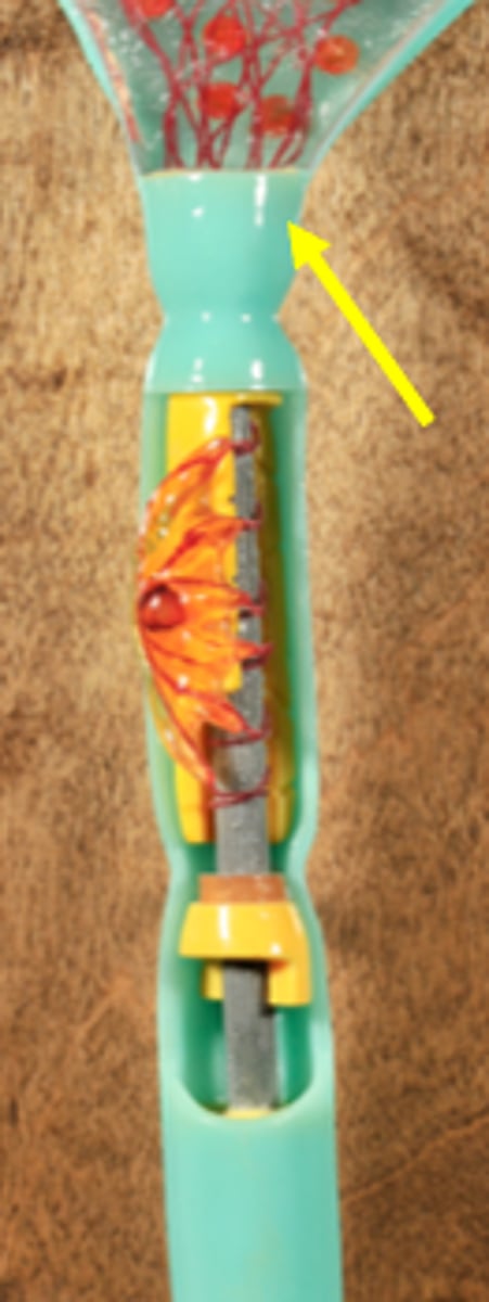
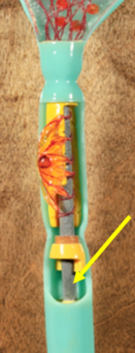
Axon
Axon (presynaptic) terminals
Myelin sheath
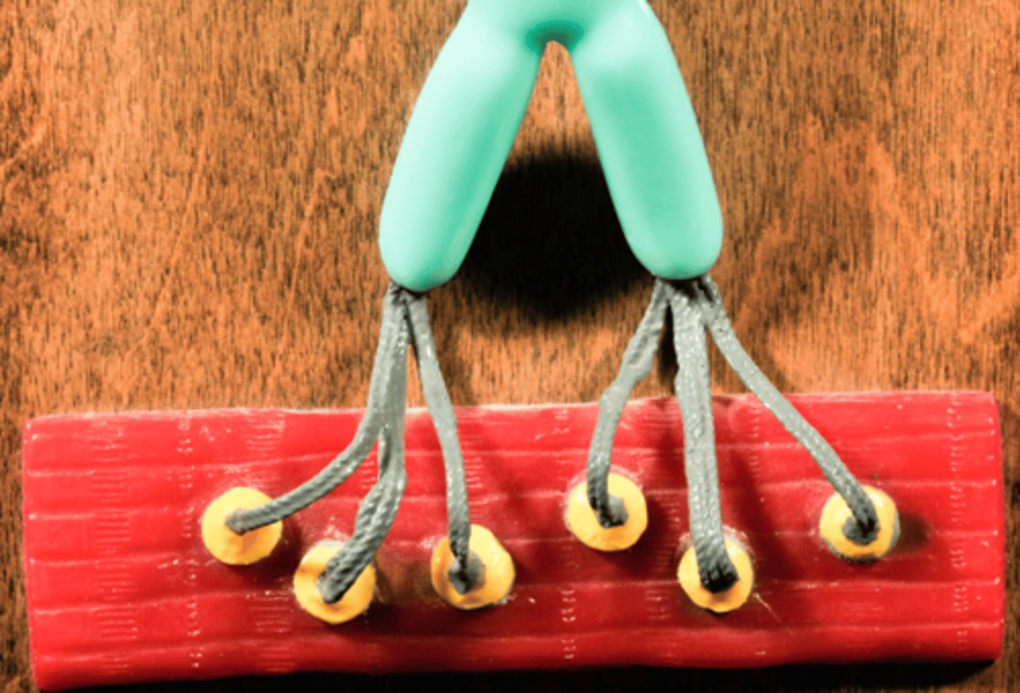
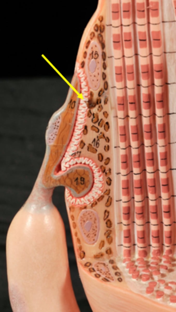
Identify all structures of Neuromuscular junction

Skeletal muscle fiber

Motor neuron
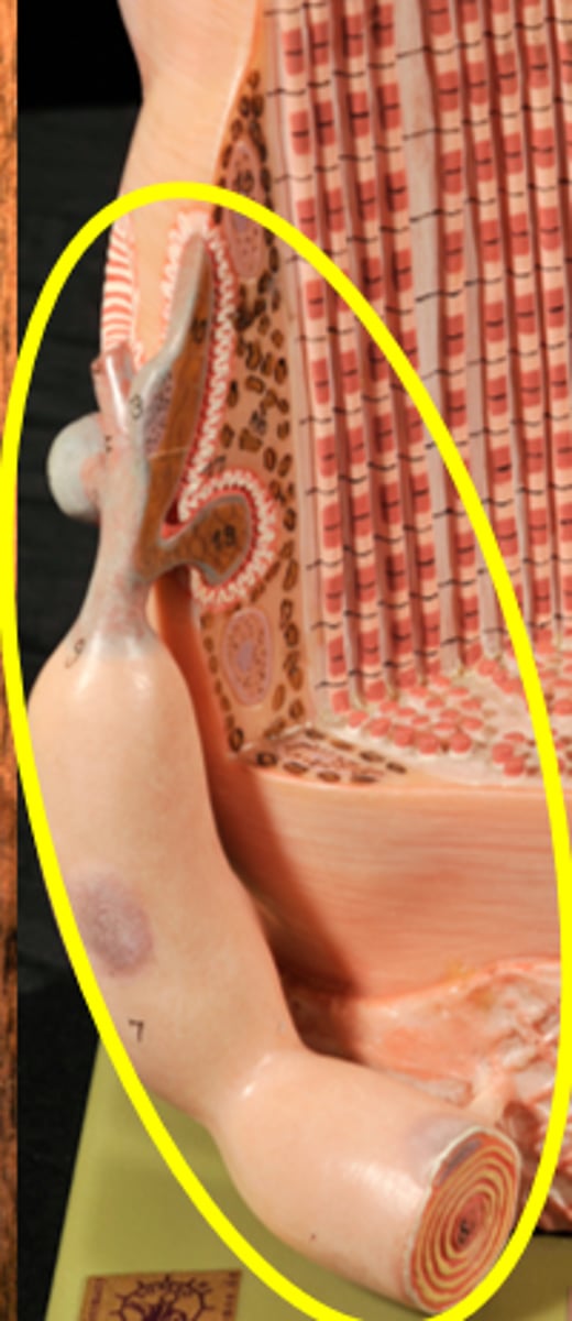
Axon
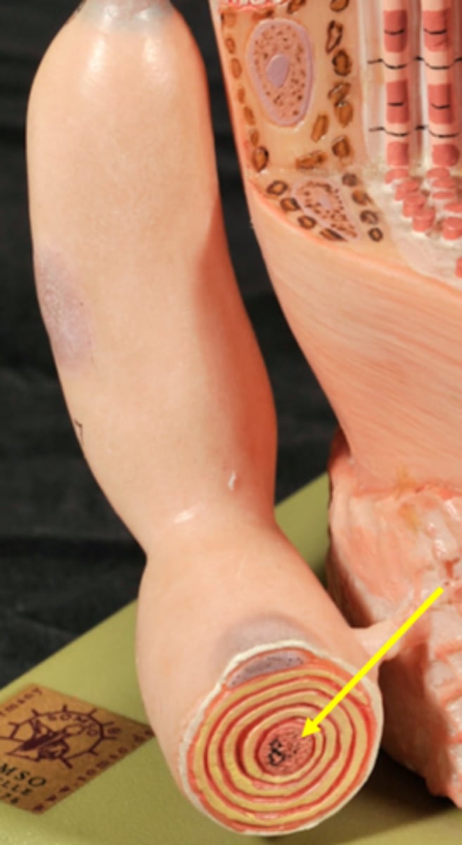
Myelin sheath
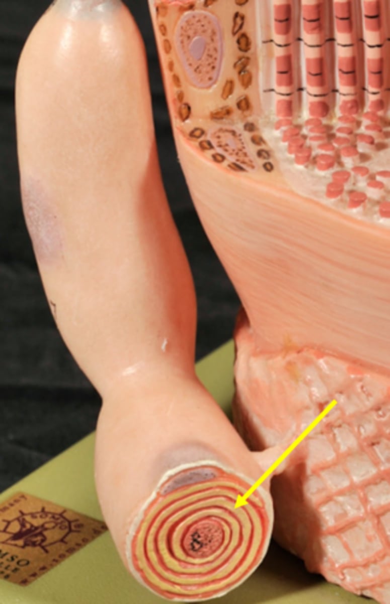
Schwann cells
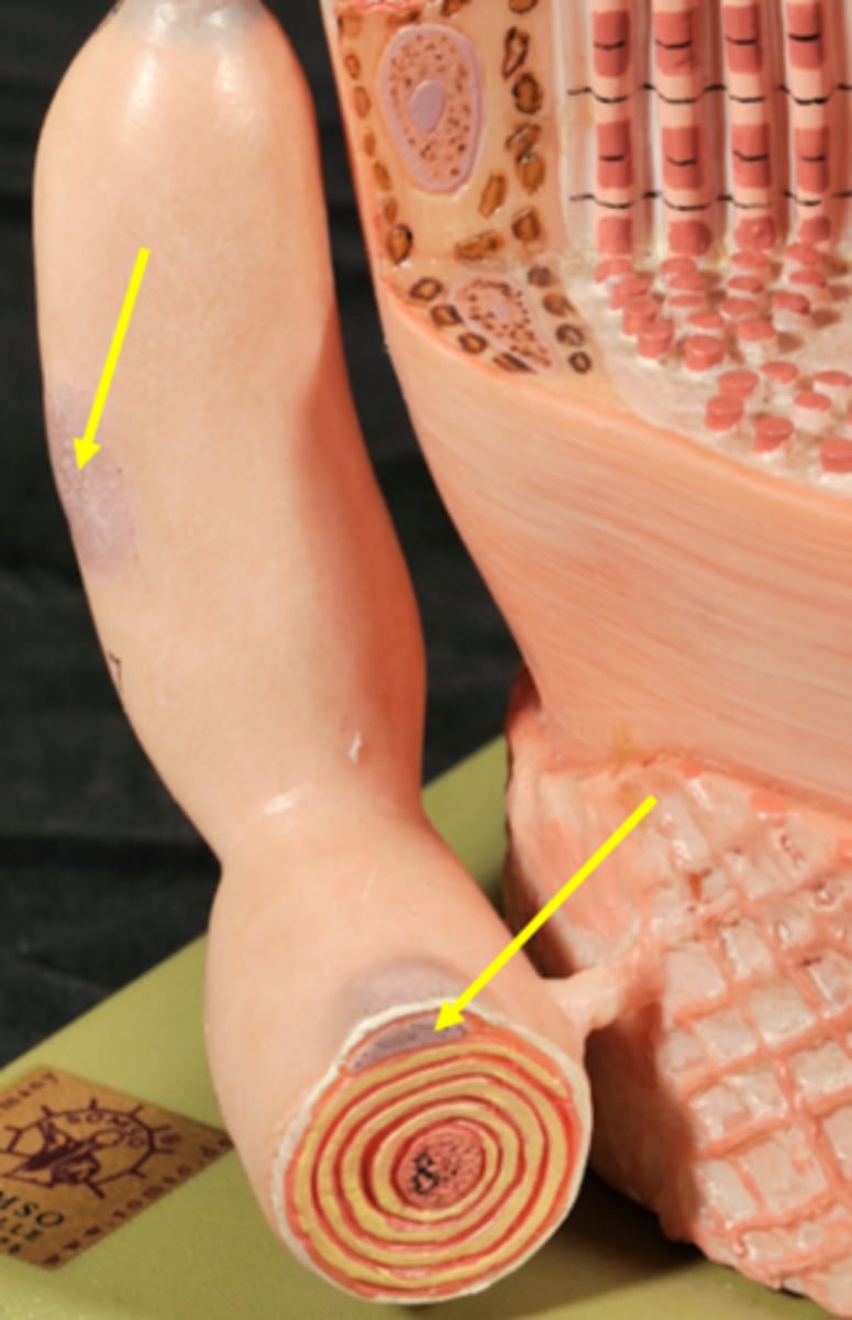
Axon (presynaptic) terminals
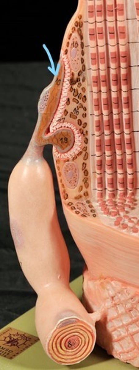
Synaptic Cleft
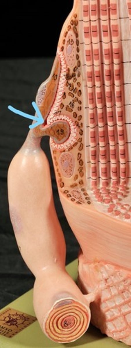
Postsynaptic membrane
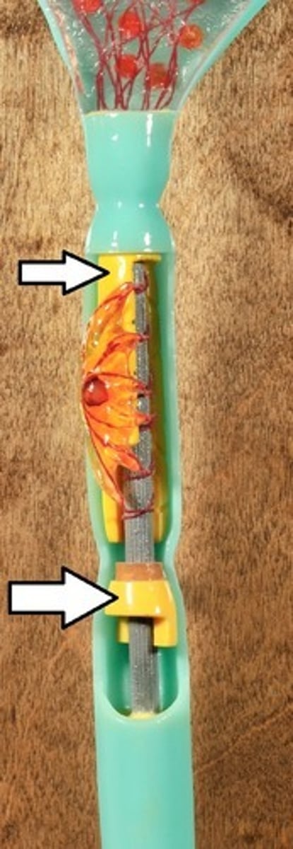
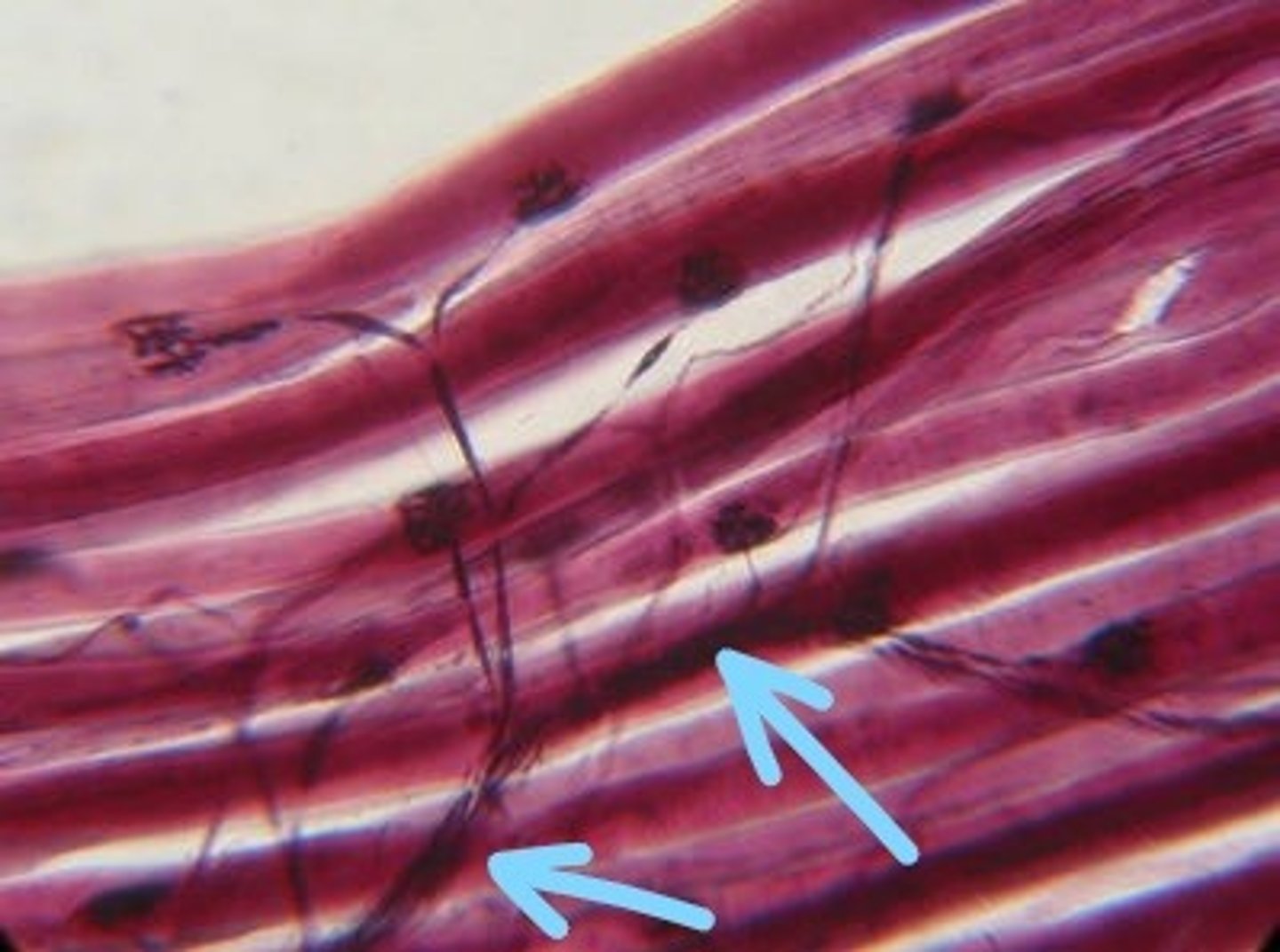
Identify all structures on the neuromuscular microscope slide
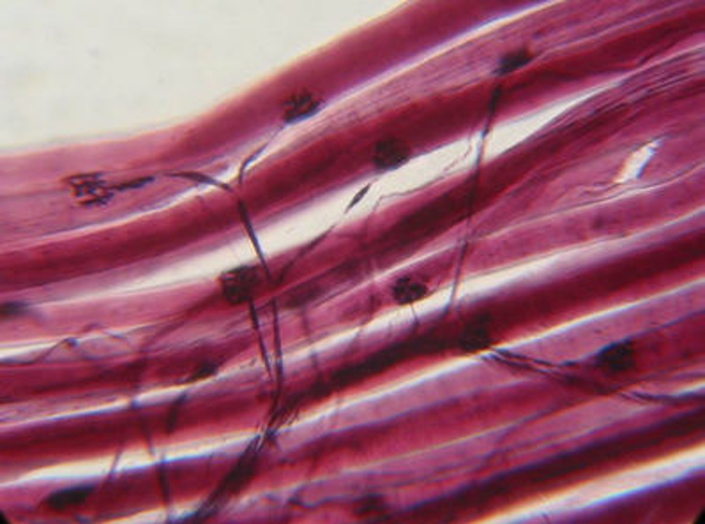
Skeletal muscle
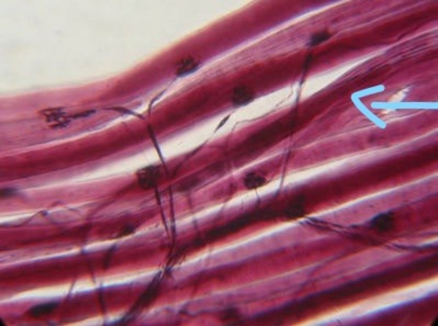
Axon Presynaptic terminals
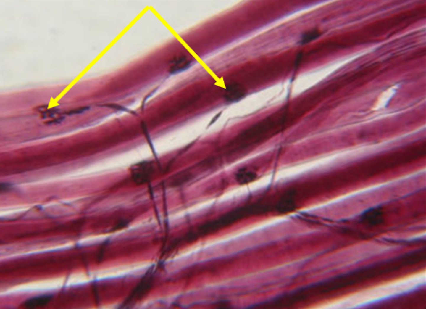
Axon
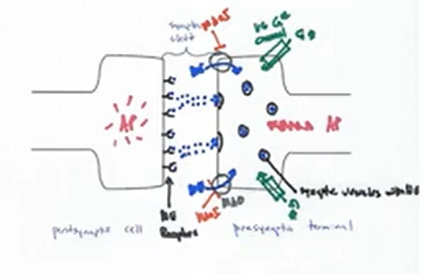
Draw and label a Synapse
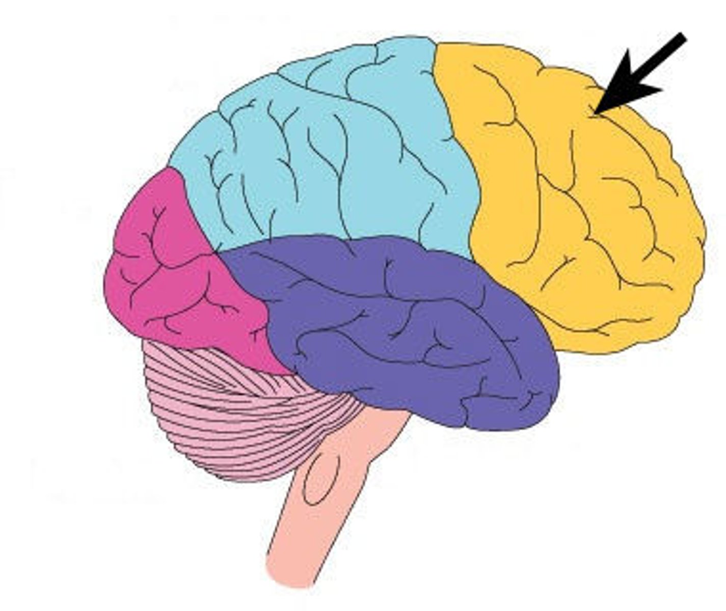
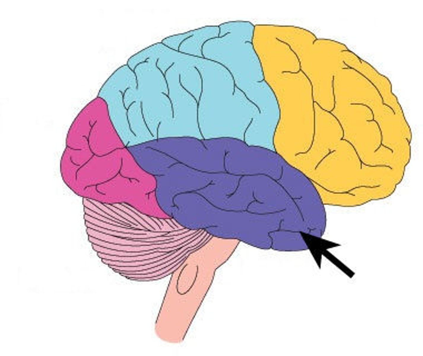
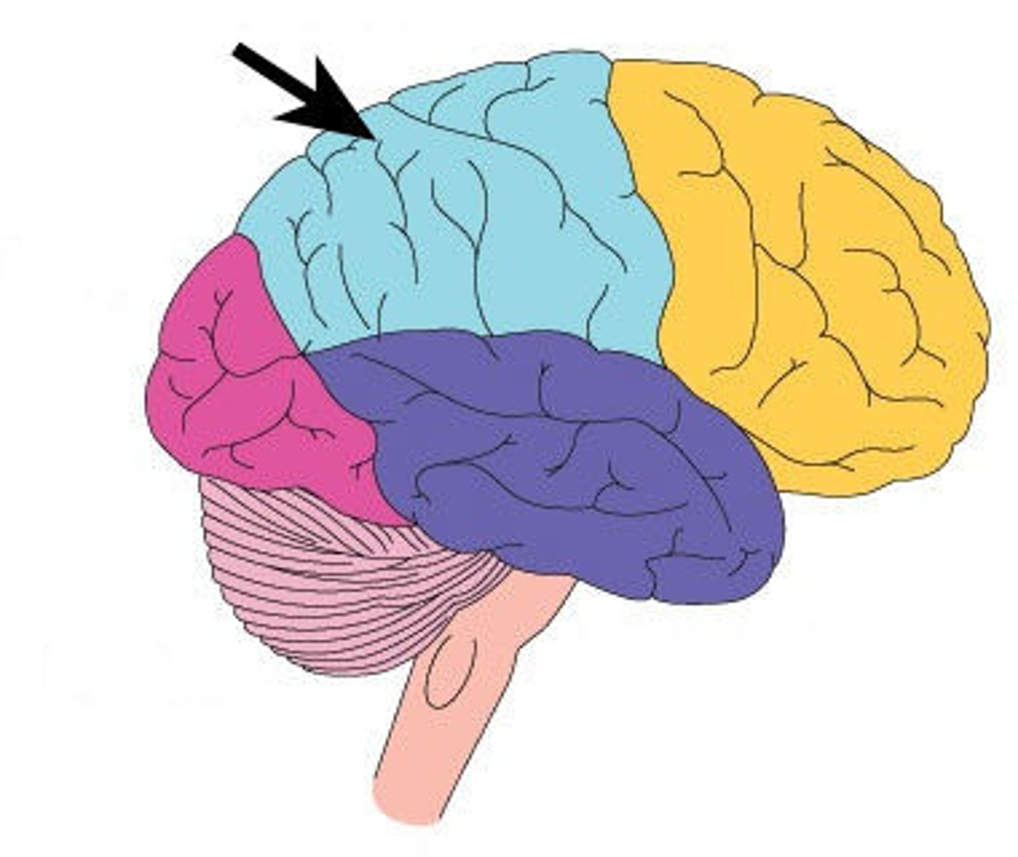
Identify structures of brain
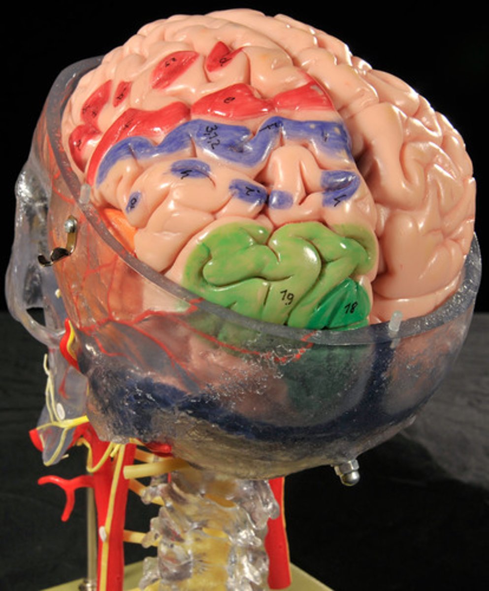
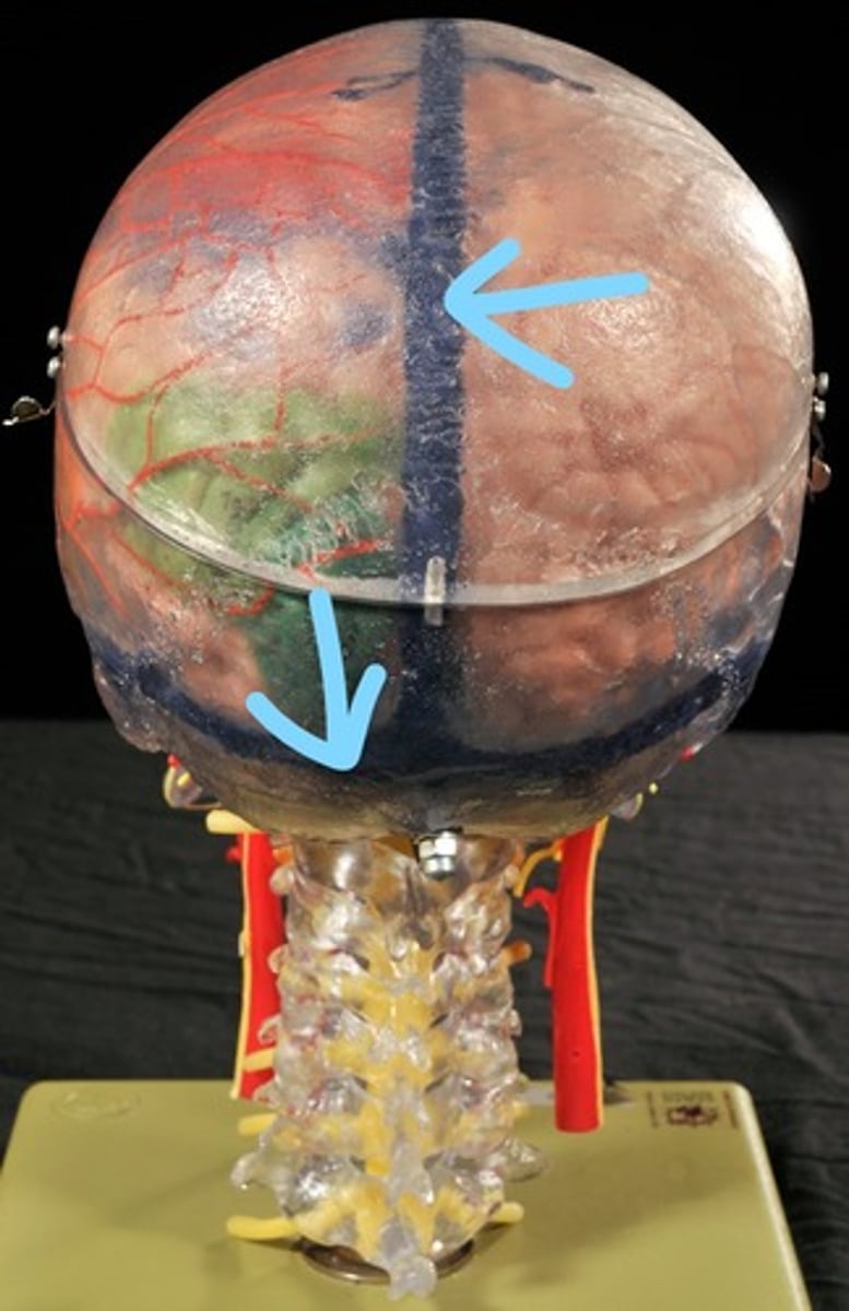
Longitudinal fissure

Cerebral cortex (gray matter)
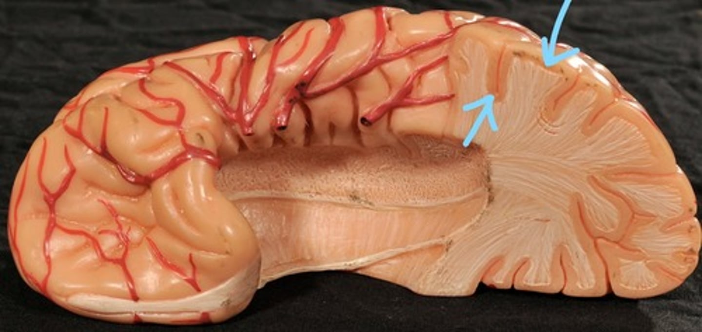
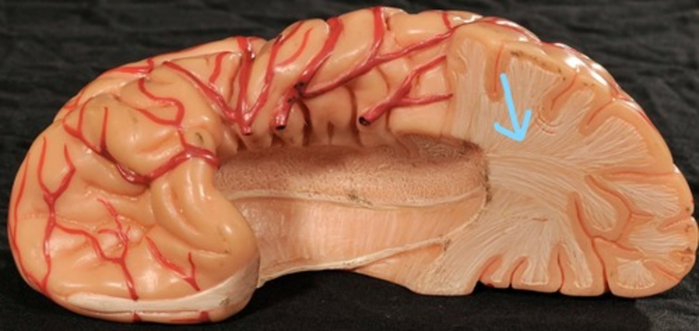
Cerebral Medulla (white matter)
Frontal lobe
Temporal lobe
Parietal lobe
Precentral gyrus
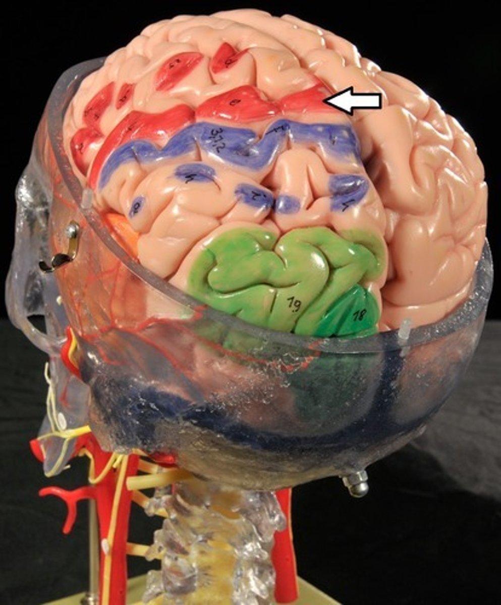
Postcentral gyrus

Central sulcus

Cerebellum
A fly landed on your hand: What area in the brain would sensory information from your hand need to travel to?
Other lobes/ Parietal lobes/ Central Cerebral Cortex
A fly landed on your hand: What area of the brain would the motor signals come from to flick the fly away?
Frontal lobe
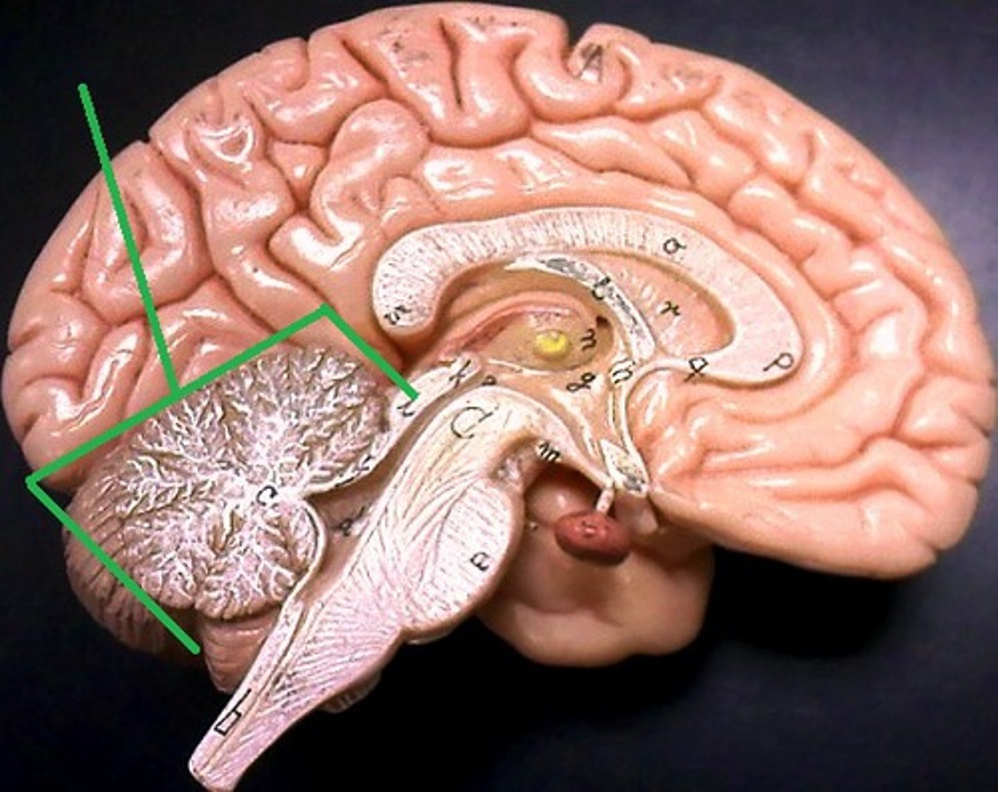
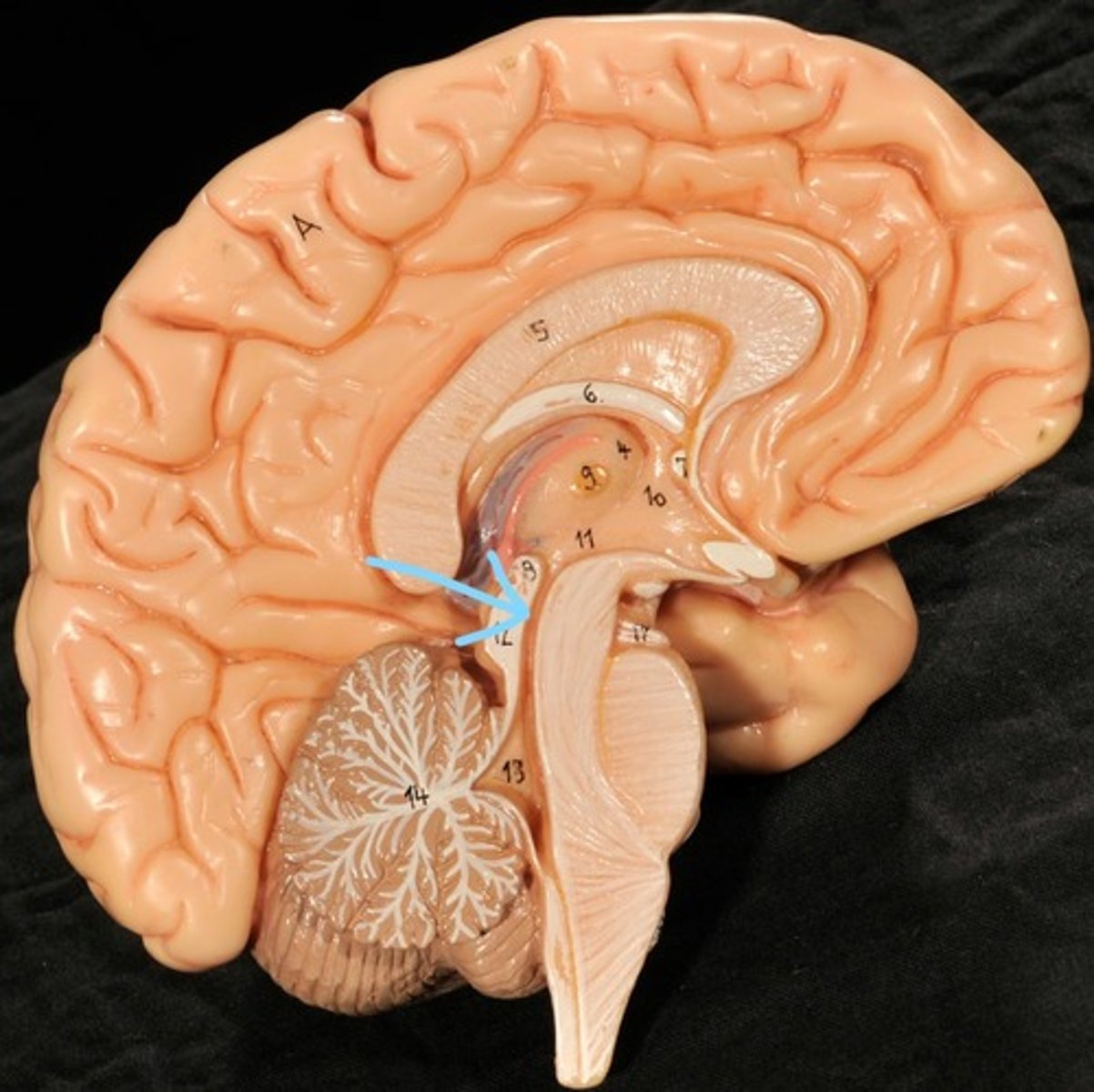
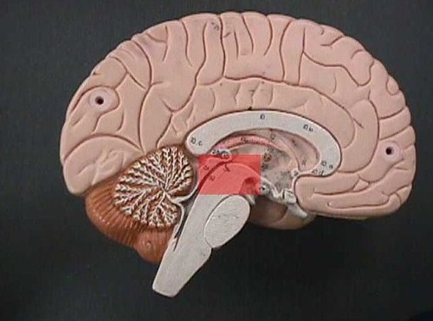
Identify structures of midsagittal brain
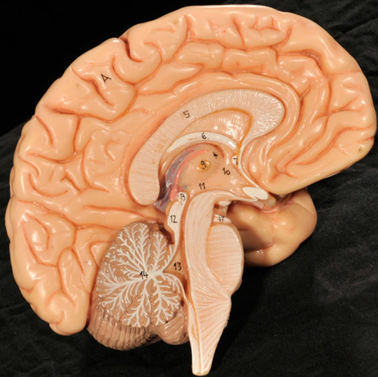
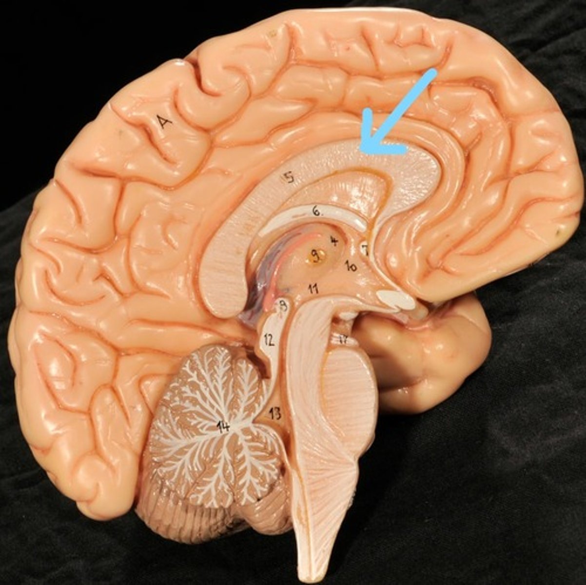
Corpus callosum
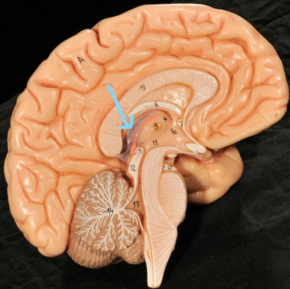
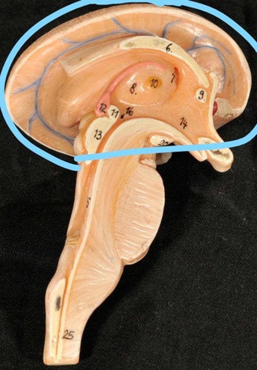
Diencephalon
thalamus, third ventricle, hypothalamus, epithalamus
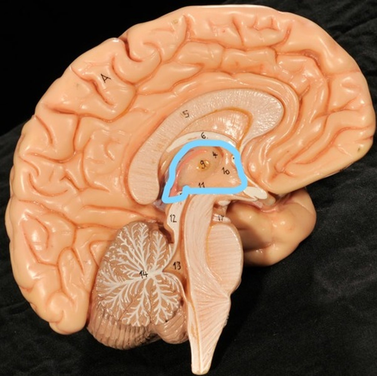
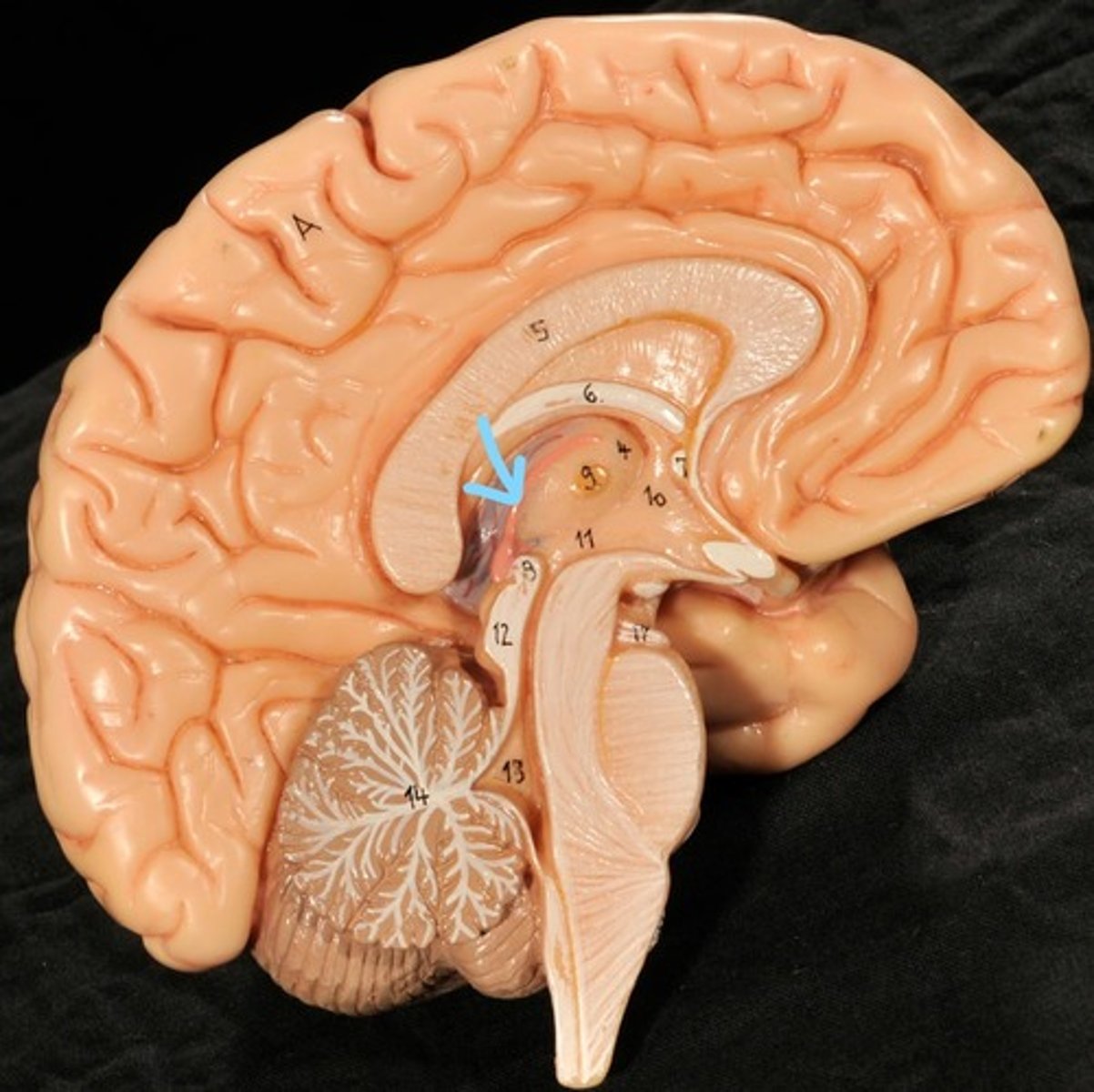
Thalamus
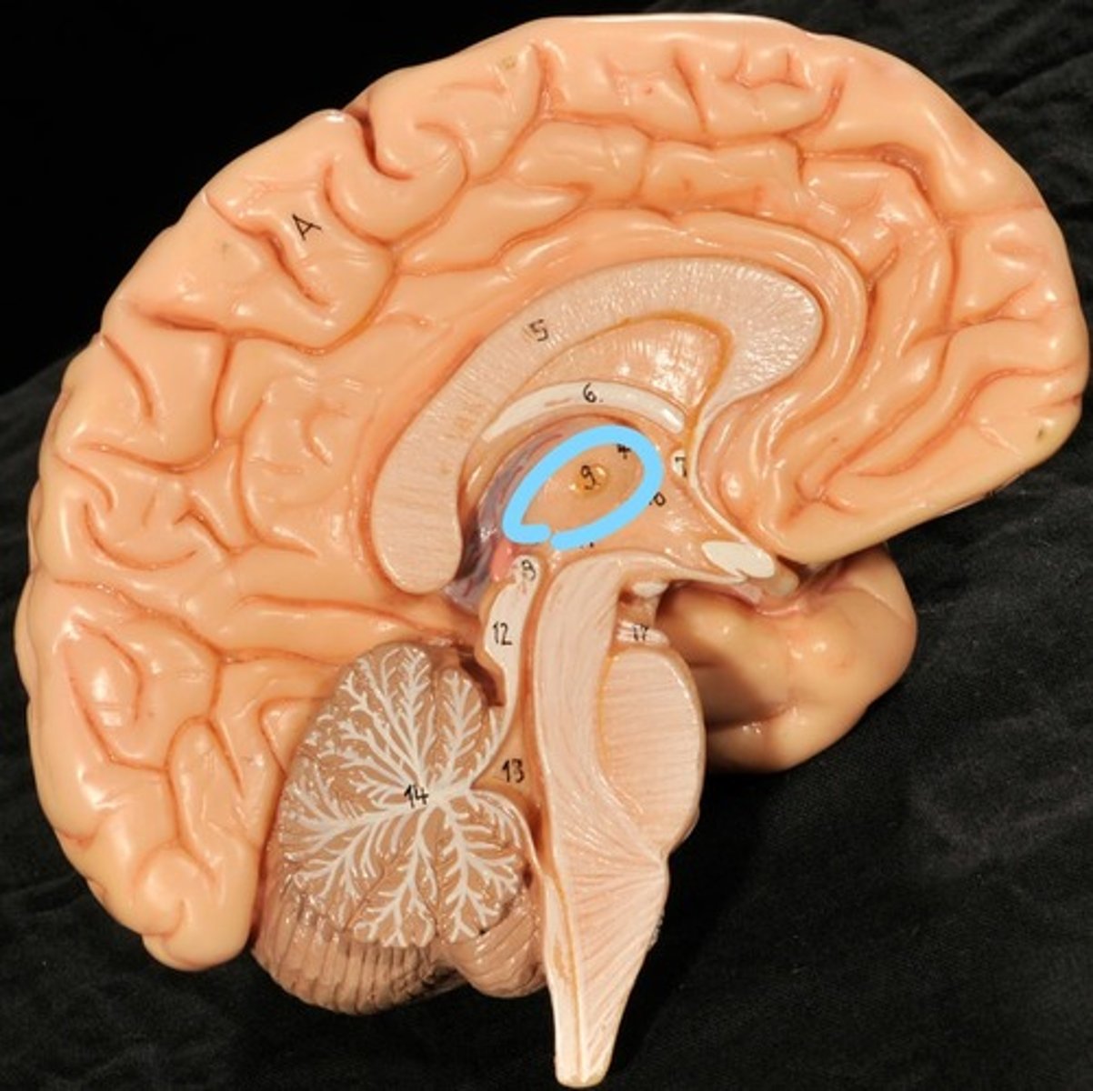
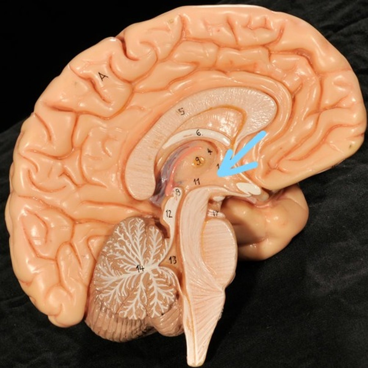
Third Ventricle
Hypothalamus
Epithalamus (pink line)
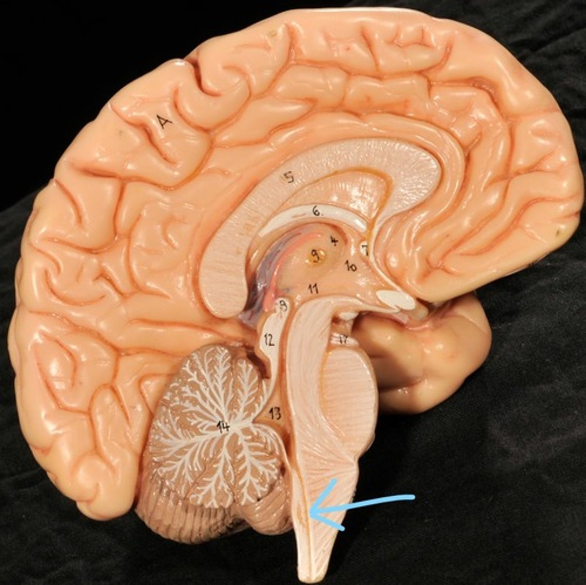
Brain stem
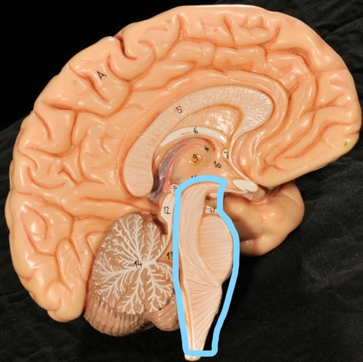
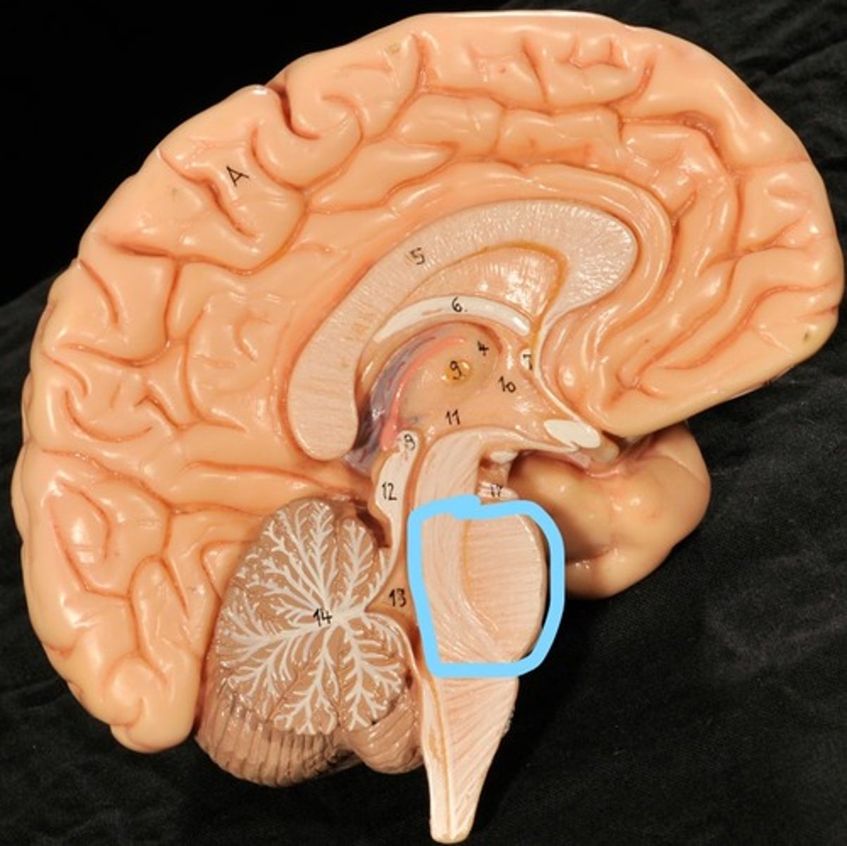
Pons
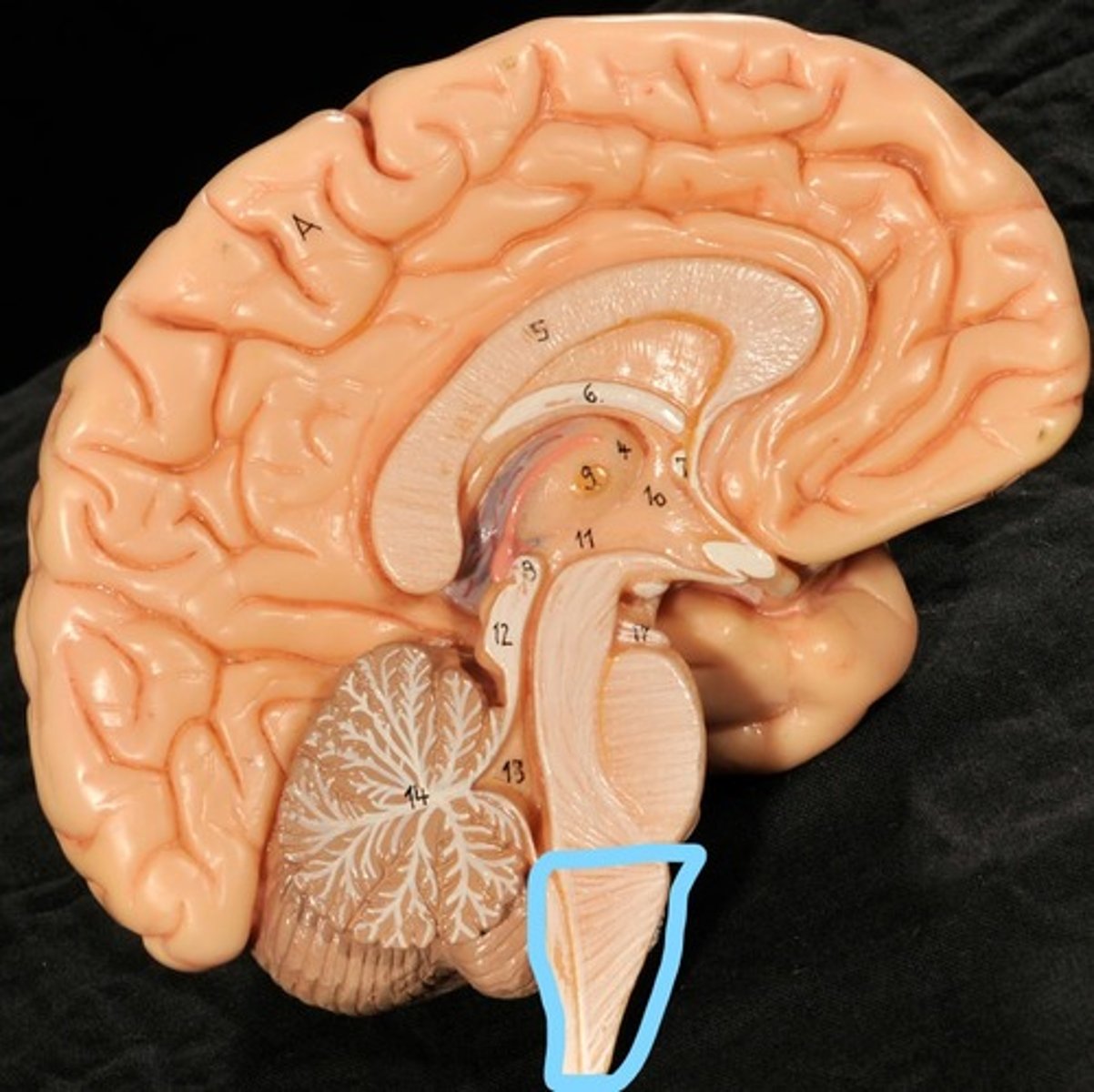
Medulla oblongata
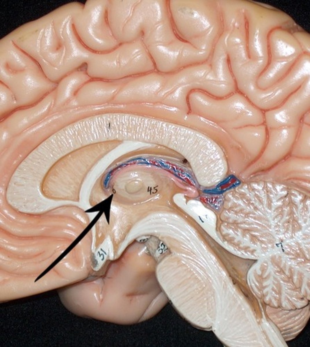
choroid plexus of third ventricle
produces cerebrospinal fluid

Third ventricle
cerebral aqueduct (midbrain)
midbrain
Fourth ventricle

Central Canal of brain
Takes Cerebrospinal fluid down to the spinal cord
Thalamus
Identify the structures
Left Lateral Ventricle
Right Lateral Ventricle
Anterior horn of ventricle
Posterior horn of Ventricle
Inferior horn of ventricle
Third Ventricle
Cerebral Aqueduct
Fourth Ventricle
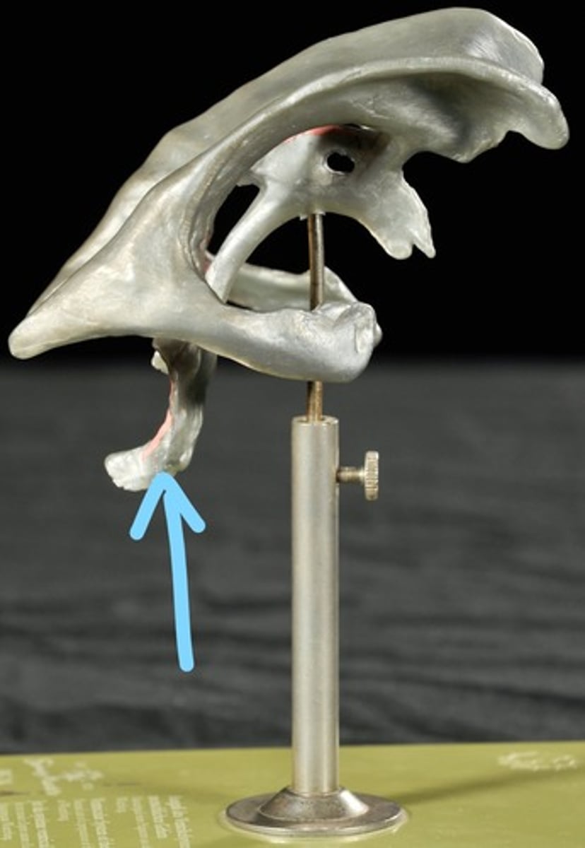
Dural Sinuses
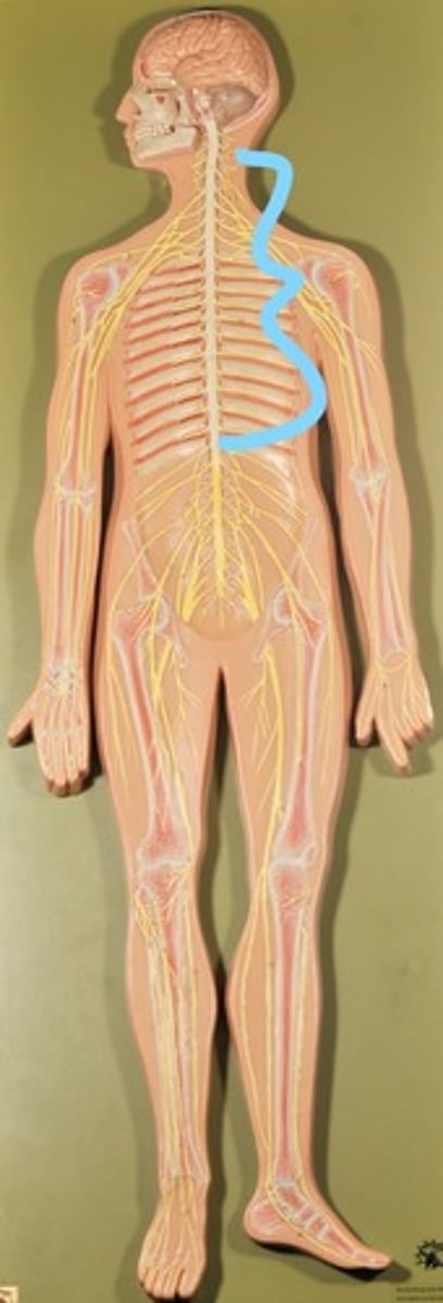
Spinal cord
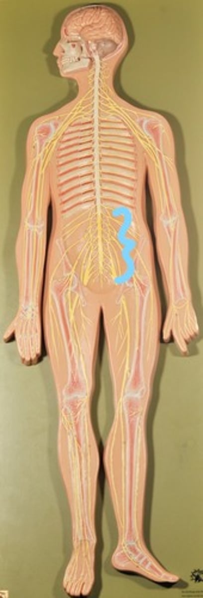
Cauda Equina
Dura Mater (outermost meninge)
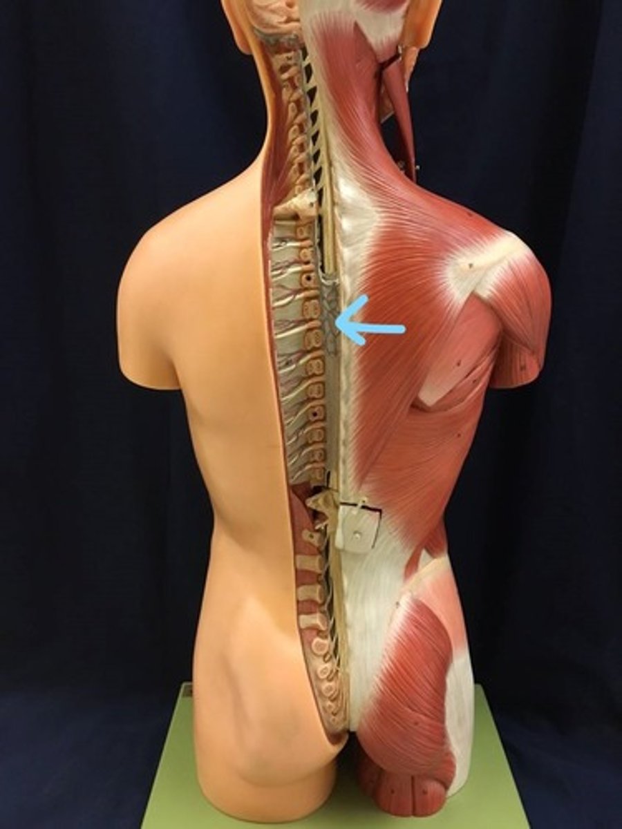
Identify all structures
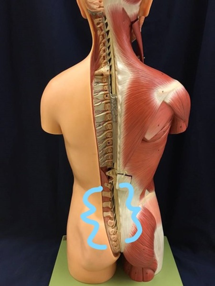
Spinal Cord (in white)
Cauda Equina
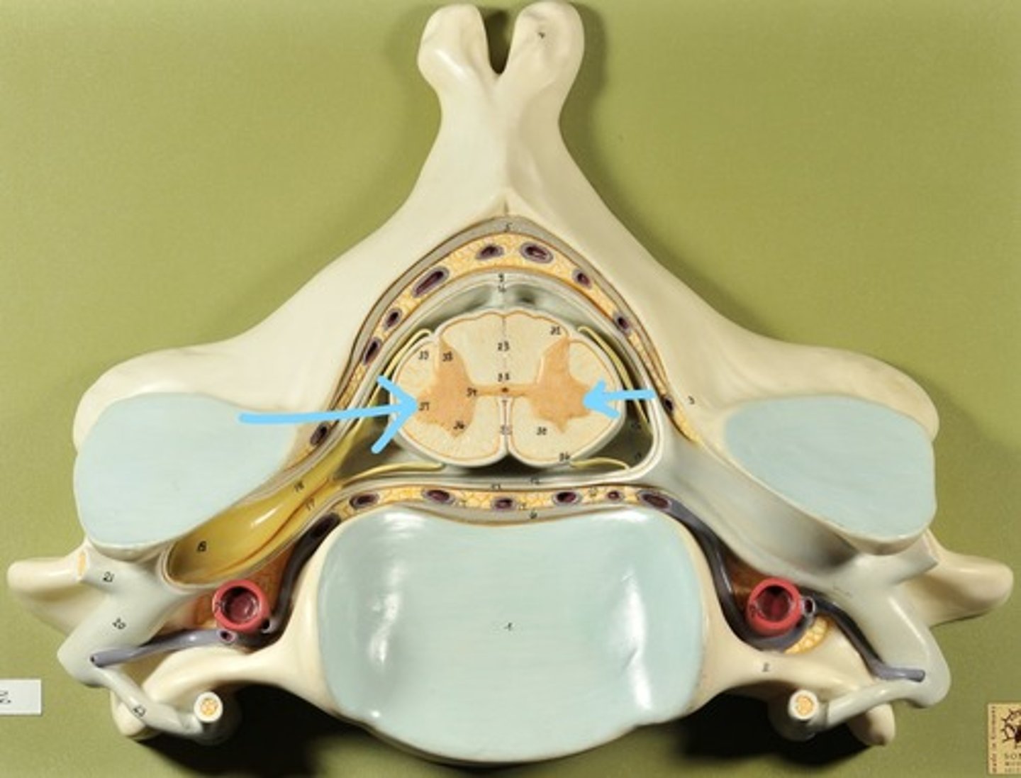
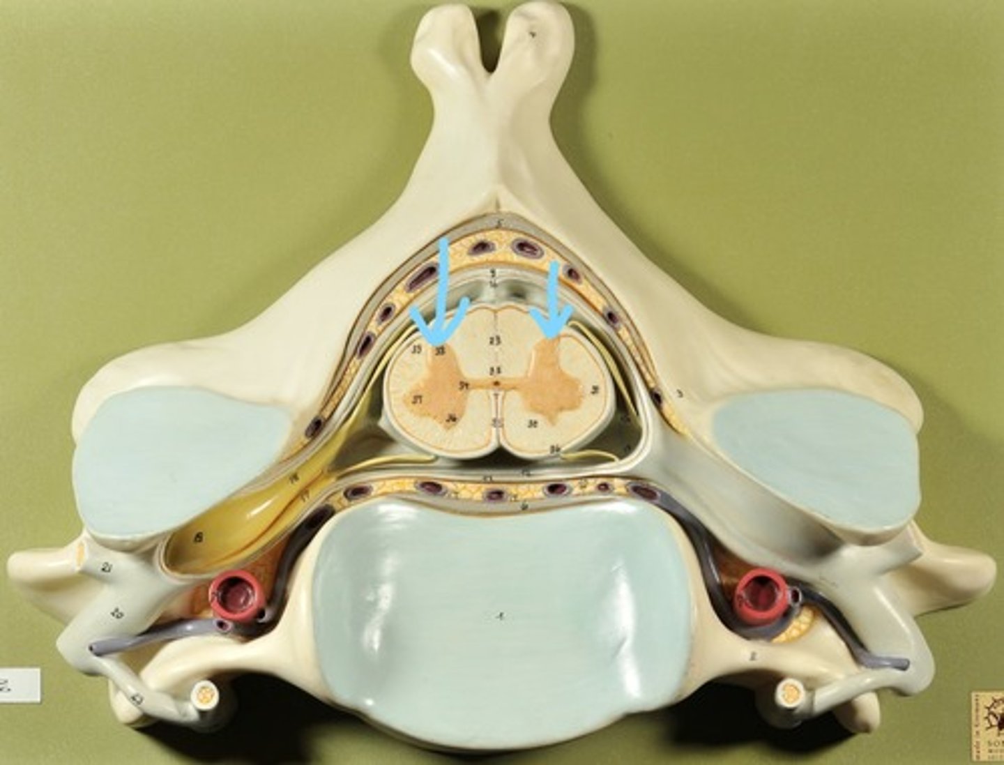
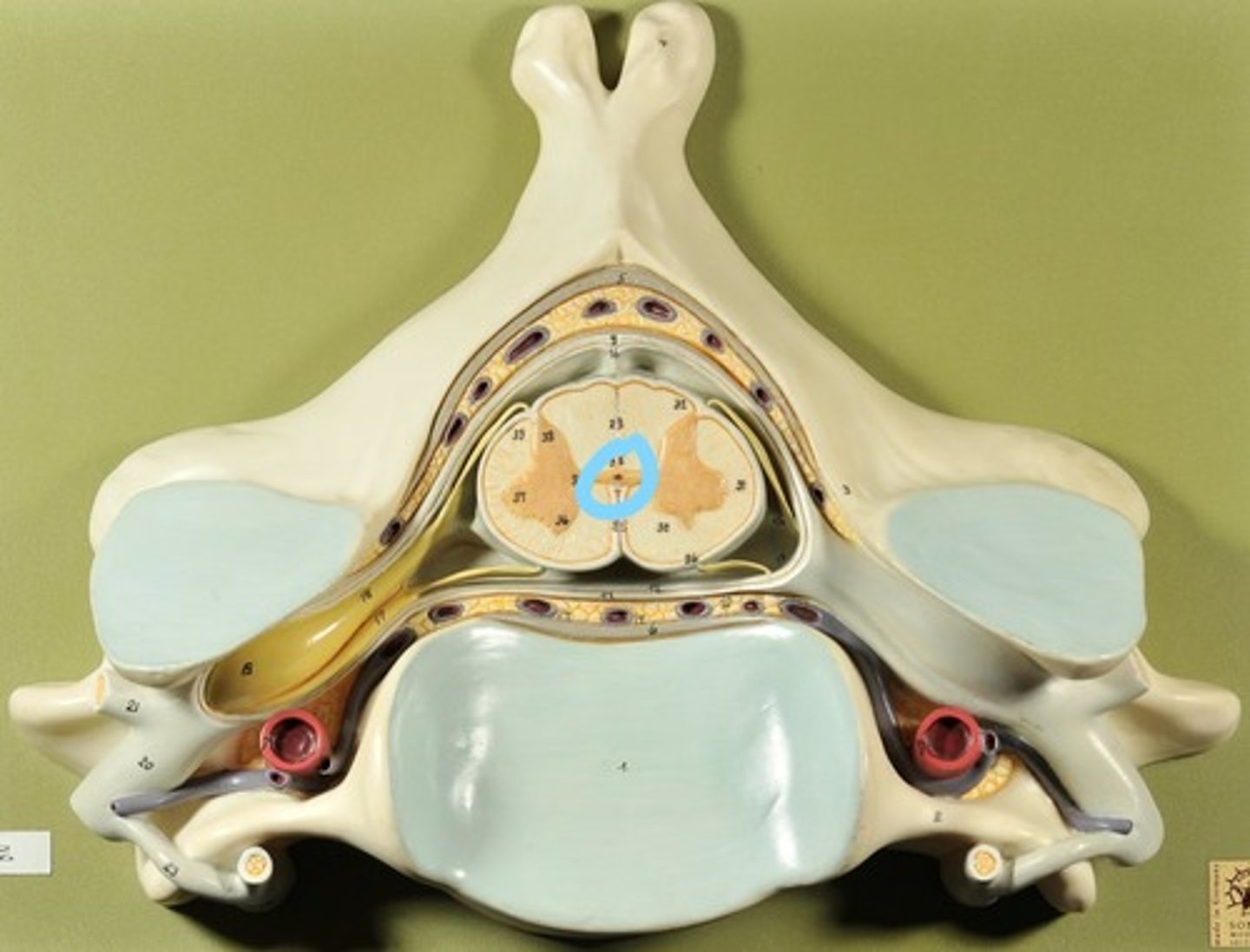
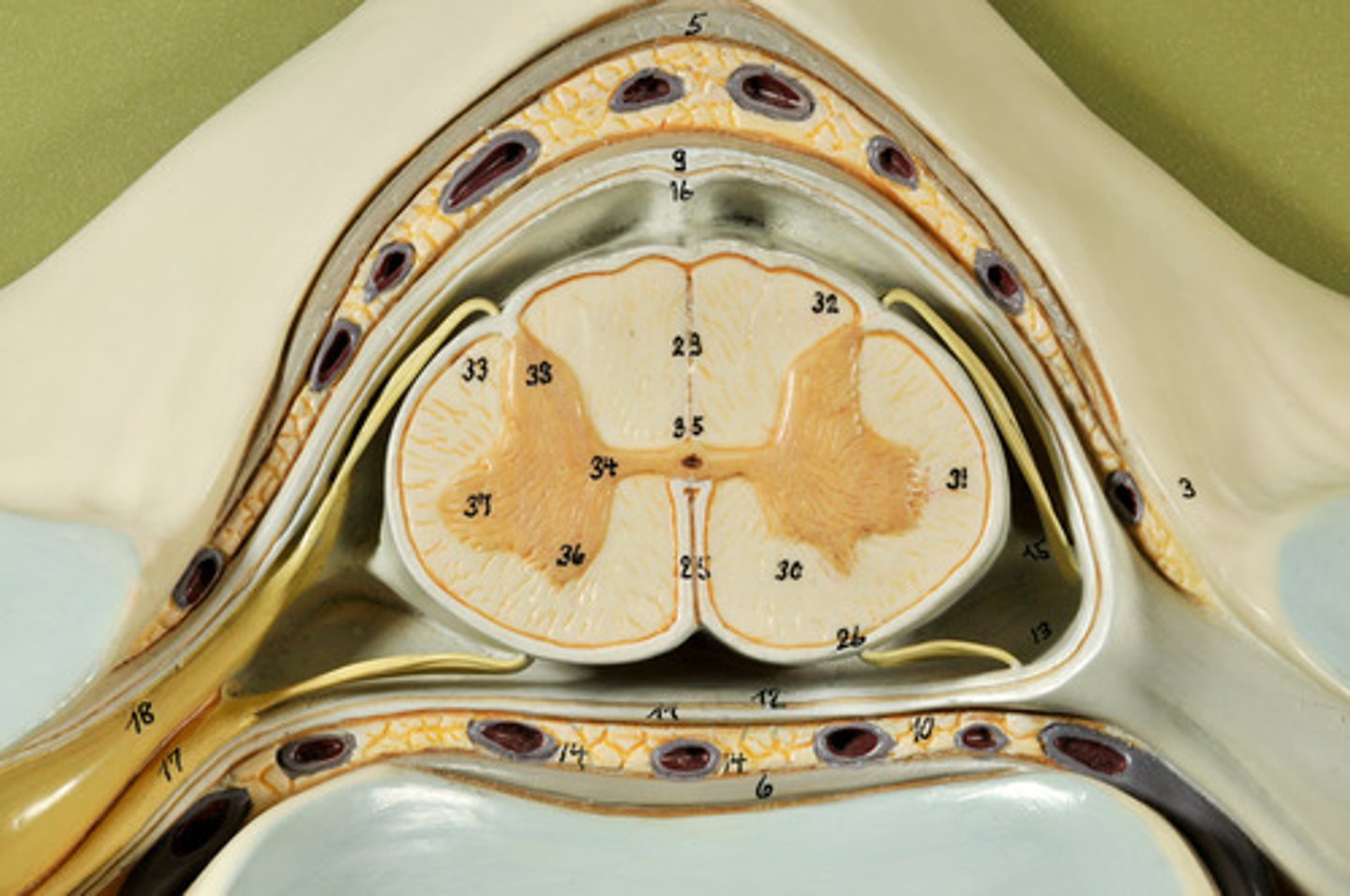
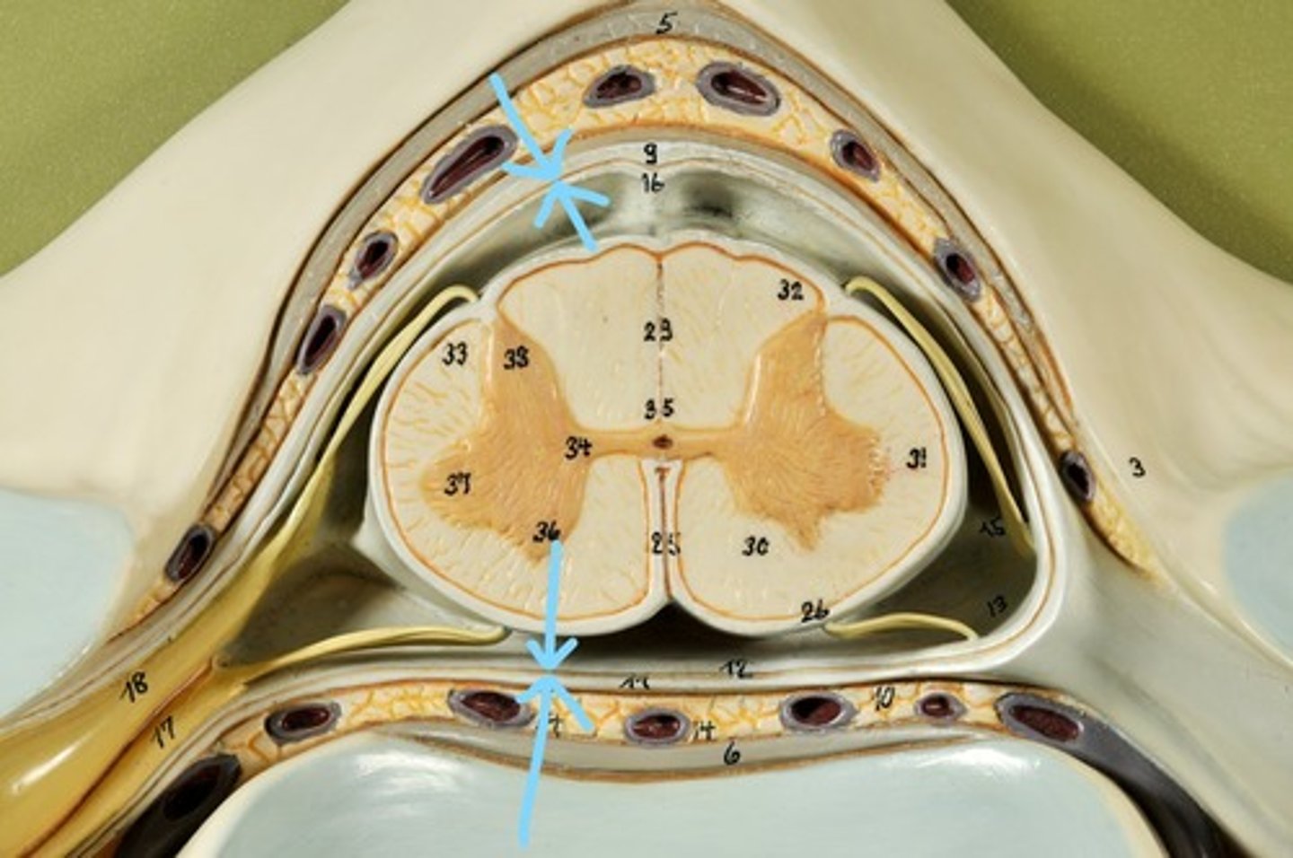
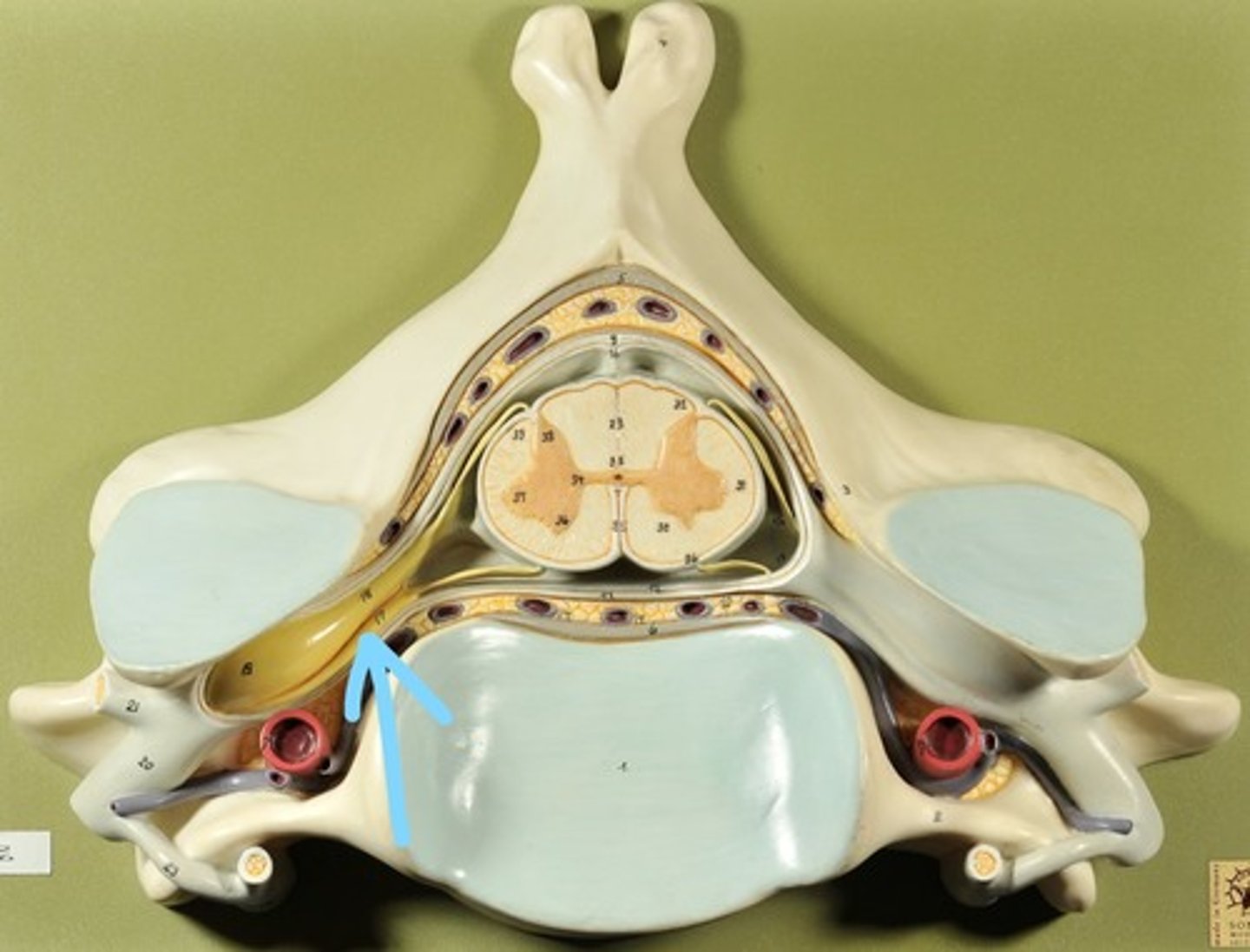
Identify all structures of spinal cord

White Matter of spinal cord
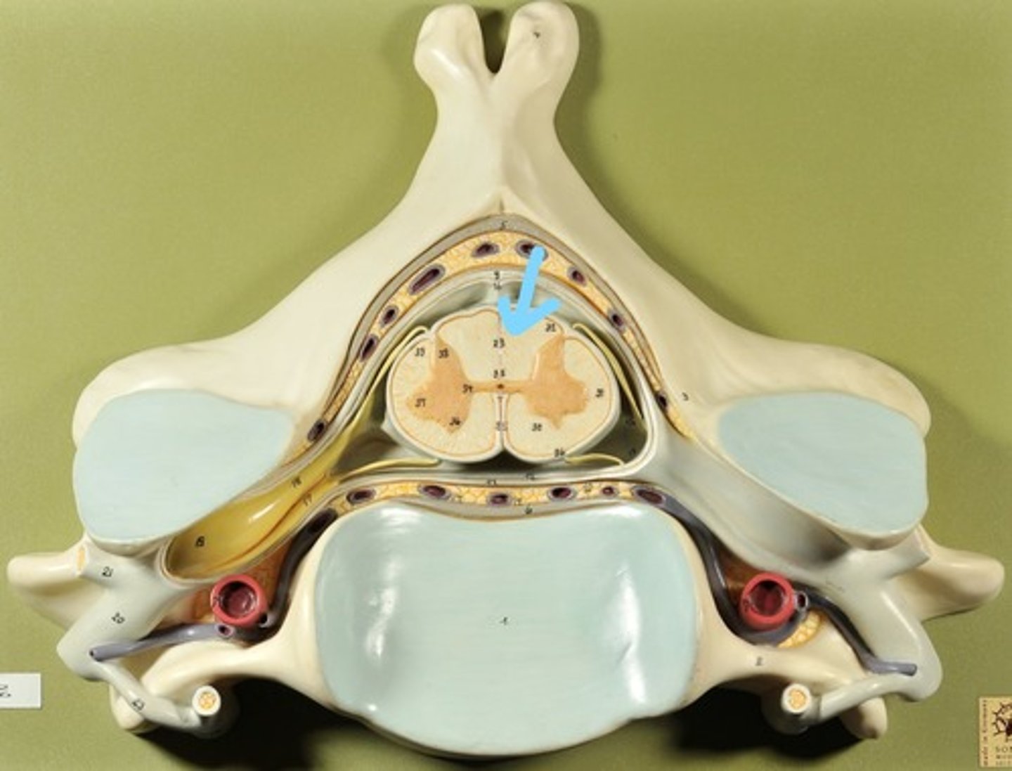
gray matter of spinal cord

Ventral Horns (gray matter)

Lateral Horns (gray matter)
Dorsal horns (gray matter)
Anterior Columns (white matter)

Lateral Columns (white matter)

Posterior Column (white matter)
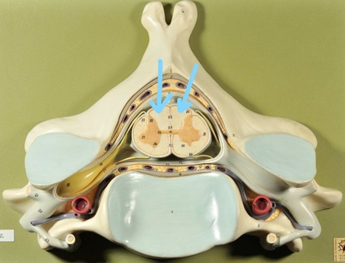
central canal of spinal cord
Identify all structures
Pia Mater

Subarachnoid Space
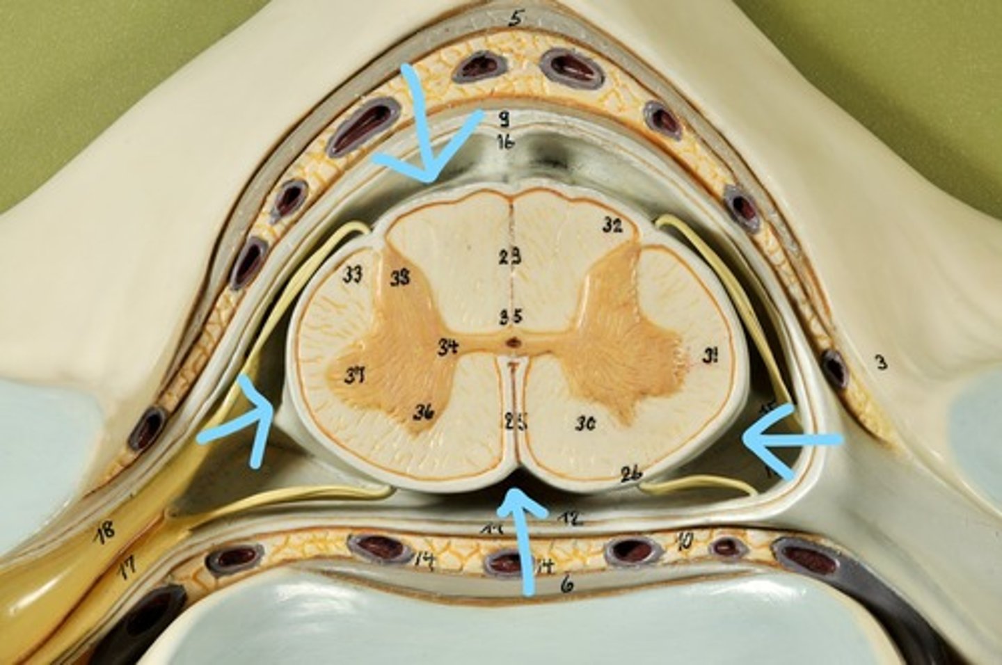
Arachnoid mater
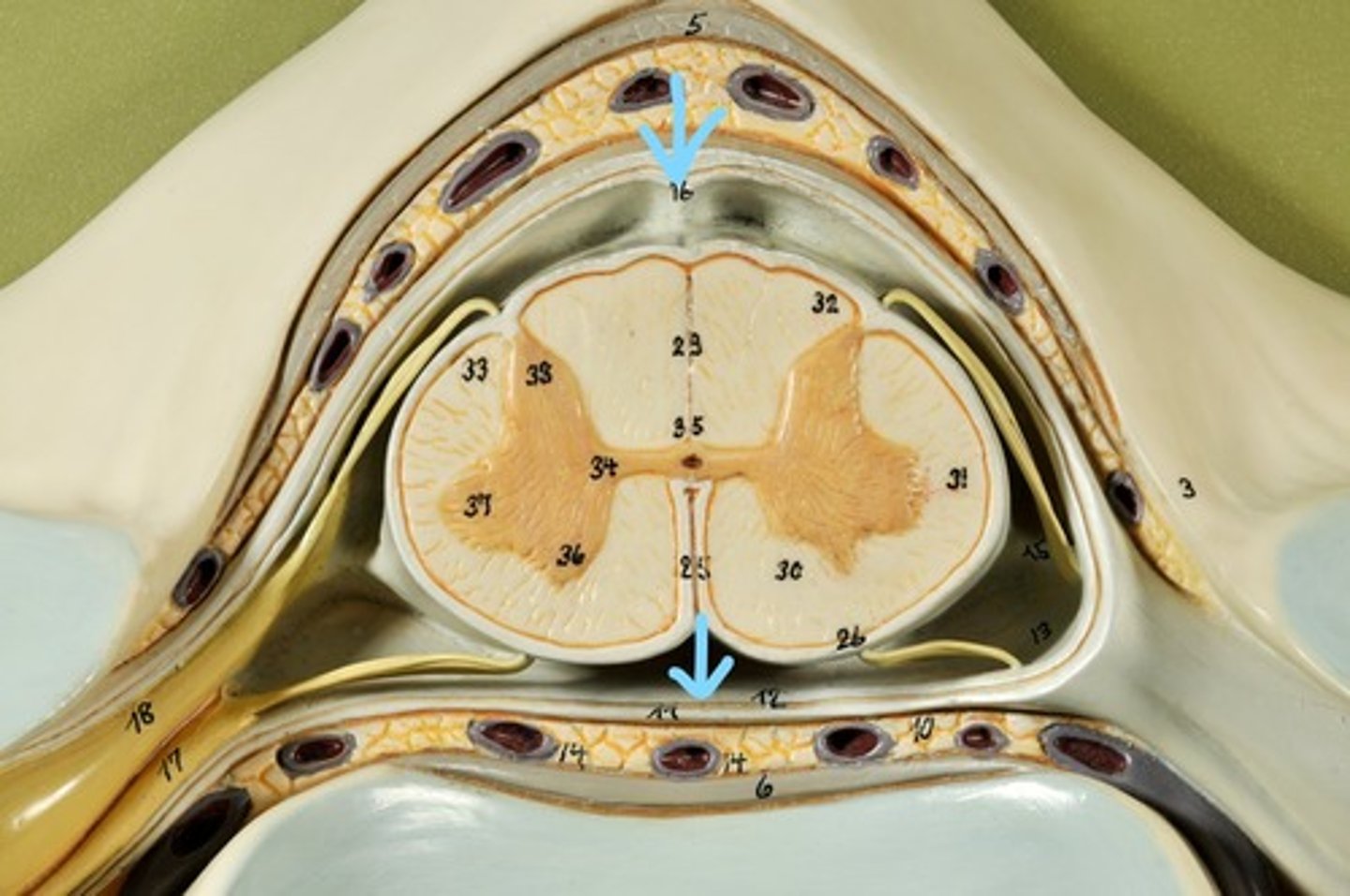
Subdural Space (brown line)
Dura mater
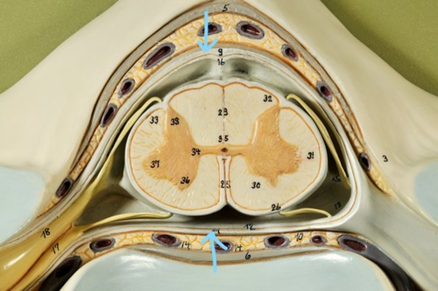
Epidural Space
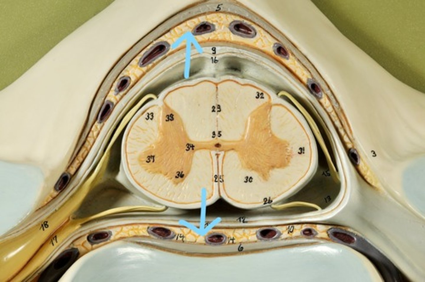
Dorsal Root Ganglion
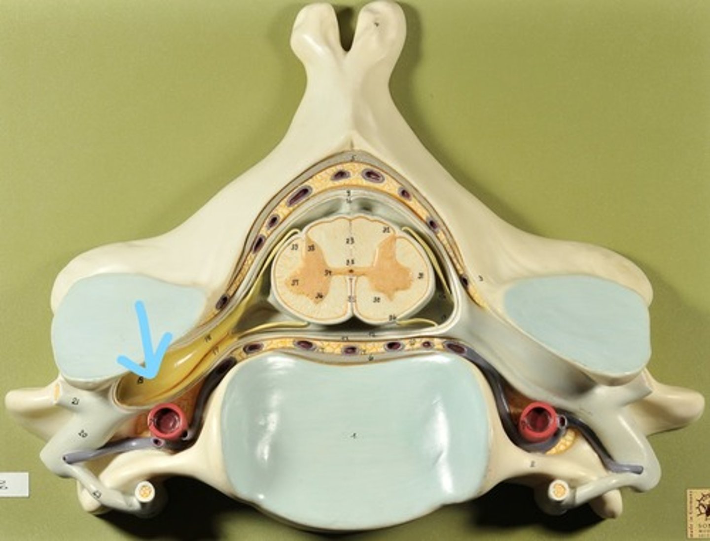
Dorsal Root
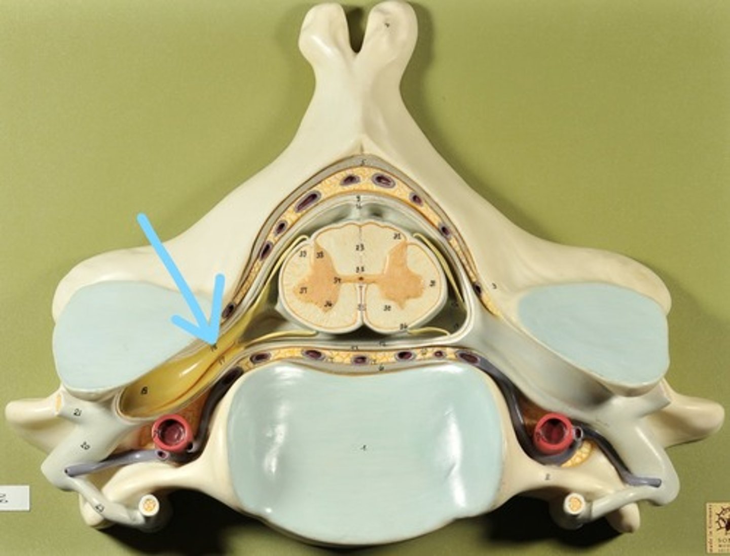
Ventral Root
Spinal Nerve

Cerebral Cortex (gray matter) (Slice 2)
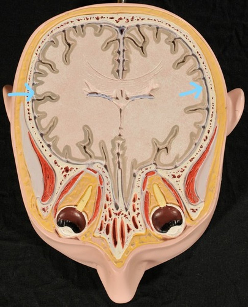
Cerebral Medulla (white matter) (Slice 2)

Right & Left Parietal Lobes (Slice 2)
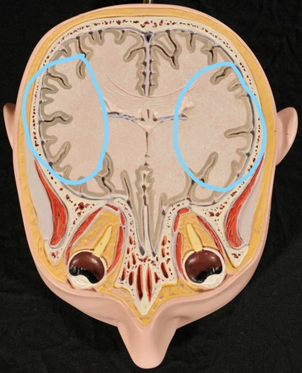
Third Ventricle (Slice 2)
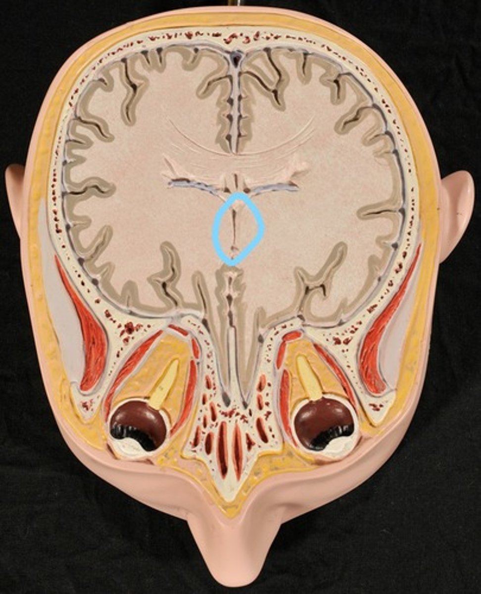
Right & Left Lateral Ventricles (Slice 2)
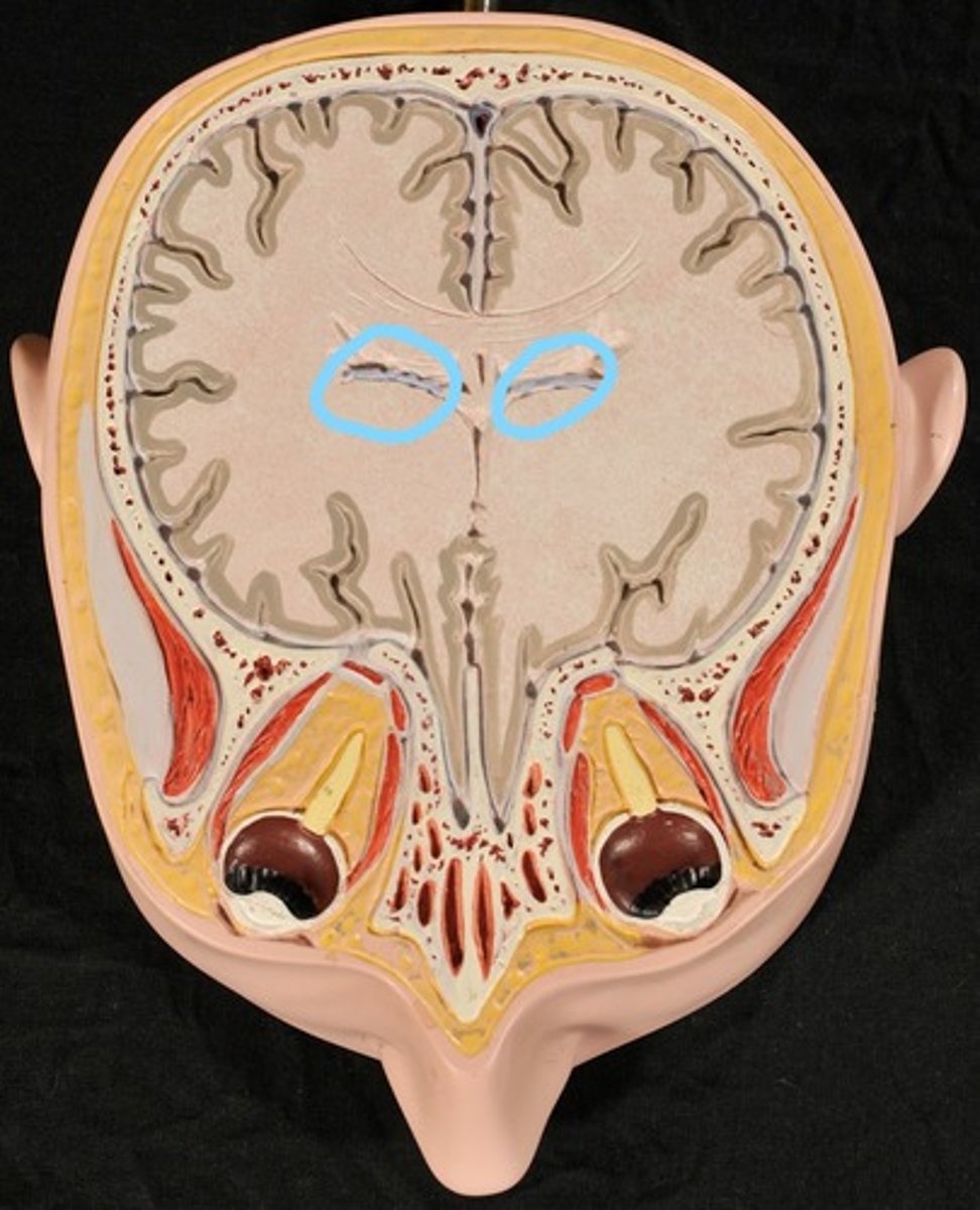
Choroid Plexus (blue) (Slice 2)
