Skull osteology
 Call Kai
 Learn
Learn Practice Test
 Spaced Repetition
 Match
Match Flashcards
 Knowt Play
1/36

There's no tags or description
Looks like no tags are added yet.
Name | Mastery | Learn | Test | Matching | Spaced | Call with Kai | Chat |
|---|
No analytics yet
Send a link to your students to track their progress
37 Terms
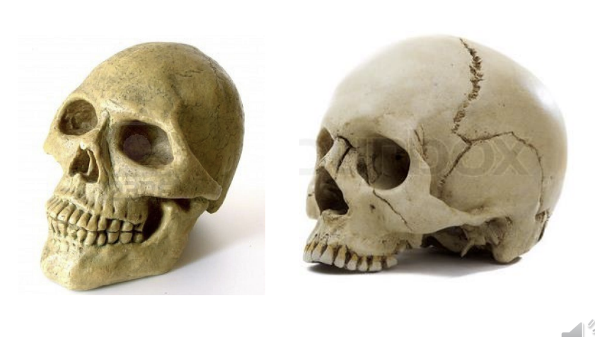
Which is the skull and which is the cranium
skull has mandible attached
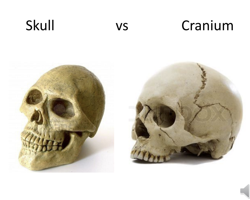
What are 4 functions of the skull?
protect the brain
upper part of the alimentary (digestive system) and respiratory system (nose and mouth)
protective capsule for organs of sight, hearing and smell
close relationship with teeth and oral soft tissue
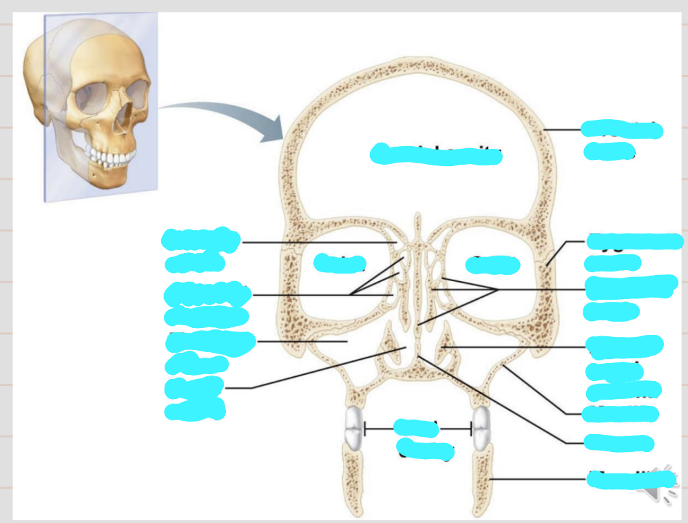
Name these structures and the cavities and role of the cavities
cranial cavity - encloses the brain
orbital cavity - encloses the eyes
nasal cavity - olfactory apparatus
passage way to respiratory system
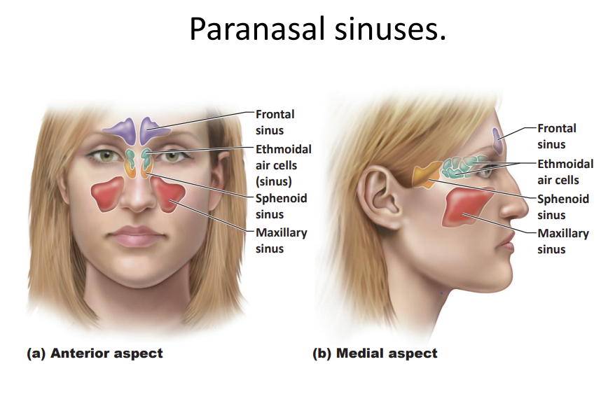
hallow - paranasal sinuses 4 of them - help lighten the weight of the skull
allow air to circulate, contain mucus and vascularised so allow air to be warmed up before reaching the lungs, provide resonance to sound
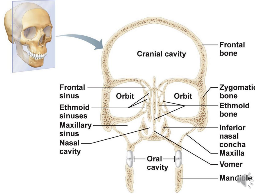
How many bones is the skull made of?
23
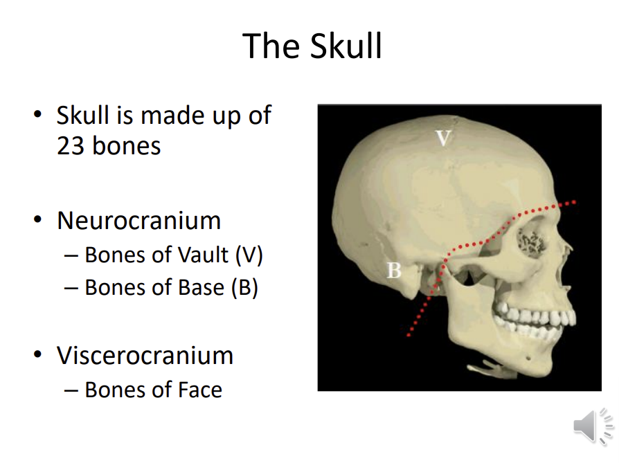
What are the bones of the face and bones that are in contact with the brain called and what aree these further split into?
neurocranium - contact with the brain
viscerocranium - bones of the face
neurocranium is split into bones of the vault - which form the roof, while the bones of the base from the base of
What are the two roles of the cranial bone?
enclose the brain in the cranial cavity
provides sites of attachment for head and neck muscles
What are the 4 roles of the facial bones?
framework of the face
cavities for special sense organs, for sight, taste and smell
openings for air and food passage
sites for attachment for teeth and muscles of facial expression and muscles of mastication
What are the names of these 3 structures
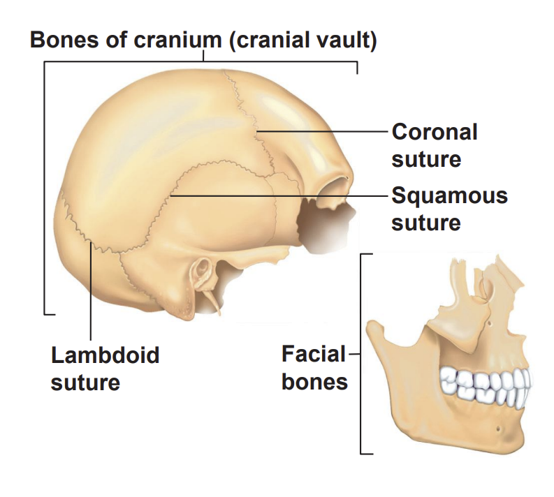
projections in anatomy are known as?
procceses
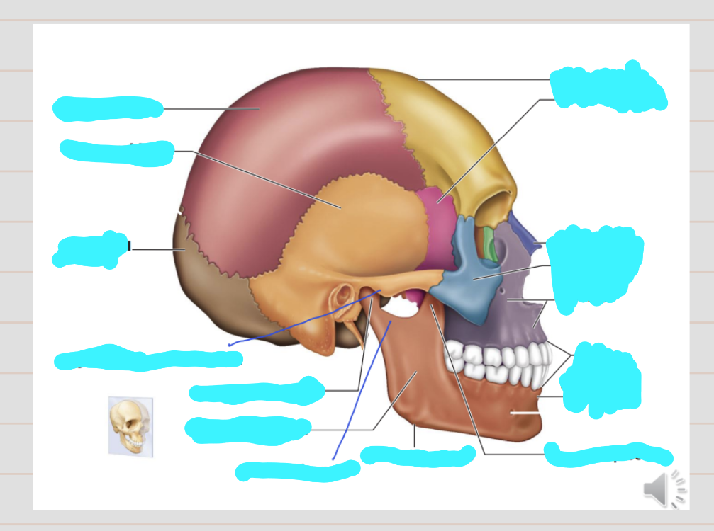
Name these structures
mandibular angle connects the mandibular ramus and body of the mandible
alveolar margin provides sockets for the teeth - present as long as teeth are present
maxilla contributes to the lateral aspect of the nose and the upper jaw, floor of the nasal cavity adn floor of the orbital cavity
temporal bone on the lateral aspect
zygomatic arch connects the occipital bone to the zygomatic bone
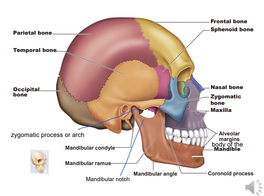
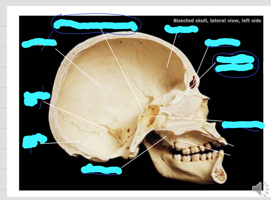
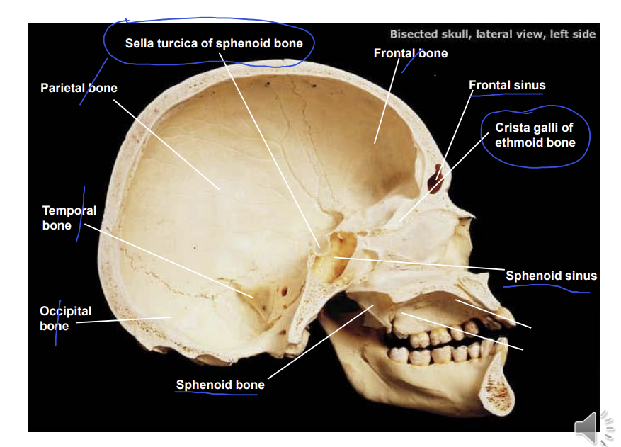
Name these structures of the skull
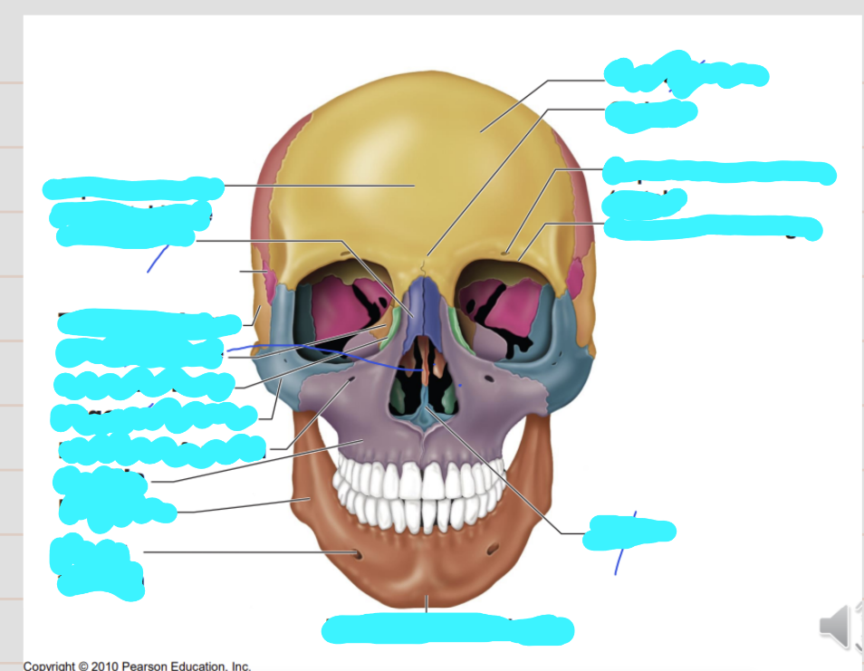
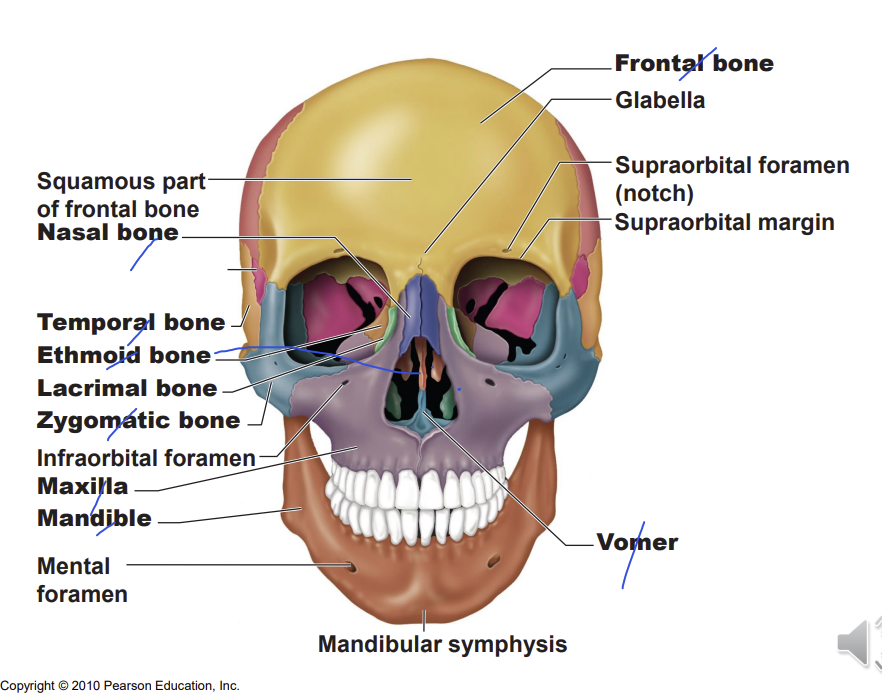
Name these aspects of the skull (anterior view)
depression - lacrimal fossa holds the lacrimal sac
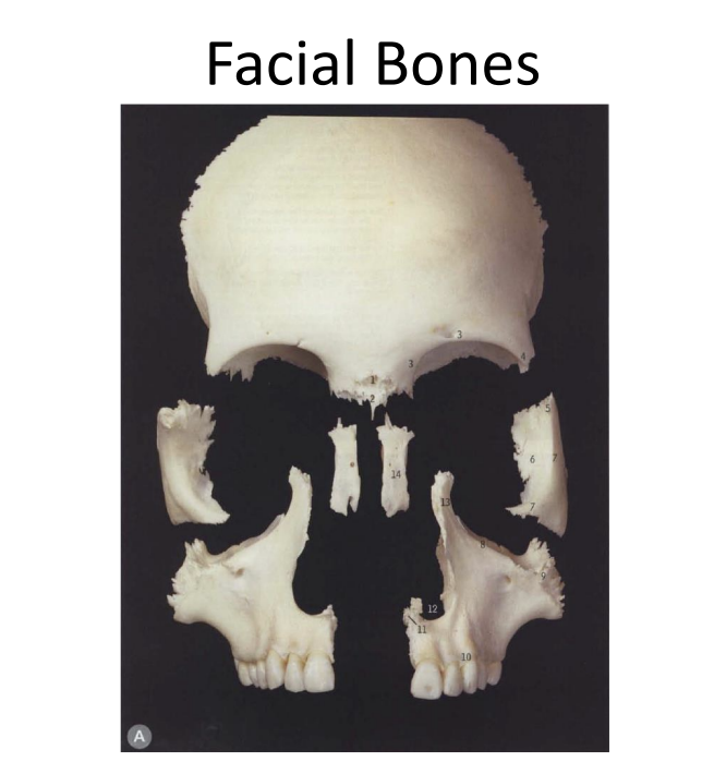
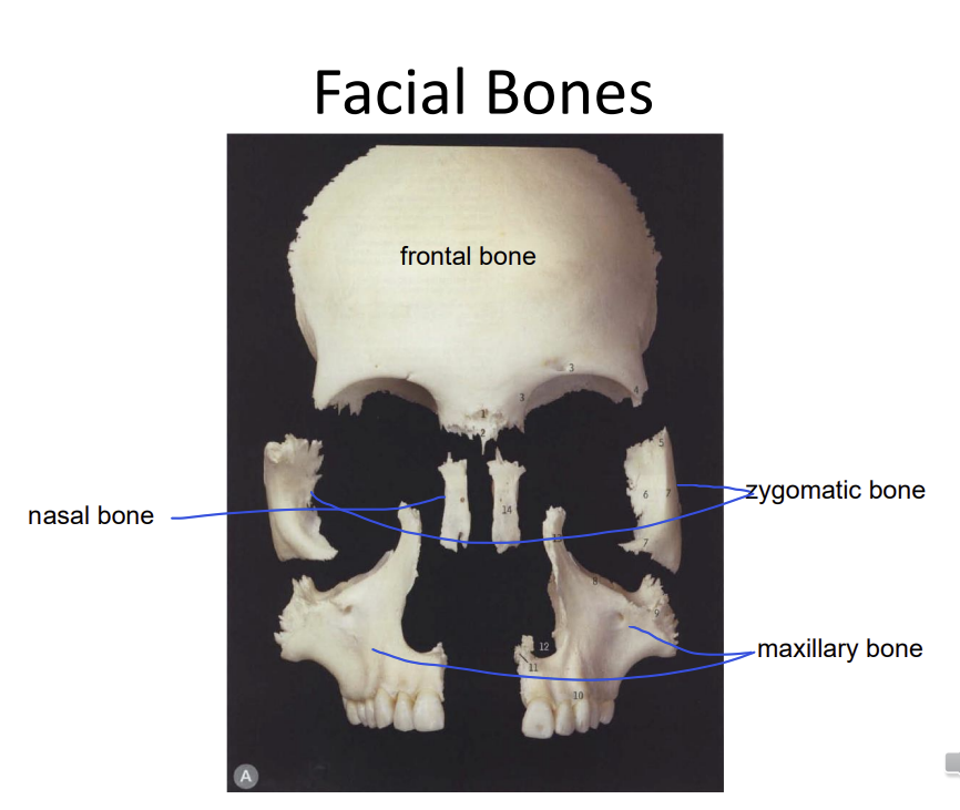
Name the facial bones
How do you describe a cavity?
roof, floor, lateral and medial wall
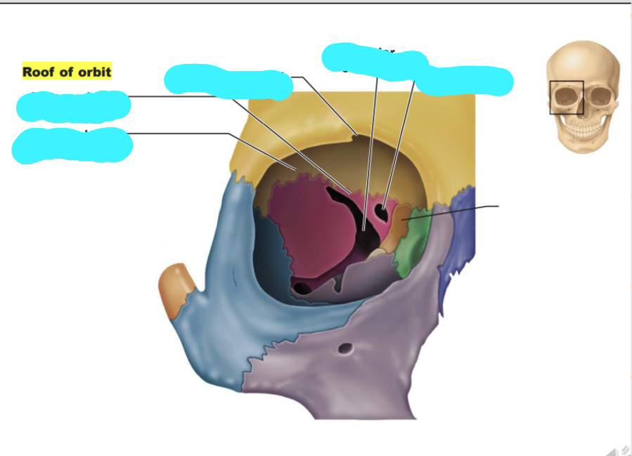
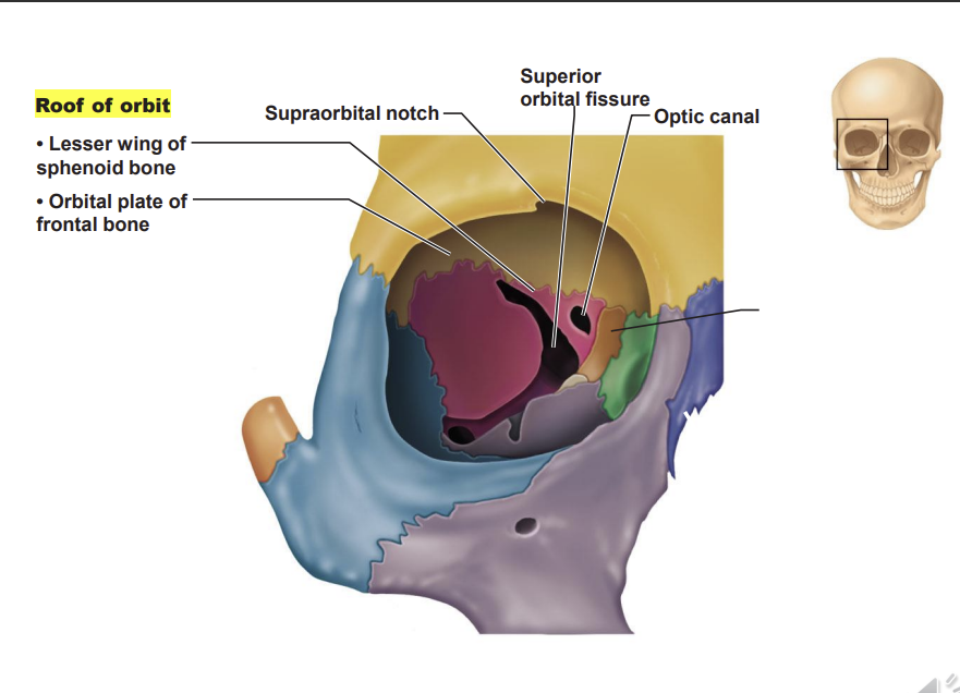
What are the structures on the roof of the orbit?
superiorly - orbital plate of the frontal bone
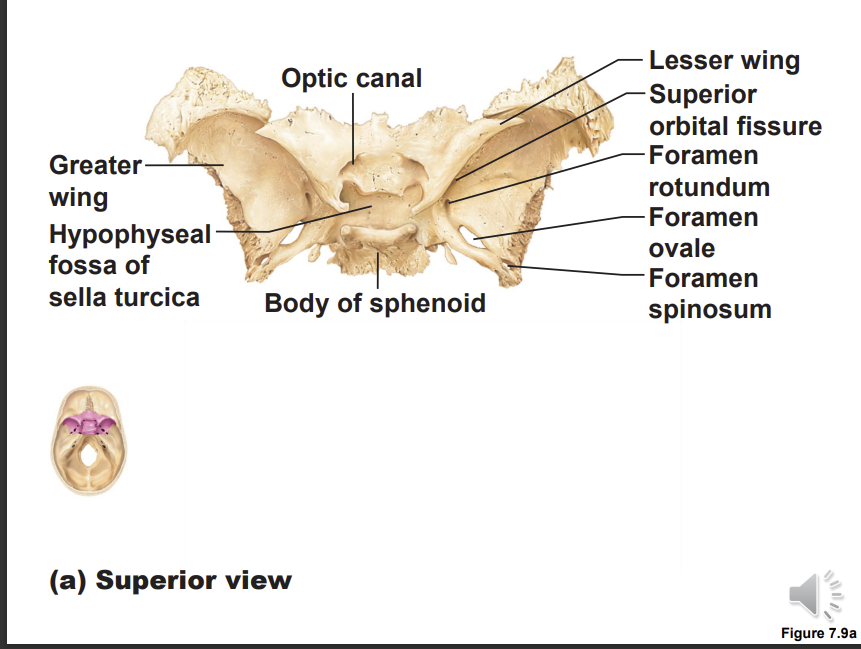
slit like structure - superior orbital fissure found in the lesser wing of the sphenoid bone, in the sphenoid bone there is another foramen - optic canal - where the optic nerve enters the retina of the eye
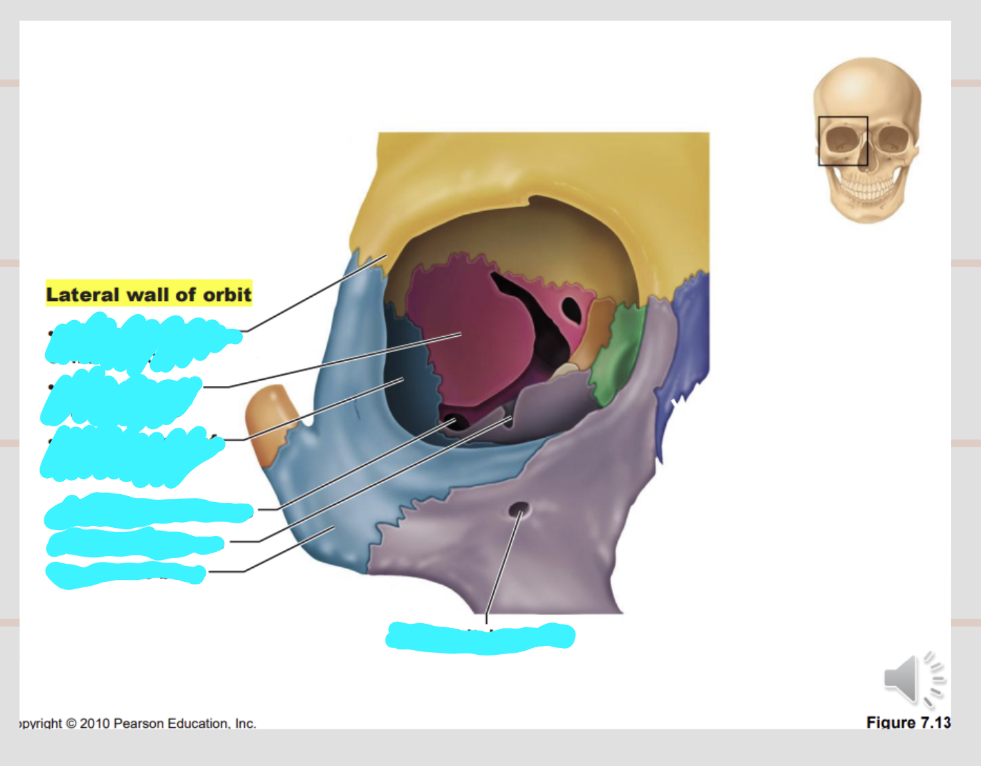
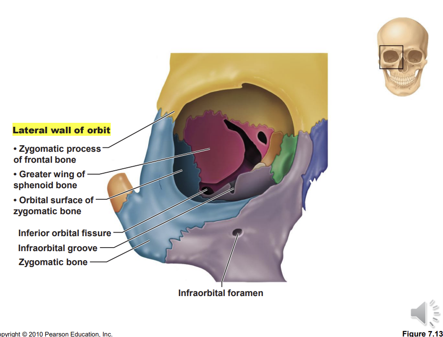
Describe the lateral wall of the orbital cavity
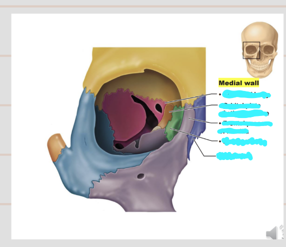
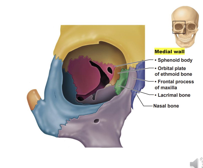
Describe the medial wall of the orbital cavity
frontal process of maxilla, the part of the maxilla joining the frontal lobe
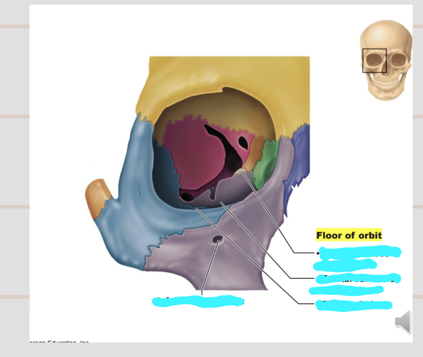
Describe the floor of the orbital cavity
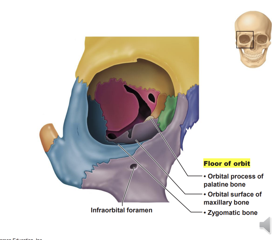
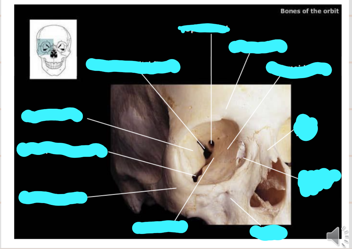
label these structures
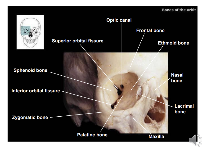
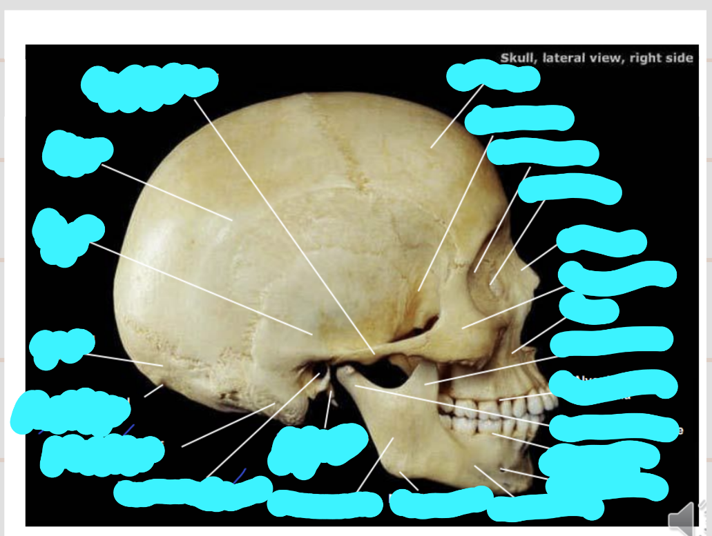
Label this structure
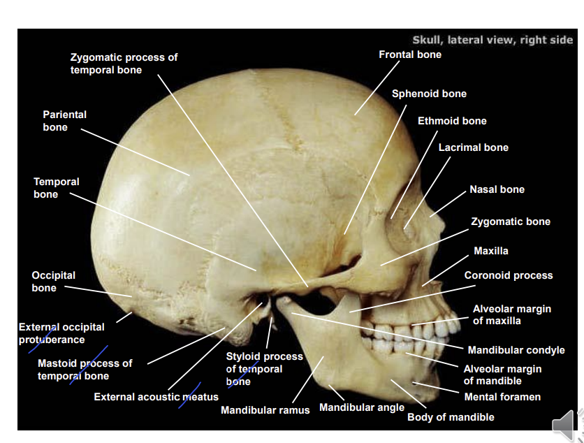
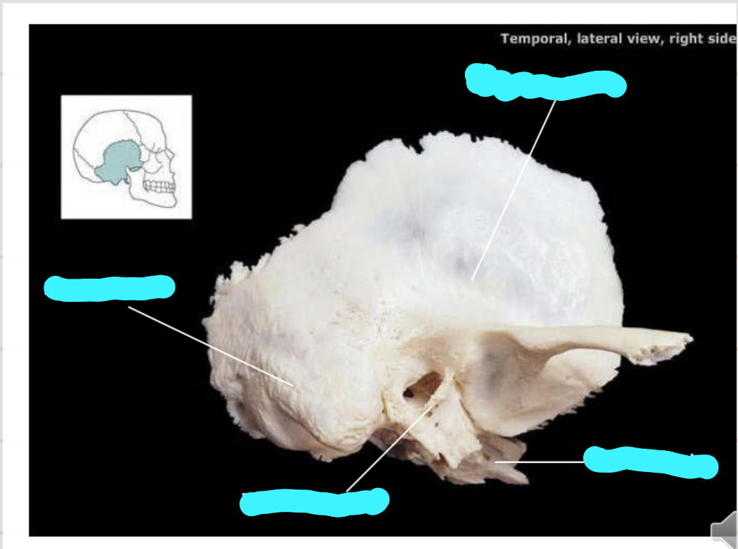
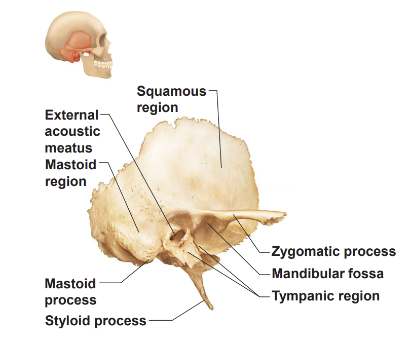
what are the 4 regions of the temporal bone?
squamous region - lateral aspect
petrous region on the base of the skull
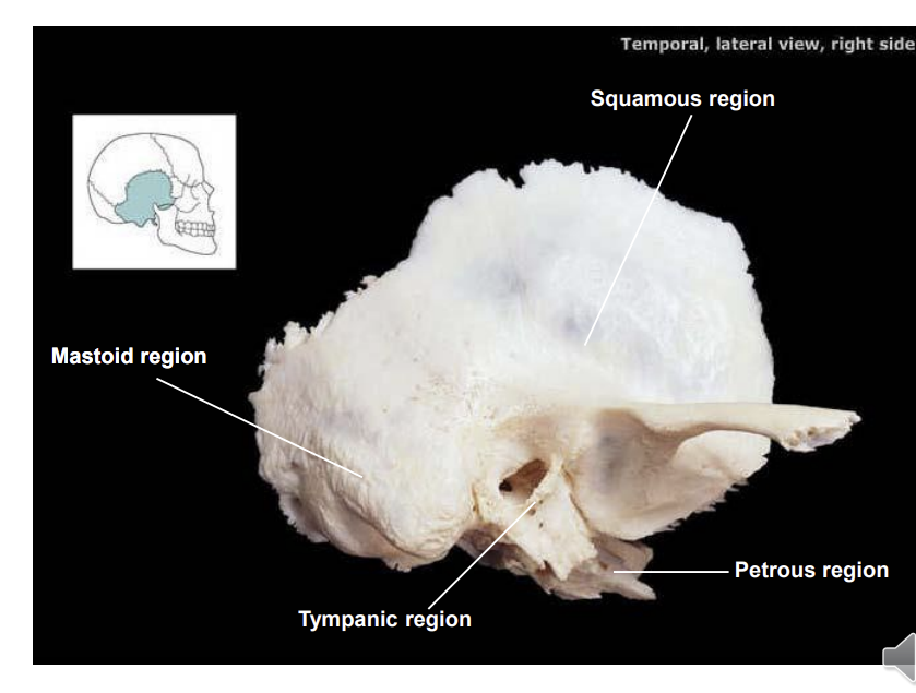
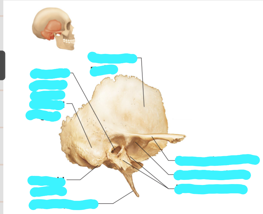
label these structures
mandibular fossa is where the mandibular condyle articulates
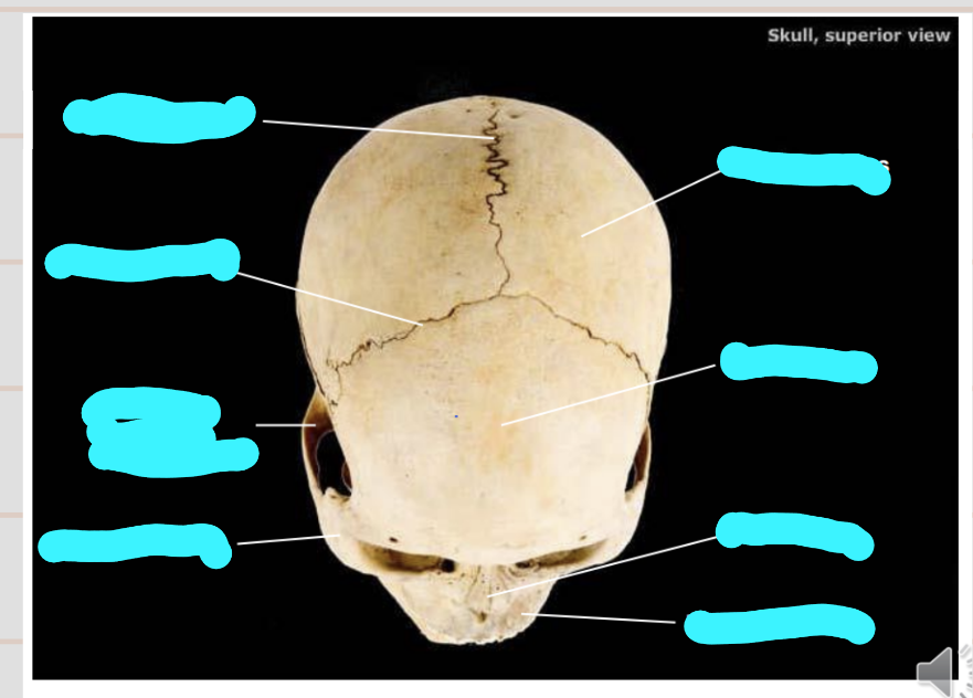
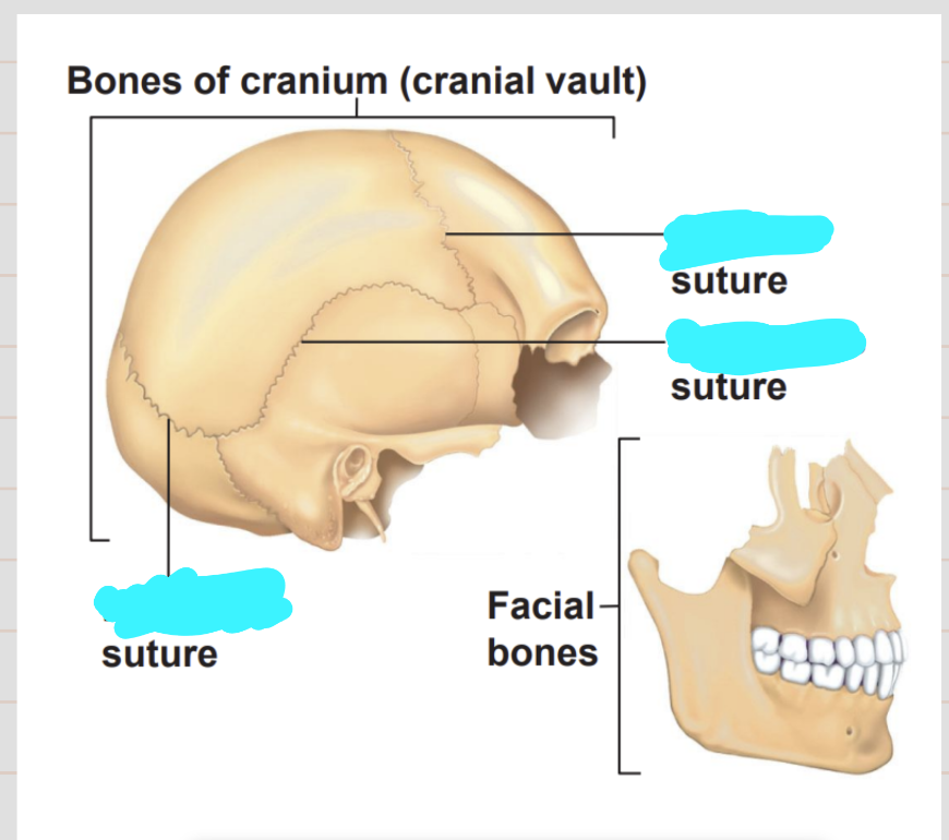
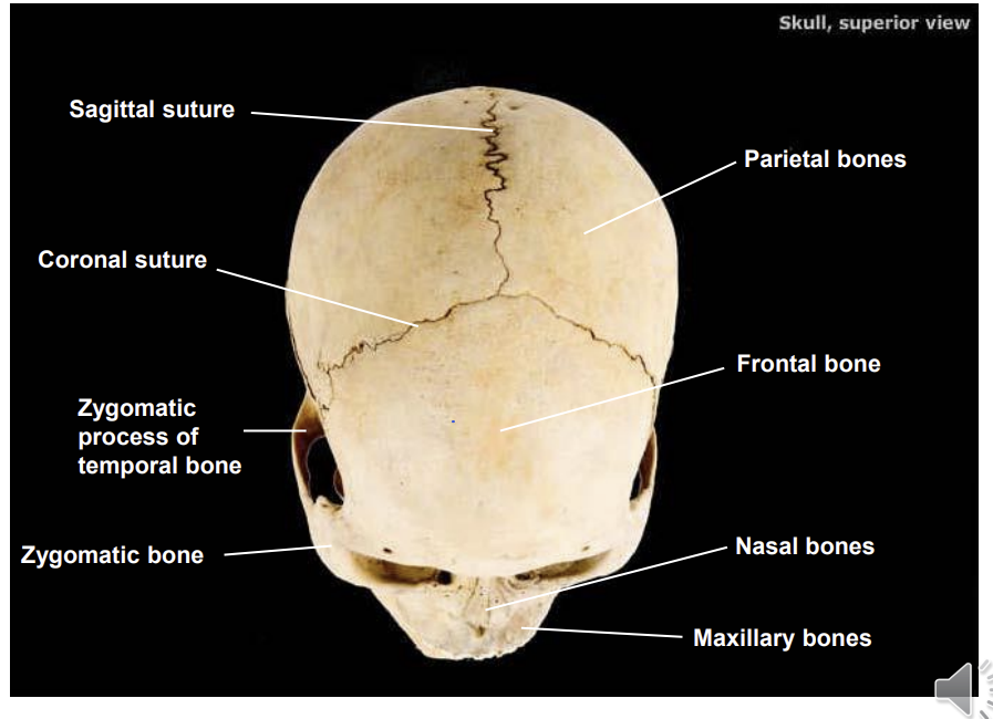
label this superior view and the important sutures
sutures are immovable joints
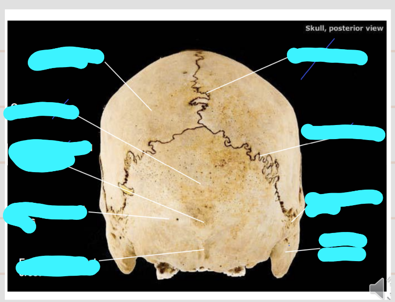
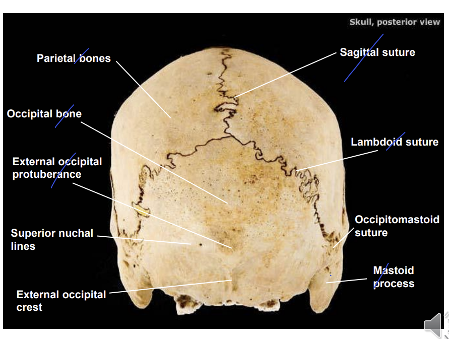
Name the structures on the posterior view?
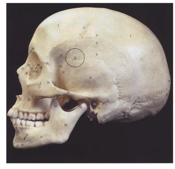
What is the circled part of the brain and what is its significance?
pterion
weaker part of the skull as many bones join here, the bone in this region is very thin, therer is also an important artery running behind it
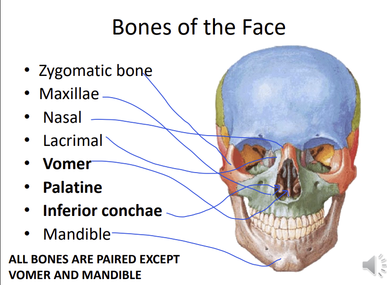
Name the 8 bones of the face?
which are paired and which aren’t
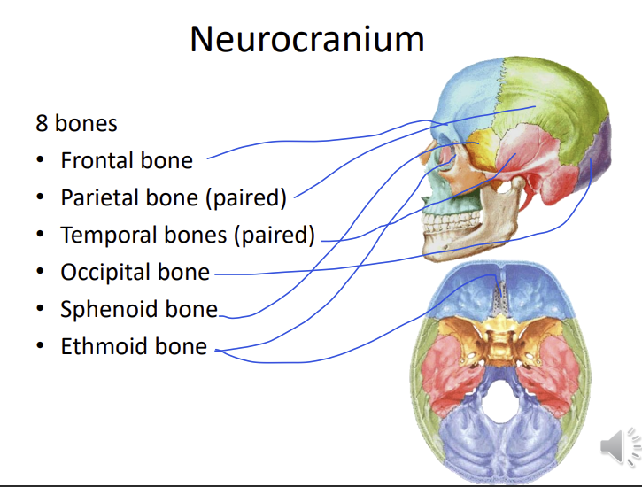
What are the 6 bones of the neurocranium?
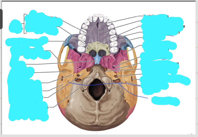
through the incisive fossa, nerves which innervate the teeth
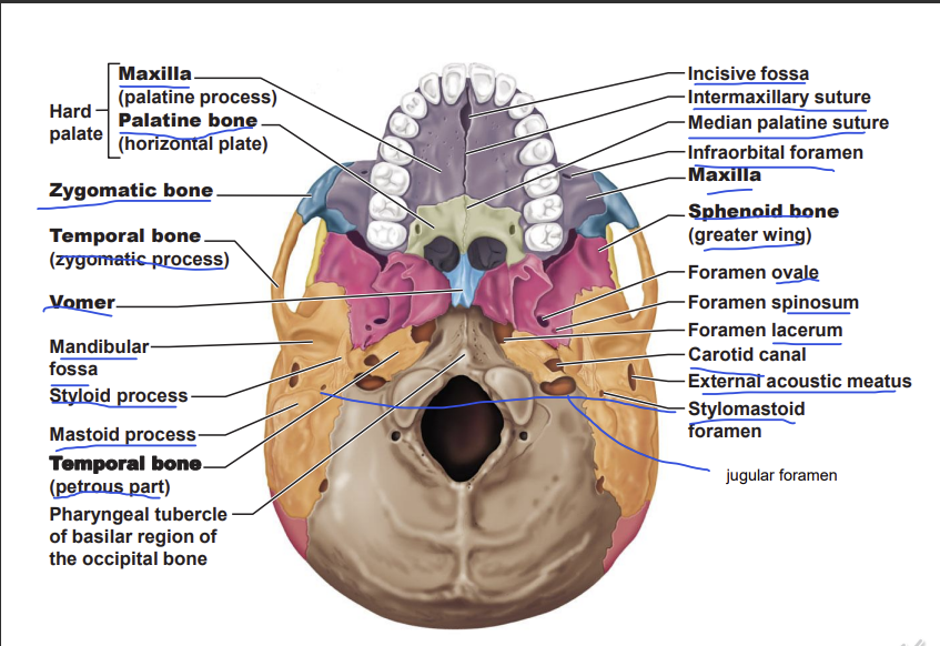
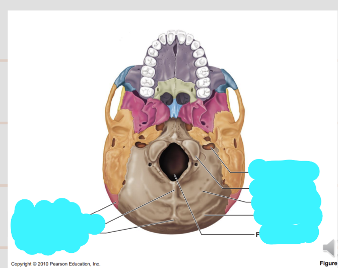
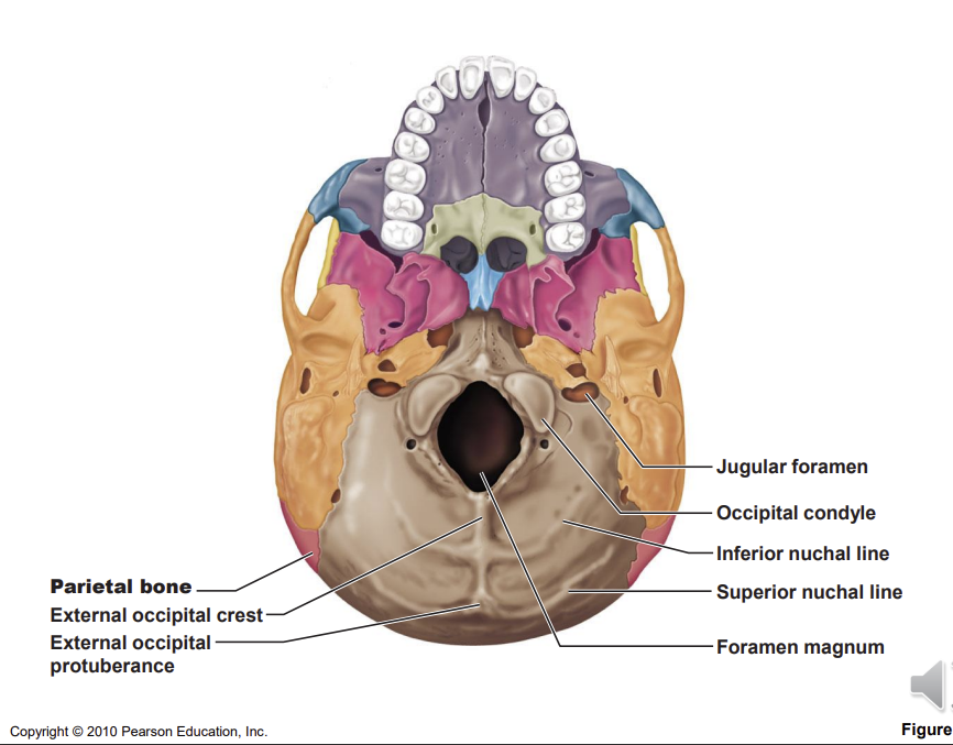
Name the labelled structures of the base of the cranium
muscles of the neck attached to the nuchal lines
foramen magnum - spinal cord
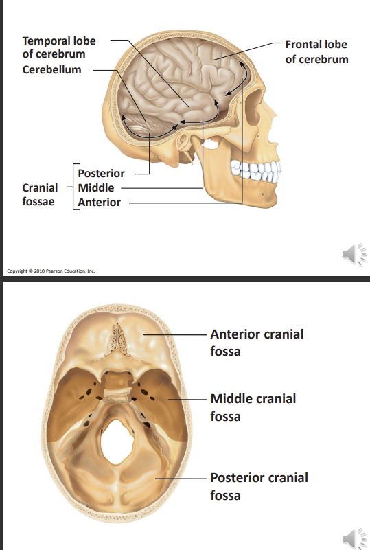
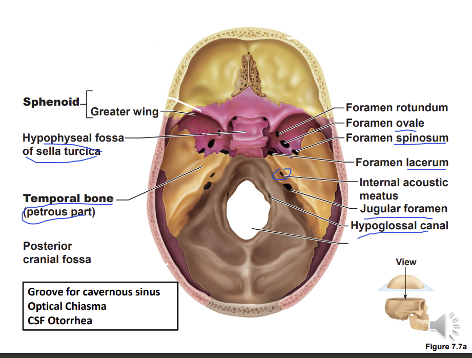
The cranial fossa is divided into what cranial cavities?
based on the part of the brain located in each cavity
anterior cranial fossa - forebrain
middle cranial fossa - midbrain
posterior cranial fossa - hindbrain
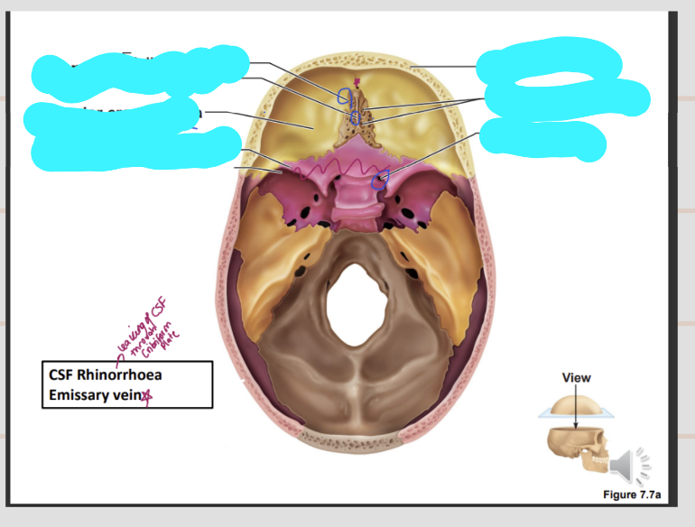
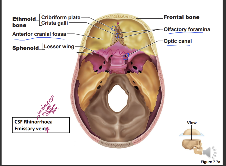
What are the structures found in the anterior cranial fossa
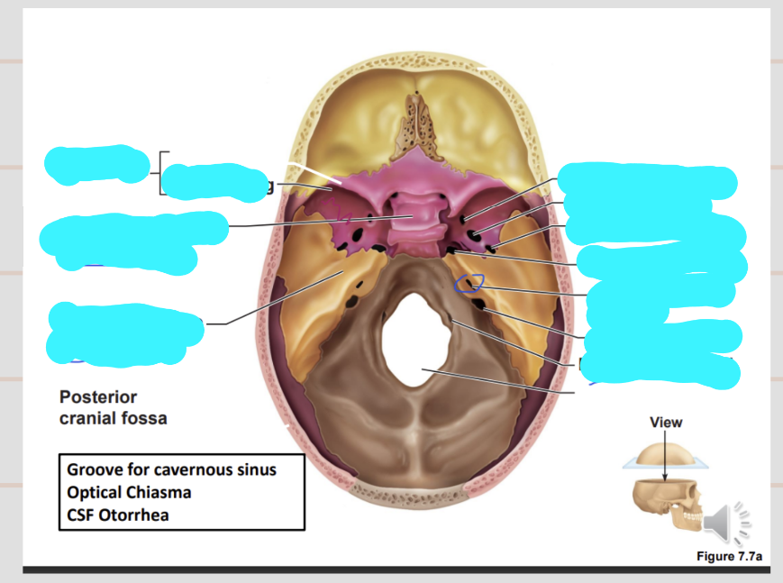
describe the structures in the middle crania fossa
sella turcica - pituitary gland is located
groove for the cavernous sinus is on the lateral aspect of the sell turcica
optical chiasma - the optic nerves cross each other leaving a groove
CFS otorrhea can happen if there is a fracture near the internal acoustic meatus
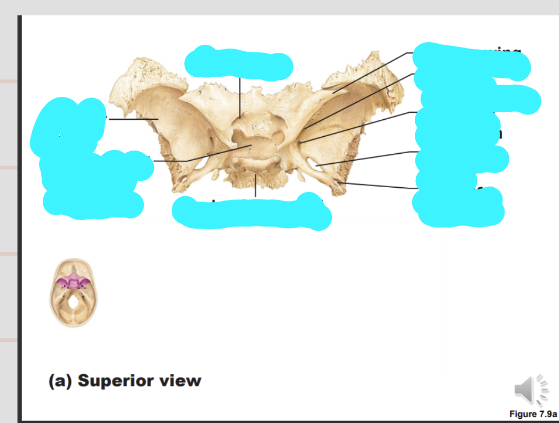
describe the sphenoid bone
body - central bit we have the sella turcica and optic canal
lesser wing
greater wing - foramen rotundum, ovale and spinosum
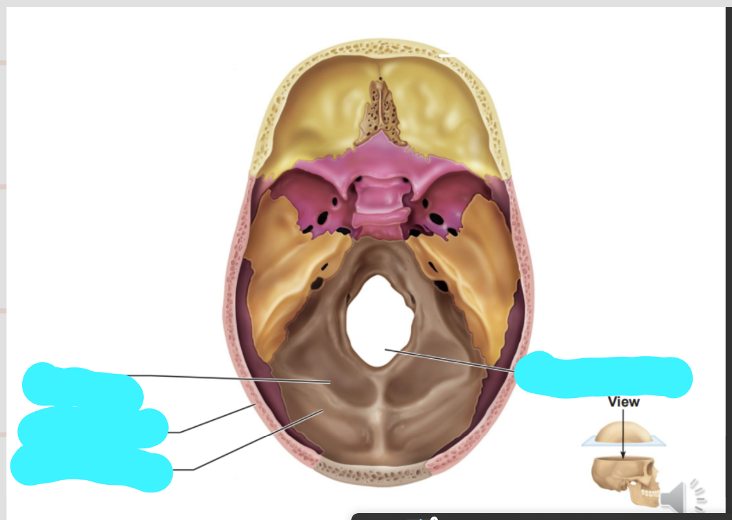
describe the structures in the posterior cranial fossa
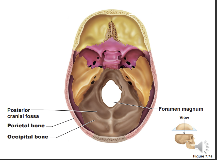
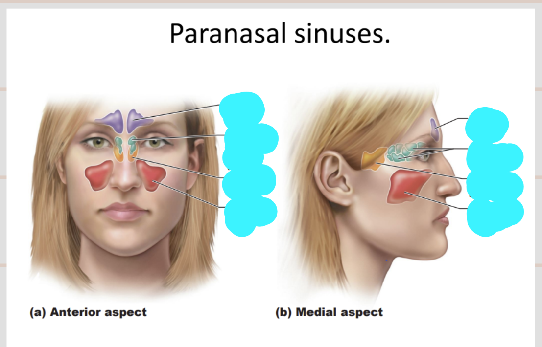
Lebel this diagram
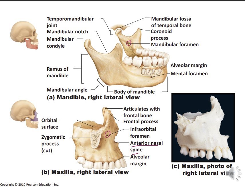
What forms the TMJ?
T MJ is formed by the mandibular condyle and the mandibular fossa
mandibular nerve enters the mandibular foramen
maxillary bone is more porous so anaesthetic can diffuse through the bone - can’t be done to the mandible
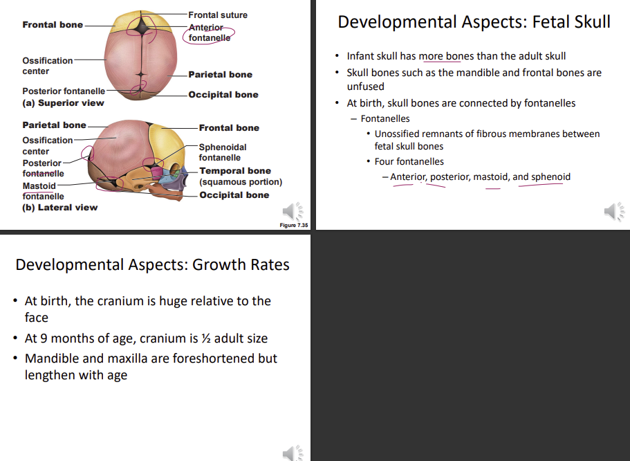
How is the neonatal skull different to the adult skull
the bones are separated from each other and not fully connected leading to fontanelle which is fibrous tissue present between bones, allows bones to overlap each other, they are growth sites
the
mastoid (left right movement and up and down) and styloid process not present - they develop during muscle attachment
skull is flat
the cranial is larger than the face
distance between mouth and orbit is small, as vertical growth of maxilla hasn’t occurred yet