BCHM: midterm
 Call Kai
 Learn
Learn Practice Test
 Spaced Repetition
 Match
Match Flashcards
 Knowt Play
1/93

There's no tags or description
Looks like no tags are added yet.
Name | Mastery | Learn | Test | Matching | Spaced | Call with Kai | Chat |
|---|
No analytics yet
Send a link to your students to track their progress
94 Terms
what are enzymes
type of molecule
actions
purpose
typically proteins (polymers of AA monomers, folded into specific 3D shapes, with different functional reigons based on different AA residues)
can be RNA (polymers of rNTPs, can form 3D shapes with specific regions to do chemistry)
eg ribosome (prot + RNA, mRNA => protein, catalysing peptide bond formation)
specific reigon to enable catalysation of a specific rxn, is the AS, informed by the residues here
enables coordination of substrates, in order to react (carefully control, and incr rate of, chemical rxns via lowering the Ea, making substrates likelier to react)
for rxns to happen alone, may take seconds - billions of years. for enzymes, rate takes less than minutes (much narrower timescale, efficiency) = key importance
eg Catalase breaks down H2O2 continuously made in our cells (Mitochondria) so it doesnt breakdown DNA & proteins

how do enzymes work
RC diagram
transition state
gibbs free energy
not entirely known, but many ideas which it may be a combo of
occurs via covalent & non covalent catalysis
(RC diagram)
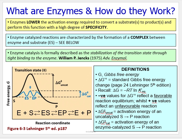
chemical rxns have G changes over time from S=>P, having to overcome Ea via a TS. higher this is, the slower the rxn (& more unlikely to occur, too high energy to happen spontaneously)
TS doesnt exist for long, it forms then either forms substrate or product. determines deltaG for rxns (deltaG ts) = uncat
enzymes lower Ea by forming a complex ES (enzyme + substrate), then EP (enzyme + product), the G energies between which have much lower energies = cat
E + S <=> ES <=> EP <=> E + P. (all reversible, at eq)
this replaces the high E TS with a much lower one. making rxn much more favorable, likely to occur, and quicker therefore
describe covalent catalysis
how it works
example
one chemical method of enzyme catalysis (the one not focused for this course)
involves forming (transient) covalent bonds between E + S => ES, to guide it towards a specific chemistry, to promote rxn to form => EP => E + P
(how it works - Protease example)
eg Chymotrypsin protease (catalyses protein breakdown)
cleaves peptide amide bonds, via the specific AS sequence it contains
such as an aromatic residue in a position before the cleavage site, which covalently links to a Serine on the protein, => Covalent intermediate
this then enables protein cleavage to occur more favorably, lower Ea barrier to overcome
(how it works - Pyridoxal Phosphate Cofactor)
co-opted by proteins into / near their AS, to promote AS chemistry between substrates, to make rxn more favorable (via chemistry the AS cannot do alone)
this cofactor specifically, via Aspartine & Lysine AS residues.
here it interacts with different substrates differently, to catalyse differet rxns (Eg Amide removal, redox chemistry) => enabling chemistry beyond the AS’s solo capabilities

describe non-covalent catalysis
what is this
what are 2 ways in which this occurs
more of the focus for this course
enzymes performing catalysis w/o transient covalent bond formaton. using other types of interactions - H bonds, ionic charge interactions, etc
(increase the chance of substrates meeting)
= entropy reduction (for joining molecules together)
these non-covalent interactions with the enzyme, position substrate functional groups nearby / in the perfect orientation, to increase their chance of reacting
this is because rxn requires subtrate meeting, and colliding at the correct orientation
so this restricts their relative motion in solution (where meeting would be rare)
(distorting the substrate)
(for breaking molecules apart)
high Ea barrier due to the unfavorable / unlikely / unstable / high E TS that must form (eg whats the chance of a stick randomly bending so it can break in half)
so to combat this, enzyme AS shapes bind the substrate in a not-exactly-complementary way - to stabilise the TS and lower the E barrier by positioning it one step closer to reaching the TS
pushing the substrate into a conformation that orientates them favorably to react
eg TS involving unstable charges together on the substrate (anion with carboxyl negatives), enzyme can use positive charge residues so that the TS can be stabilised upon formation, so it is likelier to form (Ea barrier lowered)
how does non-covalent catalysis methods to distort the substrate, link to synthetic biology?
AS is specific to the distortion on the way to the TS of the substrate - not the substrate itself (if it was exactly complementary, it isnt stabilising the TS in any way)
(Developing new / improving preexisting enzymes)
therefore when designing an enzyme to improve a preexisting one, consider stabilising / better stabilising the TS
when desigining a new enzyme, consider how the AS can stabilise the TS / redesign a preexisting enzyme’s AS to stabilise other TSs (to promote additional rxn pathways)
(Developing inhibitors)
to target a particular enzyme for inhibition, since enzymes AS mimic the TS of the substrate its rxn-specific to, design inhibitors alike the TS
this enables them to bind favorably / likely / often to the AS of target, and if designed in a way to remain attached, they block the AS from substrate binding & catalysis
what does specificity of enzymes rely on?
AS binding, which is informed by the specific way that enzyme stabilises the TS (eg distortions to make it more favorable to react, stabilising negative charges of the TS, residues for covalent linkages)
enzymes are specific to substrates, but not as much as previously thought - they bind to lots of similar molecules, but only successfully perform chemistry on those with a particular interaction
also relies on binding affinity (highest for the target substrate, it has a higher chance to form favorable interactions long enough to perform chemistry)
why are enzymes important
cell chemistry
time scale
energy
controlling cell chemistry
(cell)
organisms are made of cells, which are pouches of chemistry seperated from the environment - carried out by enzymes
enzymes enable this specific purpose
(time scale)
puts life on the same time scale in terms of chemistry
cellular rxns can take billions of years - seconds to happen spontaneously in solution, but enzyme catalysis happens in seconds
this enables life to be efficient, coordinating rxns in terms of timing (one thing relies on the next)
(energy)
enable the ultimate life cycle. taking sunlight (heat energy) => converting it to usable cellular energy via formation of biomolecules (ATP, NADH)
fuelled by this therefore. they are driven by the energy stored in the ATP phosphate bonds, coupling the release of such with formation of biomolecules (proteins, lipids, DNA - creating order non spontanteous) to fuel life
(control)
they guide essential cellular rxns pathways, to organise these into controlled metabolism for efficiency in cells
create a particular product from the pathway (guide towards a certain outcome)
guide a product onto another pathway to get to an ultimate end goal
guide away from a particular side product to increase efficiency (reduce disorder)
eg glucose, can be broken down into many possible things, but is carefully guided within the cell to be fully broken down into CO2 to release all its energy and generate maximum ATP in respiration
an example of enzyme importance
in terms of food production / industry
Cheese production requires Chymosin protease, to catalyse the breakdown of proteins, to enable milk coagulation (a key process)
it used to have to be sourced from pig stomachs (via Rennet)
but now it can be done recombinantly (transform in the gene to produce this enzyme) by bacteria (or other microorganisms) who express this enzyme, and create it via fermentation
this is isolated and mixed with cheese - enabling the product to be formed
= key enzyme for the process, important for refining efficiency of cheese production

an example of enzyme importance
in terms of Flu drug discovery
Australian Scientists knew that Flu viral particles coded for the enzyme Neuriminidase, but not why or how this related to Flu progression
they figured out, this enzyme cleaved sugars that make up host cell surface receptors, so that viral particles can bud off from infected host cells to spread infection
this is because for infection, Flu cells are envelope viruses that enter human cells via specific membrane surface receptors, that the virus has evolved to mimic its substrate
when it buds off (Exocytoses) to continue infection, it takes a bit of the host cell’s membrane so it is recognised as un-foreign by the next human cell it infects
(drugs based on Neurimindase activity)
therefore, a target for Flu drugs is Neurimindase activity - if it cannot cleave these sugars, it cannot bud off, and it cannot continue spreading infection
so these Scientists developed Neurominidase inhibitors (eg RELENZA) that tightly bind to the AS and prevent sugar cleaving & budding, so viruses are stuck in the host cell and cannot continue infection
therefore competing with the substrate and blocking AS entry
these Inhibitors were synthesised to mimic the structure of sugars at the terminal end of the Glyco receptors, that Neuriminidase AS therefore has high binding affinity to

an example of enzyme importance
in terms of cardiovascular disease
important in diagnostic & prognostic context, of heart attacks
heart attacks are often caused by artery blockages (blood clot build ups) preventing the heart from getting enough O2 via the blood to beat (some but not enough)
this causes that part of the heart muscle to slowly die (it cannot metabolise properly)
this causes certain enzymes to appear in the blood - so their presence / incr conc, can be used to diagnose a heart attack after the fact (important for getting appropriate treatment)
creatine kinase (CK), aspartate aminotransferase (AST), lactate dehydrogenase (LDH)
different half lives, so can also be used to determine when the heart attack occurred

an example of enzyme importance
in terms of PKU (Phenylketonuria)
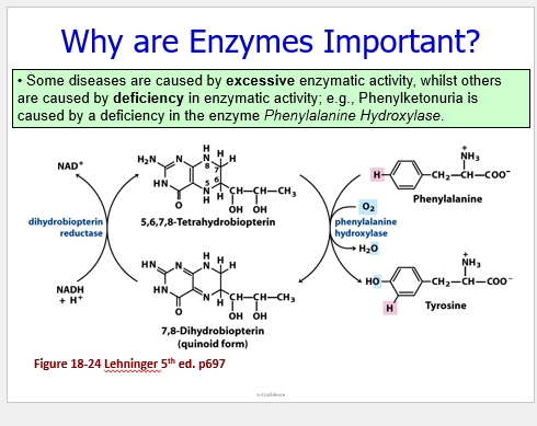
PKU / PhenylKetonUria is a disease with symtpoms of severe mental deterioration, caused by a defective Phenylalanine Hydroxylase enzyme (mutated)
this enzyme metabolises the particular AA Phenylalanine from our diets, to form Tyrosine
with this mutation, it cannot add O2 to phenyalanine (to turn the H=> OH group to form tyrosine), so we have too much phenylpyruvate (part of the metabolic pathway - the Keto Acid of Phe as a result of the build up - it cannot be converted) in our blood and urine, which causes the symptoms.
what may a mutation in an enzyme manifest as?
(beneficial)
speed up rxn
(harmful)
change structure so much it falls apart
change structure so enzyme activity occurs at a lower rate (substrate doesnt bind as well - less binding affinity)
more binding affinity so it gets stuck
different structure so it binds to other substrates (less specific) and clogs it for the particular rxn
(silent)
change AA sequence but no effect on activity
recap: metabolism
2 types
what these types involve
link to E
metabolic pathways
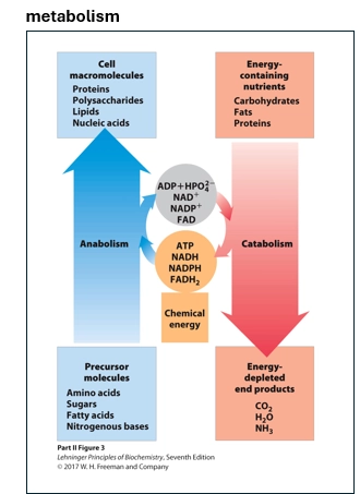
sum of chemical rxns in a cell
2 broad types - catabolism & anabolism
anabolism - building larger molecules (fatty acids, proteins, carbs) things using smaller precursor molecules (eg AAs, sugars, lipids)
catabolism - breaking down high E larger molecules, into smaller E depleted molecules
(energy)
all linked to gain & expenditure
the ultimate source of E being the sun (=> food => down the food chain to us)
anabolism - using E to add things together (E required to make order) via ATP & NADH
catabolism - gaining E from breaking down high E compound chemical bonds (increasing disorder) in the form of ATP & NADH
(metabolic pathways)
how rxns are organised
typically into distinct purposes (eg Glycolysis E extraction, Glycogenic storing of glucose, Gluconeogenesis synthesis of glucose, waste product elimintation)
however these are not linear - are all interdependent, reversible, and complex
briefly,
how are Glycolysis & Gluconeogenesis related?
Glycolysis (Glucose breakdown) Gluconeogenesis (Glucose synthesis)
these are related, being inverse / opposed pathways (reverse of eachother). glucose => pyruvate. pyruvate => glucose.
at eq, so a few main steps that drive rxn one way or another, enabling their simultaneous existance
favored rxn depends on where the cell type is (eg muscle vs brain vs liver), as different metabolic pathways are differentially important
eg muslce & brain (Glycolysis - want to breakdown glucose to fuel high E demand, so Glycolysis enzymes are highly expressed to favor this direction rxn)
eg liver (Gluconeogenesis - the liver has the purpose of storing glucose as glycogen, so we want to create glucose for glycogen storage, by highly expressing Gluconeogenesis enzymes to favor this direction rxn)
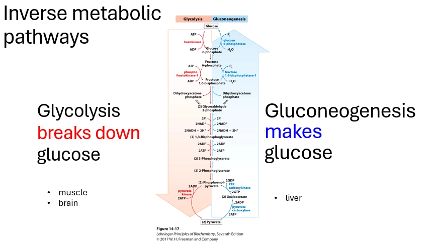
why is glucose important?
2 main reasons
(Fuel)
an excellent fuel
water soluble (upon ingestion can enter bloodstream, and circulate and enter cells around the body, for use in metabolism)
yields a good amount of E upon complete oxidation, some in GLycolysis and more later in Respiration (breakdown of high E bonds => usable cellular E)
can be efficiently stored as a polymer (Glycogen. can store E for later in chemical bonds)
a catch all E source for many organisms & tissues
(Versatile Precursor Molecule)
biochemical precurosr that the body can use for polysaccharides, proteins - in anabolism
due to glucose being a sugar
(Humans)
Where do we get glucose from?
How does glucose enter our cells?
how does this vary between cells?
how does required glucose stay in the cell?
(where does it come from)
ingested from food!
it then is transported through cells out of the GI tract, into the blood being water soluble, here it raises our blood sugar levels, and is transported around our body
but it then must get through the non-water-soluble lipid bylayer, for cells that need it
(how does it enter our cells)
via GLUTs (Glucose Transporters) membrane proteins, carrier proteins, that move Glucose across the membrane from high to low conc (no E input required)
outward open confo - binding site for gluc faces cell exterior => gluc interacts and binds => confo change
inward open confo - binding site for gluc faces cell interior => gluc can dissociate => is released into the cell
bidirectional - occurring in either direction, with one adopted preferentially based on internal / external cell conc of glucose - therefore this can change, and enable transport
highly specialised structure - having to have regions exposed to the hydrophobic lipid membrane interior AND the hydrophilic cell exterior and interior solution AND the specific hydrophilic Gluc binding cavity
(how does this vary between cells)
many different GLUTs expressed in different cell types with different requirements
essentially what differs is Km (basically Gluc binding affinity to GLUT inverse)
eg in neurons (GLUT3) Gluc has a low Km (high binding affinity) as lots of E is needed (neurons must consistently pick out Gluc from bloodstream even at low conc)
eg in small intestine (GLUT5) N/A Km as this is primarily a fructose transporter (the function is not to transport Gluc)
(how does required glucose stay in the cell)
GLUT are bidirectional, passive, conc driven - but what if the cell needs lots of Gluc and theres little in the blood - we dont want it transporting out
this is combatted by the carrying out of the first step of Glycolysis - this converts Gluc to a product along the pathway, so it no longer has GLUT binding affinity so it remains in the cell
this occurs in Cytosol (site of Gluc transport AND Glycolysis), Hexoinase adds P from ATP => Glucose-6-Phosphate
so cells readily needing E (req Gluc readily broken down - muscle, neurons) will express Hexokinase at higher levels

What is the first step of Glycolysis?
describe
enzyme involved
importance
fate of the product - what happens next?
(describe)
Hexokinase catalyses the transformation of Phosphate from ATP => Glucose => Form Glucose-6-Phosphate
important to trap Gluc in cells, so it cannot be transported out via GLUT as Gluc, when the external has a lower Gluc conc (or just due to passive movement) while Gluc is still needed in the cell
also due to the fact of adding a negative charge (via P), which repulses the GLUT bindig sites (also negatively charged) to prevent transport
rxn has a large -deltaG (very favorable, releases free E, happens spontaneously)
(enzyme)
Hexokinase
Hexose = 6C sugar like Glucose
Kinase = adds a Phosphate / catalyses the transformation of P from Donor => Acceptor (ATP => Gluc)
several forms (I-IV)
(fate of product)
Glucose-6-Phosphate
depends on which enzymes are present (at high concs so which rxn dominates) based on levels of GE of these enzymes, based on cell funciton and requirements at that time
can be broken down (continue Glycolysis)
can be shuttled to Gluconeogenesis (for Glycogen synthesis / storing Gluc / storing E)
can be used for pathways to build nucleic acids (DNA / RNA, via Pentose Phosphate pathways)
can be converted back to Glucose

Glycolysis
what is happening overall
what happens in PHASE 1
what happens in PHASE 2
(overall)
1 glucose (6C sugar) => 2 Pyruvate (2× 3C sugars)
splitting the 6C sugar ring using 2ATP to make this transformation energetically favorable
this forms 3C intermediates, which are processed into pyruvate (3C sugar), along the way forming 2ATP per 3C sugar (4 total)
(PHASE 1)
Preparatory phase / Investment phase (we pay into it. we use 2ATP, the E stored in ATP, is transferred to Glucose to split it apart)
Steps 1 & 3 each transfer 1 Phosphate from 1ATP. (Irrevesrible steps)
Step 1 transfers P to Glucose => Glucose-6-Phosphate
then a 5C sugar is formed. this is less stable.
Step 3 transfers P to Fructose-6-Phosphate => Fructose-1,6-BisPhosphate
it is then energetically favorable to be split apart
= 2× Phosphorylated 3C sugars (G3P, Glyceraldehyde-3-Phosphate)
(PHASE 2)
Payoff phase / Return phase (we create Energy stored in ATP to gain a net return on our input. 4ATP produced, 2 per 3C sugar = Net 2ATP)
achieves the point of Glycolysis, generting Energy in the form of ATP from the Energy stored in Glucose, by breaking it down - we just need the initial investment of PHASE 1
includse irreversible step 10 (final step, irreversible removal of P from PEP PhosphoEnolPyruvate 3C sugar => Pyruvate 3C sugar + 1ATP
= 2x Pyruvate 3C sugars (final product of Glycolysis)

Why is it important that some steps of Glycolysis are reversible, and some are not?
how does this relate Glycolysis to Gluconeogenesis?
(reversible)
most steps are reversible (2, 4-9)
these have deltaG of ~0, so are at eq and are favorable to spontaneosly go back and forwards (depending on susbtrate conc etc - think K)
these steps are shared with Gluconeogenesis. Essentially the inverse of Glycolysis, so these enable Glucose to go the opposite way
the directionality of these steps rely on reactant / product conc, and other more direct regulation.
(irreversible)
steps 1 & 3 (Investment phase) & step 10 (Payoff phase - final step) are irreversible
the irreversible steps are highly favorable & spontaneous (negative deltaG) which makes them thermodynamically irreversible for physiological conditions
the irreversible steps are the main means of regulating Glycolysis (eg lots of ATP in cell => inhibits enzymes involved => we dont need to generate more ATP)
these steps are NOT shared with Gluconeogenesis. their reverse rxns are thermodynamically unfavorable, therefore Gluconeogenesis involves alternate reactions to bypass these 3 irreversible ones of Glycolysis
this enables carrying out the opposite rxn, while still having both rxns thermodynamically favorable, so most of the process is the same but key steps are seperated in terms of enzyme and reaction pathways for the key substrates produced along the way

Name and describe the 3 irreversible steps of Glycolysis
inputs & outputs
enzyme involved
phase involved
(STEP 1)
Hexokinase
transfers a Phosphate (Kinase) from 1ATP to Glucose (Hexose) => Glucose-6-Phosphate
this traps Gluc in the cell so it isnt passively transported out via GLUT membrane proteins (less binding affinity being not fully Gluc, negative charge repulsing negative GLUT AS). allows gluc to keep coming in and not limited by conc gradients
large negative deltaG (spontnaoeus, thermodynamically favorable, so essentially irreversible)
(STEP 3)
Phosphofructokinase 1
transfers a Phosphate (Kinase) from 1ATP to Fructose-6-Phosphate (Phosphofructo) => Fructose-1,6-Bisphosphate
large negative deltaG, essentially irreversible
highly regulated (presence of ATP - no point carrying it out if we already have lots of ATP. maybe better to store it as Glycogen. more weight at this step to carry on into the Investment phase of another ATP - as the benefits arent as versatile as STEP 1 which also keeps Gluc in the cell and can be used for various other rxn pathways - storage etc)
(STEP 10)
Pyruvate Kinase
transfers a Phosphate (Kinase) from Phosphoenol Pyruvate (PEP) to 1ADP => 1ATP + Pyruvate
Pyruvate exists as Tautomers, so the product produced is specifically the Enol Pyruvate (C=C bond in the ring), which is able to switch to the Keto form (C=O bond, more stable) which drives eq to favor Glycolysis, as we arent getting build up of the product - it is efficiently converted to something else (Keto Pyruvate Tautomer) so that Glycolysis can continue (specifically this step where we generate ATP)
creates the less stable product (Enol) => favorable to convert to Keto => lowers product conc => drives formation of ATP (Glycolysis)
the final step of Glycolysis. large negative deltaG, essentially irreversible

why is ATP favorable as an Energy currency?
Adenine (nitrogenous base) - Ribose (sugar) - Triphosphate (3x PO43- groups)
the Triphosphate region puts 4 negative charges close together, so the bonds holding these groups on are unstable - it is more favorable sterically and for repulsion, to break these bonds
therefore, shedding a phosphate is favorable - releasing a Phosphate group via Hydrolysis, and adding it to another molecule, releases Energy stored within this bond, transferring it to another compound enabling work to be done
does require overcoming an Ea barrier (kinetically unfavorable) which aids its efficiency as it doesnt just fall apart, its Energy is released in a controlled way via ATP hydrolysis (Enzymatic interaction)
rxn catalysed via electrons on O doing Nu attack to Phosphate group - enzyme AS positions these components close by to lower Ea barrier
the ATP is then => ADP (Adenosine Diphosphate, loss one Phosphate group / bond) or AMP (Adenosine Monophosphate, loss two Phosphate groups)
ATP conc is kept high in cells (Relative to ADP & AMP) to favor ATP => ADP conversion direction of eq, which drives cells to carry out work (encourage to use ATP)
deprotonated O- in Phosphate groups (PO43-) due to physiological pH (~6.5). technically would be protonated at lower pH (H3O+ around)

how is GTP alike ATP, and why is ATP used more often?
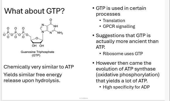
similar to ATP but with Guanine nitrogenous base instead of Adenine. otherwise the same (Ribose + TriPhosphates with neg charges)
use dominates some processes (Eg Tsln, GPCR signalling) with a similar function to ATP (releases free energy upon hydrolysis to release Phosphate)
therefore may have been an ancient precursor to ATP (utilised by the Ribosome, present early in cell chemistry?)
but now, ATP Synthase in Central Metabolic processes (may have evolved after, arising oxidative phosphorylation), has high specifity for ADP to form ATP (high yeidls, adaptive advantage), so it outweighs GTP usage
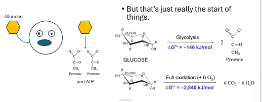
in the scheme of Glucose Oxidation, how efficient is Glycolysis?
What else can happen next?
not very efficient - yielding 146kj/mol of free Energy
compared to full oxidation (Glucose => 6CO2 + H2O) - yielding 2840kl/mol
in terms of ATP, 2ATP vs 34-36 ATP
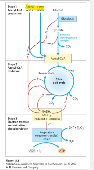
Pyruvate, the final product from Glycolysis, can be fed into the TCA cycle (first part of respiration), then into the electron transport chain, to power ATP Synthase to make 34-36ATP - where the great yield comes in
Glucose is fully broken down in these steps into CO2, so all its Energy is harnessed and made cellulary available, stored in ATP
give an overview of cellular respiration
oxidising (removing e from) biological fuels (eg glucose) to make ATP (adding e to the terminal e acceptor)
essentially shunting e from Glucose, and putting their Energy into ATP as a store
plants & animals typically use O2 as the terminal e acceptor (gives the largest deltaE0, most efficient E generation for multicellular lifestyles) = aerobic respiration
in anaerobic conditions, cells carry out Fermentation (alternate pathway after Glycolysis, converting Pyruvate => Lactic Acid, to regenerate NAD+ to feed into Glycolysis)
but ideally, in a well-fed state, the cell has enough O2 to feed enough Glucose into Glycolysis, then into Respiration (Pyruvate conversion => TCA cylce => e transport chain)

what is the first step of cellular respiration, after Glycolysis has occurred?
Pyruvate (Glycolysis output) must be cinverted into Acetyl CoA, before it can be fed into the TCA cycle (first part of Respiration) - bridging Glycolysis & Respiration
Acetyl CoA can also be formed from fatty acids (energy dense), AAs (more of a last resort, more important for building proteins than for Energy)
conversion catalysed by Pyruvate Dehydrogenase Complex. Pyruvate + Coenzyme A => Acetyl CoA + CO2 (spotaneous, TD favorable, kinetically enabled via enzyme interaction)
essentially removing CO2 from Pyruvate, and linking Coenzyme A (important for enzyme interaction later in the cycle), via some Disulfide bond

describe the TCA cycle
overview
inputs, outputs
where does this fit into respiration
(where does it fit)
occurs after Glycolysis, and after Pyruvate => Acetyl CoA conversion, before the e transport chain
(overview)
a cycle to break down glucose (Acetyl CoA) further, and remove more e, and add these to e carriers, to be shuttled downstream to the e transport chain (where they are used to generate a proton gradient, which is coupled with the formation of ATP via ATP Synthase - so the energy from Glucose is added to ATP bonds via electron power)
via reducing NAD+ & FAD+
depending on an enzymes isoform, GTP or ATP is generated in a rxn with Succinyl-CoA
(inputs, outputs)
=> 3NADH
=> 1FADH2
=> 1ATP / 1GTP
per Acetyl CoA

where does Glycolysis, and the serperate steps of Respiration, occur - and how are products / substrates transported between?
Glycolysis = Cytosol (Gluc => pryvuate, NAD+ => NADH)
this pyruvate must then be shuttled into the Mitochondrial Matrix, crossing 2x memrbanes (inner & outer) via a specific transporter
Pyruvate Oxidation = Mitochondrial Matrix (Pyruvate => Acetyl CoA + CO2)
co-localised to the location of the TCA cycle, to incr effieicny & regulate Acetyl CoA to be used in Respiration rather than for alternate pathways (not effieicntly able to breakdown glucose fully if it goes on to do another process)
TCA cycle = Mitochondrial matrix (=> 3NADH + 1FADH2)
e carriers produced here, move e towardds the e transport chain (e cant just move around in solution. reactive. and they wouldnt do so in a controlled way. to maintain this - e carriers are used which can be regulated)
Electron Transport Chain = Inner Mitochondrial Membrane embedded proteins (=> 36-38ATP + H2O)
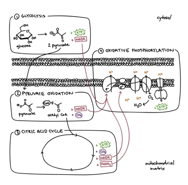
describe the Electron Transport chain phase of respiration
overview
e movement in more detail
shuttling e through Inner Mitochondrial Membrane complexes, via passing between them and various e carriers (Redox rxns - reduced species reduces another and becomes oxidised, where it can be reduced again - passing along electrons)
protein complexes = I, II, III, IV
each e movement, is coupled with / powers H+ pumps to release H+ into the Intermembrane space (against their conc gradient), eventually generating a Proton Motive Force / electrochemical gradient (high H+ ion conc in the Intermembrane space, relative to the Mitochondrial matrix)
ATP Synthase then couples the passive movement of H+ back into the Mitochondrial matrix, with ADP Oxidative Phosphorylation w Pi, to form ATP (harnessing the E of glucose via its e)

when glucose isnt around, how is ATP generated?
what are the drawbacks to this in certain cell types?
what is the strategy to overcome this?
we still need E, in the form of cellular E in ATP to do work, even w/o the presence of Glucose
(Fatty Acids)
turned to first. Beta Oxidation breaks them down into Acetyl CoA, which can be fed into the TCA cycle
then as before, this adds e to e carriers which power the e transport chain to generate ATP via ATP synthase
(AAs)
can be broken down to form Acetyl CoA, which can go into the rest of respiration as before
(drawbacks?)
some cells require glucose for ATP generation
eg Red Blood Cells lack mitochondria, so require gluc for Glycolysis to generate ATP
eg Nerve Cells highly prefer gluc use
how do we maintain gluc in times of deprivation, especially for cells that require it for ATP genreation (RBCs and Nerve cells very important in the body! we need to carry O2 for cellukar function, we need brain to function!)
(solution)
Gluconeogenesis, the inverse pathway of Glycolysis occurring primarily in the Liver (vs Glycyolsysi occurring primarily in the muscles & brain), to create Gluc instead of breaking it down
is costly (4ATP + 2GTP + 2NADH) and generates NO energy or e carriers. BUT is physioologcially essential in the times of requirement
overall, required for Gluc generation, in intense times of deprivation / glycogen depletion, for specific cell types that require Glucose for ATP generation
eg starvation, vigourous excerise, inability to genreate glucose form fatty acids (and must use AAs which is harmful - degrading proteins)
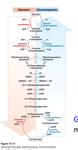
how do Glycolysis & Gluconeogenesis coexist?
the problem
the solution
regulation
(the problem)
they are opposing pathways (create glucose vs break it down) but are both TD favvorable.
they cannot be exactly the same but opposite, as otherwise all steps would be reversible. no TD driving force.
(the solution)
not directly opposite. their reversible steps are shared (Glycolysis steps 2,4-9), but their irreversible steps (deltaG negative, TD unfavorable steps) differ
Gluconeogenesis carries out additional steps to bypass Glycolysis’s 3 Irreversible rxns. so they can coexist and both be TD favorable
(regulation)
Gluconeogenesis IS opposing. instead of creating ATP (as in Glycolysis), it uses ATP / GTP, but this is a worthwhile investment in cell types which need a steady glucose supply even in times of deprivation
regulation occurs differently between the two, preventing a futile cycle - we want pathways to be completed in one direction (GLycoysis OR Gluconeogenesis. not both)
eg if we need Glucose, we dont want intermediate components of Gluconeogenesis to be reversing at eq to undergo Glycolysis and breakdown Glucose overall.
a level of regulation occurs with enzyme GE. Liver = Gluconeogenesis prominent = Gluconeogenesis enzymes expressed preferentially. Muscles / Brain = Glycolysis prominent = Glycolysis enzymes expressed preferentially
Name and describe the Irreversible steps of Gluconeogenesis
(STEP 1)
the bypass taken instead of direct Pyruvate => Phosphoenol Pyruvate (PEP) conversion as in Glycolysis (final step, PEP => Pyruvate, irreversible STEP 10)
Pyruvate Carboxylase, converts Pyruvate => Oxaloacetate, via carboxylation (adding a Bicarbonate’s Carboxyl, via Biotin cofactor)
requires ATP (using Energy stored in ATP, makes it TD favorable)
requires transport into the Mitochondria
(STEP 2)
the second part of the bypass, to convert Oxaloacetate => Phosphoenol Pyruvate (PEP)
Phosphoenol Pyruvate Carboxylase, removes the Carboxyl added in STEP 1
it then adds a Phosphate group (in another position) transferred from GTP
requires GTP (using Enegry stored in GTP, makes it TD favorable)
releases CO2 (forms from added Carboxyl)
completes the bypassing of Glycolysis STEP 10
occurs in Mitochondira or Cytosol (dpdnt on org)
(IRREVERSIBLE STEP)
Fructose Bisphosphatase 1 (opposite of a Kinase, removes a Phosphate), removes the Phosphate from Fructose 1,6-Bisphosphate => Fructose 6-Phosphate
catalyses the Phosphate hydrolysis (use of water for cleavage) to remove the group
ATP is NOT generated, even though P is removed
irreversible, and is the differential converse rxn from Glycolysis STEP 3 (F6P phosphorylation => F16BP)
(FINAL STEP)
Glucose-6-Phosphatase removes the Phosphate group from Glucose-6-Phosphate => Glucose. it catalyses the hydrolysis of this group (use of Water for cleavage)
= we have synthesised Glucose!

how can Brain & Nervous System cells run off alternative fuels to Glucose?
what do they use
how does this work
what scenarios
(overview)
Brain cells rely on Glucose for energy generation (ATP generation Respiration) preferential to Fatty Acids & AAs - but they can utilise Ketone Bodies as an alternative fuel
(scenarios)
in severe glucose scarcity (eg starvation, last resort after days), ketone Bodies can support 60-70% of the Brain’s Energy demands
Ketone Body formation is driven by the buildup of Acetyl-CoA in the Liver cells (entry point to TCA cycle, Glucose => Pyrvate => Acetyl CoA). is counter intutive (you’d think more Acetyl CoA = more glucose = more respiration) why is it not used if the cell is deprived?
due to the maintenance of stable blood sugar levels in the Liver!!! its more of an intearction between blod sugar (Liver - site of gluconeogenesis & fatty acid metabolism) and brain cells (Req glucose)
in low blood glucose situations, Oxaloacetate required for Acetyl-CoA to combine with in the TCA cycle to continue Respiration, is shunted into Gluconeogenesis (as this is happening in Liver cells). (both rxns occurring in the Mitochondria, so diversion can occur)
if Oxaloacetate is all diverted, there is none there for AcetylCoA to combine with to carry out the TCA cycle, therefore it builds up despite low glucose
eg Prolonged Fasting, Starvation, Exhaustive Excersie, Type 1 Diabetes / other conditions (Pathology)
(formation of Ketone Bodies)
built-up Acetyl CoA is converted => 3x Ketone Bodies (Acetone, Acetoacetate, D-B-Hydroxybutyrate)
these can leave the Liver, and be used as fuel for the brain & nervous system cells - even with low Glucsoe (and their pickiness for Glucose!)
Acetone = breathed out. HOWEVER Acetoacetata & D-B-Hydroxybutyrate are soluble => circulate in bloodstream => cross the Blood Brain Barrier via Monocarboxylate Transporters
in Brain cells, they are reconverted to Acetyl CoA => fed into TCA / whatever metabolic pathways => Do repsiration & generate ATP even with low Glucose!

how may the Keto diet be a helpful disease treatment?
a diet hypothesised about since the 1920s. favoring ketone body production for use as fuel for the brain & nerve cells rather than glucose.
low carb (glucose), high fat (Fatty acids), moderate protein (AAs). rather than replicating starvation, they are replicating ketogenesis requirements
(childhood epilepsy)
diseases like childhood epilepsy that are treatment-resistance, may be reduced with a Ketogenic diet
hypothesised to work due to increasing GABA levels, which is an Inhibitory Neurotransmitter in the brain, reducing neurotransmission in the brain - as Epilepsy is cause by heightened neurotransmission
(diabetic medical emergencies)
unintentional Keteogenesis can be involved in Type I most commonly (no insulin transporters produced, no insulin can be released into the bloodstream)
if you miss doses of insulin, no glucose from the bloodstream (from food, from store release from liver) can be taken into cells to do work (carry out survival processes)
this heigthens blood glucose & lowers intracellular glucose - so cells are starved, as no insulin is released to help their uptake of glucose
this replicates the starvation situation for cells, and gluconeogenesis + fatty acid metabolism heigtens - along with ketone body production!
ketone body production however can have harmful effects when put alongside this high blood glucose. they acidify, decreaes blood pH => metabolic acidosis blood + hyperglycemia in blood
w/o insulin, ketone body levels are not regulated
may even seem drunken! acetone breathing off (smell of alcohol on breath) weird symptoms (Acifiied blood & hyperglycameia!)

what are metabolic pathways?
give an example
how are these helpful for synthetic biology?
(metabolic pathways)
a series of linked rxns in the cell
one substrate => converted to various intermediates via enzyme catalysed rxns => ultimately producing an end product
that end product, can then be converted to various others - in other metabolic pathways
starting material => byproducts => end product (eg Energy, Biomolecules, Building block to sustain life)
each enzyme has a different role in the process, to modify products along the way to eventually make the final one
(Example)
Glycolysis
Glucose (starting product) => via hexokinase => Glucose-6-Phosphate (Byproduct / Intermediate) => via Phosphoglucose Isomerase => Byproduct / intermediate
(synth bio)
if we understand these metabolic pathways, we can potentially modify them for our own benefit
this is the fundamental idea of synth bio

what is synthetic biology
how does genetic engineering relate
(synth bio)
engineering principles, in combination with biology, to focus on constructing biological systems / redesigning existing ones, from in the lab
for the purpose of using these for our benefit - various applications (health, environment, manufacturing, agiruclture)
(genetic engineering)
a method that falls under synth bio
carrying out synth bio by manipulating an organism’s genetic material, using techniques in the lab
eg inserting foreign genes, removing native genes, modifying foreign or native genes
in order to purposefully create an organism with new or desired properties, fit for a particular purpose (Depending on the application)
=> Produces a GMO (Genetically Modified Organism)

what is the general opinions around Genetic Engineering (particularly for the general public)
in NZ
in other countries
overall stigma
(NZ)
heavily regulated over its development in the past 30 years - via the HSNO act (Hazardous Substances and New Organisms act 1996)
this rules against GMO commercial crops, GMO field testing - any gene modification is considered a “New Organism”, even single nuc changes
it also ensures strict border control for GMOs
currently - new act trying to pass to make GE easier (Gene Technology Act)
aims to reap its benefits (make commercial crops more efficient by ctrling pathogens, aiding CC by modifting environmental conditions, speeding up health & medical developments)
(other countries)
most other countries allow, and have allowed for a while, Genetic Engineering and commercial growing of GMO crops / food crops
increases their efficiency, yield, profit, etc - refining / adding genes for pest resistance, herbicide resistance, drought resistance, etc
eg USA Soybeans, corn, sugar beets, canola, cotton
(stigma)
seems scary and unknown to non-scientists, as they dont understand the process, its simplicity, and its potential benefits
however, GE is mostly used for beneficial applications, and falls under heavy regulation to ensure so
eg we arent creating glowing animals. we are beneficially making efficient crops

how and when was Genetic Engineering first carried out in history?
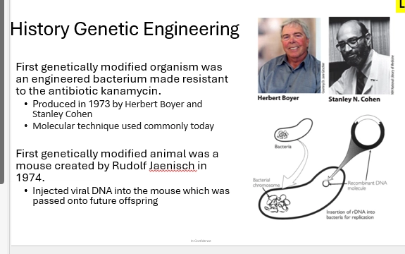
1973, Boyer & Cohen
done so first with a Bacterium - easy to work with.
used a recombinant DNA molecule (a plasmid / vector), which can exist seperately to the BActerial chromo
they added the gene of interest to the recombinant DNA outside of the cell => added this to the bacteria => resulted in the bacteria gaining Kanamycin Antibiotic resistance = phenotypic transformation
this technique is still commonly used today
(next steps)
1974, the first GE animal was developed - a mouse by Jaenisch
used viruses => injected viral DNA into mouse => passed onto future offspring = genetic change = GMO mouse
describe the example of Insulin, as GE to insert Foreign Genes into an organism
(Insulin)
an important regulator of Gluc synthesis / Glycolygen synthesis & breakdown - many important pathways for regulating blood suar levels
is secreted by Beta cells in the Pancreas in high Gluc conditions, and inhibited in low Gluc conditions
in Diabetes, Insulin is either not produced / reduced / prevented from secretion (no transporters) - so for their body to properly function they must recieve it from another source
(original gathering)
originally, they derived this from dog (1922), then large scale production with cows & pigs (1923-1930s) -
however, these animals have different AAs to humans so their Insulin was recognised as foreign and attacked - rejected from the body - by some diabeteics (allergy effects)
this was also costly and had questionable animal ethics (60 pigs req to provide for a diabetic person’s insulin for a year)
(the refined, current, method)
so GE enables recombinantly produced insulin, the intuitive strategy, inserting the unmodified Insulin gene (Human, avoid allergy), into bacteria, which then can produce enough insulin commercially, with their cell machinery, to treat diabetics
1982, insulin was the first large scale application of this method
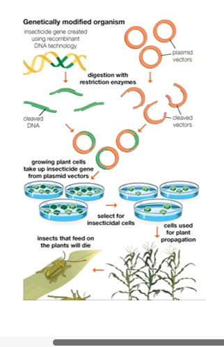
DNA of interest identified, Human Insulin Gene => excised from human DNA => vector cleaved open => gene inserted and sealed = recombinant vector (plasmid)
add plasmid to host (bacteria, E.Coli) => as it replicates, each offspring gains a copy of this plasmid => is translated with host machinery, alongside host genome = recombinantly produces insulin that is harbested
with this method, 1L bacteria = 4g insulin = 170 pig Pancreases

describe the example of BT Corn, as GE to insert Foreign Genes into an organism
Commercial corn growing had a problem, crops kept getting infected by European Corn Borer, damaging the plant and decreasing yield
they knew that Bacillus thuringiensis Bacteria produced a toxin toxic to Borer larvae, but not to humans - it was typically used in an organic insecticide spray
(genetic engineering)
however, this is the perfect setup to use GE to make the corn itself, Borer resistant, rather than having to apply a spray!
typical recombinant method was uesd, to add the toxin-producing BT gene to corn, to provide it resistance from these insects = better commercial output
plasmid vectors opened => BT toxin gene inserted and sealed => produces a recombinant plasmid => is added to plant cells => plant cells are selectd for with the plasmid => they pass this onto offspring => these cells are used for plant propagation
= these have the plasmid so will translate the gene and produce the resistance toxin = harmful insects feeding on the plant will be killed!
how can GE modify native or foreign genes?
name the 3 methods
(modify?)
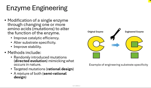
via Enzyme Engineering! to change the AAs in a genetic sequence, to modify the protein it encodes for, in a meaningful way that benefits our problem
introducing mutations, but specific to what we want it to do
eg improve catalysis efficiency (make a less efficient enzyme more efficient)
eg change specificity to substrate (eg if we want it to favor one rxn pathway more for efficiency or to reduce another process, if we want to introduce a new rxn pathway)
eg increase stability (increase efficiency, amount carried out in cell)
(3 methods)
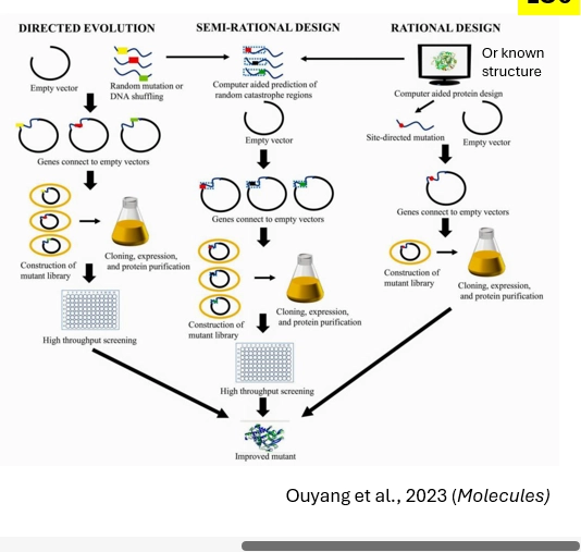
directed evolution - randomly introduce mutations, mimicing what occurs in nature (hence the evolution term. directing evo.)
rational design - targetted mutations, designing the enzyme structure to be specific for the purpose, requiring specific mutations to achieve
semi rational design - a combination of the two.
describe the Rational Design method of Enzyme Engineering
targetted, intentional, method to redesign a protein, rationally mutating based on the AAs u want to change, to make the Enzyme change in the way u need it to
changing AAs / DNA genetic sequence, in the particular area of interest for the enzyme / protein, specific and purposeful (not mutating the rest of the protein structure)
requires in depth knowledge on the Enzyme (structural, mechanisms)
to test it, insert the mutated DNA for the enzyme into a plasmid => recombinant => add to organism to do tsln => see whether the modified enzyme does what you want
(pros)
only generating a few mutant variants, which can easily be tested to see if the mutation has done what you want
not requiring a broad screening, we can individually asses each variant - so less effort computationally
(cons)
must know lots about the enzyme / protein structure before we begin - but quite often we dont know this
without previous knowledge, more work will be required to figure this out

describe the Directed Evolution method of Enzyme Engineering
Nobel Prize 2018. revolutionary at this time, so still quite a new idea
the fundamental idea is directing / promoting evolution to occur for an enzyme’s genetic sequence
natural selection, mutations will be accumulated and may become beenficial or hamrufl as conditions change, so are selected for or against
but in a lab, on a time scale of weeks.
(Process)
(Generating the variants) take the DNA of interest (eg region coding for an Enzyme) => inducing mass mutations to happen => generate a library of DNA variants (up to millions!)
(Screening the variants) these DNA variant genes are added to plasmids => recombinants into bacteria => cultured bacteria will grow and use a random one of tehese variants as a template to make the enzyme
(Screening the variants) those with more efficient enzyme activity / ability to catalyse the desired rxn (Gene mutated in a beneficial way) will be selected for (those who can survive efficiently, will survive, we can culture these, and determine what variant of the Gene they contain)
(Repeat the cycle) then from this, the cycle repeats with the more-ideal-DNA enzyme mutant, in another bacteria, to try accumulate more beneficial mutations = beneficial activity for purpose
(pros)
we dont need to know in depth prior knowledge about protein structure
can use the mutation to solve for protein structure, if we know where it is and how it helped
(cons)
screening is large to look at all the variants , must sort through heaps - due to the fact that there is a high chance for the mutations to be useless / silent / harmful rather than beneficial (& to the purpose we want)
may need many rounds, to enable slow compounding changes to the enzyme via each mutation, to get the desired changes

describe the Semi-rational Design method of Enzyme Engineering
whats done in reality, rather than a defined Rational or Directed Evo approach
making do with the combination of info u start with
also aided by the up and coming advances in AI tools, gotten better and getting better (Eg Alphafold to predict protein structure, may not be entirely accurate but can provide a good guess )
helps figure out AS enzyme structure from DNA sequence - so helpful for these examples where we want to change the AS to do a purpose we want, whether rational or directed to hope it happens ranomly
AI can then help see what mutations will give effects we want (reverse)
Semi = higher chance of a variant with the desired function
how is DNA mutated in the lab to carry out Enzyme Enginering? (Directed Evo approaches)
can do PCR but with less specific / accurate Polymerases = epPCR / Error Prone PCR
here they replicate the target gene, but have a higher chance of adding in incorrect nucs => higher chance of a mutation forming
describe the Enzyme Engineering process that generated Roundup Ready Crops
premise
engineering process
controversy / cons
(the premise)
roundup is glyphosate, weed killer, used in farming so is beneficial for commercial crops - increasing yield & efficiency
Glyphosate inhibits EPSP Synthase enzyme, in the Shikimate Pathway, which converts Shikimate => Trp / Phenal / Tyr (essential AAs)
inhibiting this pathway therefore, causes plant death as the synthesis of essential AAs is prevented
however, alongside killing Weed plants, Glyphosate also kills commercial crops if sprayed - so spraying must be done very carefully
to make this more efficient - could we create Glyphosate-Resistant commercial crop seeds? so the spray will only kill the weeds?
(the engineering)
Directed Evolution method of EE - as they didnt know enough about EPSP Synthase protein structure to Rationally Design AS via genetic sequence
EPSP Synthase Gene was of interest => Generated 200,000 variants with error prone Pol to introduce mutations
variants added to plasmids => transformed into bacteria plated with Glyphosate => therefore those who survived and grew, were of interest to screen!
these had the particular mutation in EPSP to make it Glyphosate resistant => 13 surviving colonies were screened => found to have a particular mutation shared among (1AA chagne)
this mutation was selected, as this shows it does the particular function of interst!
(the controversy / cons)
may enable spread of herbicide resistance to other plants (eg deterimental if spread to a weed plant) via pollen, as we are creating this super enzyme
may contaminate neighbouring farms where resistance could arise (via seed contamination with the crops)
this also brings up points on intellectual property claims - unintentional possession, is it right to have these growing in areas where they could spread to neighbouring farms that dont want it?
needs more overall spraying
risk assessment of the mutation - what if it affects the enzyme in another way (Eg another pathway) or an interconnected pathway, or costs the cell more energy (worse for commercial plants)

what must you consider with Enzme Engineering in terms of Metabolism?
we must consider potential effects in our testing, before releasing it for commercial use
(metabolic pathways are connected)
metabolism is a large series of interconnected pathways - if we modify one enzyme to be better at our process of interst - what if it has knock on effects to other pathways?
these mutations / engineerings arent occurring in isolation. how else may it be affecting the cell?
(potential metabolic burden)
recombinantly added proteins (our modified enzymes) may introduce a high metabolic burden with the changes we engineer
consider the speed & flux of rxns we are promoting

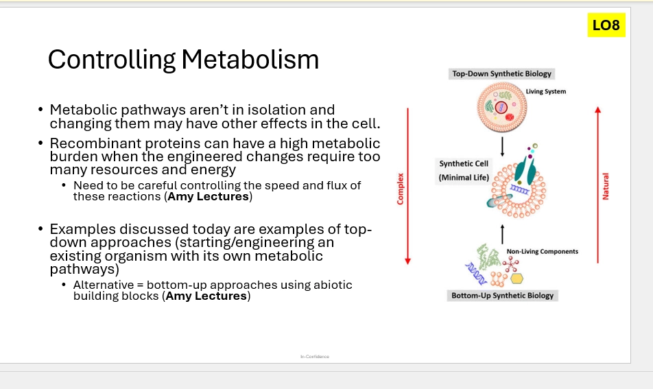
for Enzyme Engineering, what are the Top Down & Bottom Up approaches?
(Top down)
starting with a living system, and engineering its metabolic pathway
may have problems with preexsisting, interconnecting pathways / metabolic balance in the cell (Eg may be energy burden)
(Bottom up)
starting with chemical compounds in the lab, and building these up to engineer a new metabolic pathway from scratch
rather than interferring with existing pathways - we make our own
however we lack the template as in top down (we have to build something from nothing)
what was Science like at the time of the Michaelis-Menten theory
what was the purpose / premise behind these scientists developing it?
DNA & genes were unknown, and proteins werent really known about either
we knew cells, and that they were made up of polymers - how they worked was where there was lacking knowledge
(the premise)
the Michaelis Menten duo (Husband & Wife) worked on among the first enzymes to be studied - Invertase
Invertase, like other first enzymes, isolated from animal guts & fungus (readily available, those at high conc were the ones extracted) - breaks down Glucose
they wanted to find out how enzymes worked kinetically, how quickly they do chemistry, and how they actually do chemistry
(the equation)
from this they determined a Mathematical Framework - the Michaelis Constant & Michaelis-Menten Theory
MM theory
what is it
what are the parameters and what do each represent
(what?)
relating the chemical equation that underpins Enzyme rxns, with the rate of these rxns - to put into a useful framework
(Enzyme Rxn Equation) E + S <=k1=> ES -k2→ E + P
(MM Equation) V0 = V max [S] / Km [S]
(parameters)
[S] = Substrate conc
Vmax = Maximum rate of Enzyme Catalysis for a rxn, the point where all Enzymes are saturated and rate cannot increase further therefore
maximum Enzyme Velocity - the amount of S used / P formed, per unit time
pleatueas on curves where rate incr with incr [S], where incr [S] cannot phsyically incr rate
Km = Michaelis constant. the [S] at half the [S] required to reach Vmax
essentially represents E+S binding affinity. Lower Km means less [S] is required to saturate the enzymes (Reach Vmax) = higher binding affinity
V0 = the Enzyme Velocity (S used per unit time) at the very start of the rxn - where [S] is at peak, [P] is at minimum, and only the forwards rxn is occurring
(assumptions)
Steady state conditions
[S] > > [E]. Substrate conc much higher than Enzyme conc
Initial conditions of [S] > > [P], essentially no Product at V0, only Substrate
![<p>(what?)</p><ul><li><p>relating the chemical equation that underpins Enzyme rxns, with the rate of these rxns - to put into a useful framework</p></li><li><p>(Enzyme Rxn Equation) E + S <=k1=> ES -k2→ E + P</p></li><li><p>(MM Equation) V0 = V max [S] / Km [S]</p></li></ul><p>(parameters)</p><ul><li><p>[S] = Substrate conc</p></li><li><p>Vmax = Maximum rate of Enzyme Catalysis for a rxn, the point where all Enzymes are saturated and rate cannot increase further therefore</p></li><li><p>maximum Enzyme Velocity - the amount of S used / P formed, per unit time</p></li><li><p>pleatueas on curves where rate incr with incr [S], where incr [S] cannot phsyically incr rate</p></li><li><p>Km = Michaelis constant. the [S] at half the [S] required to reach Vmax</p></li><li><p>essentially represents E+S binding affinity. Lower Km means less [S] is required to saturate the enzymes (Reach Vmax) = higher binding affinity</p></li><li><p>V0 = the Enzyme Velocity (S used per unit time) at the very start of the rxn - where [S] is at peak, [P] is at minimum, and only the forwards rxn is occurring </p></li></ul><p>(assumptions)</p><ul><li><p>Steady state conditions</p></li><li><p>[S] > > [E]. Substrate conc much higher than Enzyme conc</p></li><li><p>Initial conditions of [S] > > [P], essentially no Product at V0, only Substrate</p></li></ul><p></p>](https://assets.knowt.com/user-attachments/05088032-7598-49eb-baee-01fe0c851363.png)
what are the assumptions of the MM theory
why are each important
(importance)
if we ensure these assumptions are met to the best of our ability experimentally, we can simplify & derive the MM equation to only contain the 3 simple parameters ([S], Vmax, Km) that we can easily / realistically, measure and figure out
so set up experiments well, to ensure these are mostly true
from this, we can use experimental data to figure out MM parameters, despite its complexities
can vary [S] etc, can vary enzymes, can use to compare
we use MM equation = v, the v term being initial rxn velocity (v0), which ensures we are meeting these assumptions as best as possible
([S] > > [E])
Substrate conc much higher than Enzyme conc
this enables us to make measurements experimentally and accurately, by changing only [S]
(Initial conditions of [S] > > [P])
essentially no Product at V0 / start of rxn, only Substrate
means we only have to account for the forwards Enzyme Catalysis S&P rxn, in terms of rate - simplifying the equation AND there cannot be any reverse rxn possible, fiurther simplifying it
(Steady state conditions)
where the rate of ES formation (E+S => ES) = rate of ES breakdown (ES=> E+S OR ES→E+P)
recall E+S <=k1, k^-1=> ES -kcat→ E+P
this is assuming that k1 = k^-1 + kcat
this is done to simplify the equation into measurable quantities - we can never know [ES] in solution, it is so quickly forming and deforming, but we know the initial [S] & [E] & [ES] )(will = 0), where we can assume that [ES] will go up quickly, and be made stable (formation = breakdown)
steady state is therefore achieved experimentally - but with caveats. formation = breakdown does change a bit, but not alot. we try our best to ensure this assumption is true - but relies on the other ones that we can do for sure
![<p>(importance)</p><ul><li><p>if we ensure these assumptions are met to the best of our ability experimentally, we can simplify & derive the MM equation to only contain the 3 simple parameters ([S], Vmax, Km) that we can easily / realistically, measure and figure out</p></li><li><p>so set up experiments well, to ensure these are mostly true</p></li><li><p>from this, we can use experimental data to figure out MM parameters, despite its complexities</p></li><li><p>can vary [S] etc, can vary enzymes, can use to compare</p></li><li><p>we use MM equation = v, the v term being initial rxn velocity (v0), which ensures we are meeting these assumptions as best as possible</p></li></ul><p>([S] > > [E])</p><ul><li><p>Substrate conc much higher than Enzyme conc</p></li><li><p>this enables us to make measurements experimentally and accurately, by changing only [S]</p></li></ul><p>(Initial conditions of [S] > > [P])</p><ul><li><p>essentially no Product at V0 / start of rxn, only Substrate</p></li><li><p>means we only have to account for the forwards Enzyme Catalysis S&P rxn, in terms of rate - simplifying the equation AND there cannot be any reverse rxn possible, fiurther simplifying it</p></li></ul><p>(Steady state conditions)</p><ul><li><p>where the rate of ES formation (E+S => ES) = rate of ES breakdown (ES=> E+S OR ES→E+P)</p></li><li><p>recall E+S <=k1, k^-1=> ES -kcat→ E+P</p></li><li><p>this is assuming that k1 = k^-1 + kcat</p></li><li><p>this is done to simplify the equation into measurable quantities - we can never know [ES] in solution, it is so quickly forming and deforming, but we know the initial [S] & [E] & [ES] )(will = 0), where we can assume that [ES] will go up quickly, and be made stable (formation = breakdown)</p></li><li><p>steady state is therefore achieved experimentally - but with caveats. formation = breakdown does change a bit, but not alot. we try our best to ensure this assumption is true - but relies on the other ones that we can do for sure</p></li></ul><p></p>](https://assets.knowt.com/user-attachments/c48dc566-cf16-424b-aaef-c0fef64a5657.png)
what is the Kinetics context behind the MM equation?
Kinetics of E & S
relation to MM & derving the equation
(kinetics of E & S)
the rxn rate of E + S → ES is first order with respect to [E] & [S], and second order overall
therefore rxn rate of Enzyme Catalysis, depends on [E] & [S] (how they decrease over time) & [ES] (how it increases over time) - along with the rate constant
the rates in the equation were simplified to only formation & breakdown (in two directions from ES), as the MM equation takes only initial rate (V0), theres no backwards conversion from products, and the ES => EP can be considered very quick)
this means what only has to be looked at, is K1, k-1, kcat
(simpligying the equation)
to figure out [ES] (difficult to measure), we just use the [S] & [E] (known at the start of rxn), along with the initial rate measured (k1, E+S => ES)
with the steady state assumption, rate of ES formation = rate of ES breakdown (with this knowledge, relating k1 (formation rate) to k-1 & kcat (breakdown rate)along with [E] & [S])
simplifying this equation, we can solve for ES, and simpligy the k rates of rxn, to Km ([S] when V is half Vmax)
we combine this with v = kcat[ES], and are even closer to the MM equation (expressing the equation using v)
(next)
here we can relate Kcat & Vmax by [E] - since max velocity occurs when [ES] = [E] (this is when all Enzymes AS are full - fuly occupied by S, maximum rate! the amount of enzyme put in = amount of ES complex)
this simplifies the equation, instead of having both kcat&[E], we link the two into Vmax.
we put this back into the MM equation, which simplifies it to the final one
![<p>(kinetics of E & S)</p><ul><li><p>the rxn rate of E + S → ES is first order with respect to [E] & [S], and second order overall</p></li><li><p>therefore rxn rate of Enzyme Catalysis, depends on [E] & [S] (how they decrease over time) & [ES] (how it increases over time) - along with the rate constant</p></li><li><p>the rates in the equation were simplified to only formation & breakdown (in two directions from ES), as the MM equation takes only initial rate (V0), theres no backwards conversion from products, and the ES => EP can be considered very quick)</p></li><li><p>this means what only has to be looked at, is K1, k-1, kcat</p></li></ul><p>(simpligying the equation)</p><ul><li><p>to figure out [ES] (difficult to measure), we just use the [S] & [E] (known at the start of rxn), along with the initial rate measured (k1, E+S => ES)</p></li><li><p>with the steady state assumption, rate of ES formation = rate of ES breakdown (with this knowledge, relating k1 (formation rate) to k-1 & kcat (breakdown rate)along with [E] & [S])</p></li><li><p>simplifying this equation, we can solve for ES, and simpligy the k rates of rxn, to Km ([S] when V is half Vmax)</p></li><li><p>we combine this with v = kcat[ES], and are even closer to the MM equation (expressing the equation using v)</p></li></ul><p>(next)</p><ul><li><p>here we can relate Kcat & Vmax by [E] - since max velocity occurs when [ES] = [E] (this is when all Enzymes AS are full - fuly occupied by S, maximum rate! the amount of enzyme put in = amount of ES complex)</p></li><li><p>this simplifies the equation, instead of having both kcat&[E], we link the two into Vmax.</p></li><li><p>we put this back into the MM equation, which simplifies it to the final one</p></li></ul><p></p>](https://assets.knowt.com/user-attachments/ad9ca4ce-fa60-4449-b484-cc0a9aa44ab3.png)
describe the steps of deriving the MM equation, briefly / considering the overall purpose of each step
(step 1)
determining the rate of ES formation & breakdown (in terms of k1, k-1, kcat, [E] & [S])
(step 2)
then using the steady state assumption to have these two equations equalling
(step 3)
simplifying the equation begins. we multiply across, etc
we rearrange the equation to solve for [ES]
we then add in Km to replace k-1+kcat / k1 (rate of breakdown/formation), as by definition, Km = rate of ES breakdown / formation - as it is directly half of Vmax (max rxn rate)
(step 4)
we then insert the equation v = kcat[ES]. v being initial rxn velocity (we measure here due to the assumptions fitting)
finally, we insert the equation Vmax = kcat[E]
this provides the final MM equation
v0 = Vmax [S] / [S] + Km
![<p>(step 1)</p><ul><li><p>determining the rate of ES formation & breakdown (in terms of k1, k-1, kcat, [E] & [S])</p></li></ul><p>(step 2)</p><ul><li><p>then using the steady state assumption to have these two equations equalling</p></li></ul><p>(step 3)</p><ul><li><p>simplifying the equation begins. we multiply across, etc</p></li><li><p>we rearrange the equation to solve for [ES]</p></li><li><p>we then add in Km to replace k-1+kcat / k1 (rate of breakdown/formation), as by definition, Km = rate of ES breakdown / formation - as it is directly half of Vmax (max rxn rate)</p></li></ul><p>(step 4)</p><ul><li><p>we then insert the equation v = kcat[ES]. v being initial rxn velocity (we measure here due to the assumptions fitting)</p></li><li><p>finally, we insert the equation Vmax = kcat[E]</p></li><li><p>this provides the final MM equation</p></li><li><p>v0 = Vmax [S] / [S] + Km</p></li></ul><p></p>](https://assets.knowt.com/user-attachments/fd607a36-3d45-4eca-97f6-7241da16e925.png)
How do we approach Enzyme Kinetics & solving the MM equation, experimentally?
How was this figured out in history?
(what do we want)
we want to experimentally determine Km ([S] at half max velocity) & Vmax (maximum velocity / rxn rate) - however these cannot be directly measured
to figure these out, we must find other aspects of the MM equation, and rearrange to solve for what we want
eg [S], [E] (known at the start). [P] (can be measured), V0 (initial catalysis rate with no backwards rxn - so the model fits)
(first attempts in history)
Invertase was looked at first (abundant in nature) - which takes Sucrose (S) => breaks a Glycosidic bond (ES) => Forms Glucose & Fructose (P)
the Catalysis Rate V, increased with [S] - upto a certain point (Osullivan & Tompson & Brown) = a Rectangular Hyperbole Curve
not expected in chemistry - incrasing [S] should increase with rxn rate forever!
from this, they determined the E+S <=> ES <=> EP <=> E + P, model of Catalysis rxn
however - each step had its own rate. 6 rates, made it complex - how would this be described on a single curve?
Henri & MIchaleis Menton came in - experimentally setting up the rxn in a way without P at the start, so by only measuirng V0, there cannot be any back rxn from Products
can also assume, that the ES<=>EP step is very fast (non covalent step)
this provides only 3 rate parameters, that can be described by the curve (easy to model, easy to experimentally determine)
(MM equation)
fits the invertase experimental data well. provides evidence for it being suitable.
![<p>(what do we want)</p><ul><li><p>we want to experimentally determine Km ([S] at half max velocity) & Vmax (maximum velocity / rxn rate) - however these cannot be directly measured</p></li><li><p>to figure these out, we must find other aspects of the MM equation, and rearrange to solve for what we want</p></li><li><p>eg [S], [E] (known at the start). [P] (can be measured), V0 (initial catalysis rate with no backwards rxn - so the model fits)</p></li></ul><p>(first attempts in history)</p><ul><li><p>Invertase was looked at first (abundant in nature) - which takes Sucrose (S) => breaks a Glycosidic bond (ES) => Forms Glucose & Fructose (P)</p></li><li><p>the Catalysis Rate V, increased with [S] - upto a certain point (Osullivan & Tompson & Brown) = a Rectangular Hyperbole Curve</p></li><li><p>not expected in chemistry - incrasing [S] should increase with rxn rate forever!</p></li><li><p>from this, they determined the E+S <=> ES <=> EP <=> E + P, model of Catalysis rxn</p></li><li><p>however - each step had its own rate. 6 rates, made it complex - how would this be described on a single curve?</p></li><li><p>Henri & MIchaleis Menton came in - experimentally setting up the rxn in a way without P at the start, so by only measuirng V0, there cannot be any back rxn from Products</p></li><li><p>can also assume, that the ES<=>EP step is very fast (non covalent step)</p></li><li><p>this provides only 3 rate parameters, that can be described by the curve (easy to model, easy to experimentally determine)</p></li></ul><p>(MM equation)</p><ul><li><p>fits the invertase experimental data well. provides evidence for it being suitable.</p></li></ul><p></p>](https://assets.knowt.com/user-attachments/ff0f7183-e682-49a8-bee6-0edf6d074117.png)
in practice, how would we carry out an experiment to solve the MM equation?
(the goal)
determine V0 (initial rate) with a known [E] & [S], to figure out Vmax (max catalysis rate) & Km ([S] at half max catalysis rate)
(step 1)
determing V0 at varying [S]
mix the S at varying concs, while keeping [E] constant (we are determining a propety of the enzyme, keep it consistent), while measuring [P]
eg use the Beer-Lambert law to use Absorbance & Molar Absorptivity to find [P]
plot this [P] as a function of time
measure the initial run, the linear increase in rate before it plateaus (the high [S], the rate goes up quicker in an observable way), which = V0
express this in units of [P] / unit time (eg mmol / minute)
(step 2)
plot V0 as a function of [S], at the varying levels (collection of experiments - collect lots of data to reduce error, to make the modelled parameters more accurate)
this nicely fits the MM euqation
Vmax will be represented at the Asymptope (the catalysis rate plateau)
1/2Vmax will be half this
then Km will be the [S] at ½Vmax
![<p>(the goal)</p><ul><li><p>determine V0 (initial rate) with a known [E] & [S], to figure out Vmax (max catalysis rate) & Km ([S] at half max catalysis rate)</p></li></ul><p>(step 1)</p><ul><li><p>determing V0 at varying [S]</p></li><li><p>mix the S at varying concs, while keeping [E] constant (we are determining a propety of the enzyme, keep it consistent), while measuring [P]</p></li><li><p>eg use the Beer-Lambert law to use Absorbance & Molar Absorptivity to find [P]</p></li><li><p>plot this [P] as a function of time</p></li><li><p>measure the initial run, the linear increase in rate before it plateaus (the high [S], the rate goes up quicker in an observable way), which = V0</p></li><li><p>express this in units of [P] / unit time (eg mmol / minute)</p></li></ul><p>(step 2)</p><ul><li><p>plot V0 as a function of [S], at the varying levels (collection of experiments - collect lots of data to reduce error, to make the modelled parameters more accurate)</p></li><li><p>this nicely fits the MM euqation</p></li><li><p>Vmax will be represented at the Asymptope (the catalysis rate plateau)</p></li><li><p>1/2Vmax will be half this</p></li><li><p>then Km will be the [S] at ½Vmax</p></li></ul><p></p>](https://assets.knowt.com/user-attachments/d49d97bd-535c-4fc3-b71a-e304df41064e.png)
how can we transform the experimentally determined MM plot? (V0 as a function of [S])
why might we do this?
(transforming)
use the same MM plot (V0 as a function of [S]) of experimental data, but take the reciprical of each axis, and plot these (=Lineweaver-Burk Analysis)
1/V0 as a function of 1/[S]
this provides a straight line, rather than a Rectangular Hyperbola
at the Y intercept (where the line meets the Y axis) = 1/Vmax (the recipricol version)
at the X intercept (where the line meets the X axis) = 1/Km
(why)
makes it easier to measure the 3 paraneters of the MM equation, from the experimental data - accurately
with a straight line, can simply get a ruler and make the line of best fit, and determine Vmax & Km based on where it intercepts with Y & X
What do Vmax & Km actually represent, considering Enzyme Activity in the cell itself
(Vmax)
maximum velocity = maximum rate of enzyme catalysis = enzyme rate at infinte substrate conc
the hypothetical idea that S immediately binds to E, catalyses the rxn, then immediately dissoaicates as P and another S immediately binds
= AS always occupied = ES conc always known to be equal to E conc = first order rxn = modelled with MM (the assumptions)
(Km)
the [S] at 1/2Vmax = the [S] where half AS are full, and half are enmpty, for the Enzyme
therefore is an approximation for k-1 + kcat / k1 (the rate of breakdown / the rate of formation, for ES). the simplified rate constants for the enzyme rxn, achieved when measuring V at time0
therefore this is also an approximation of the Enzymes binding affinity for the substrate, which would alter the ratio of the rate of formation vs breakdown (will breakdown at a higher rate, if lower binding affinity)
so with a higher Km = lower binding affinity (higher breakdown rate / lower formation rate = larger number)
with a lower Km = higher binding affinity (lower breakdown rate / higher formation rate = smaller number)
definitely is an approximation - as some ES breaksdown to PRODUCT rather than SUBSTRATE (characeterstic of a lower binding affinity) but Km appraoches this
![<p>(Vmax)</p><ul><li><p>maximum velocity = maximum rate of enzyme catalysis = enzyme rate at infinte substrate conc</p></li><li><p>the hypothetical idea that S immediately binds to E, catalyses the rxn, then immediately dissoaicates as P and another S immediately binds </p></li><li><p>= AS always occupied = ES conc always known to be equal to E conc = first order rxn = modelled with MM (the assumptions)</p></li></ul><p>(Km)</p><ul><li><p>the [S] at 1/2Vmax = the [S] where half AS are full, and half are enmpty, for the Enzyme</p></li><li><p>therefore is an approximation for k-1 + kcat / k1 (the rate of breakdown / the rate of formation, for ES). the simplified rate constants for the enzyme rxn, achieved when measuring V at time0</p></li><li><p>therefore this is also an approximation of the Enzymes binding affinity for the substrate, which would alter the ratio of the rate of formation vs breakdown (will breakdown at a higher rate, if lower binding affinity)</p></li><li><p>so with a higher Km = lower binding affinity (higher breakdown rate / lower formation rate = larger number)</p></li><li><p>with a lower Km = higher binding affinity (lower breakdown rate / higher formation rate = smaller number)</p></li><li><p>definitely is an approximation - as some ES breaksdown to PRODUCT rather than SUBSTRATE (characeterstic of a lower binding affinity) but Km appraoches this </p></li></ul><p></p>](https://assets.knowt.com/user-attachments/f60f4e87-c25d-49f3-b030-d3959bc573c8.png)
what can comparing Km tell us about different enzymes?
their relative substrate binding affinities
enzymes with a higher Km will have lower binding affinity than enzymes with a lower Km
they require a higher concentration of Substrate to have half of their AS occupied
provides a standardised way of confrmation
How does the MM equation relate to Inhibitors?
why may this be helpful?
different parameters of the MM equation (Km, Vmax), change with different types of inhibition
(helpful)
can determine the mechanism of how a certain inhibitor inhibits an Enzyme
eg looking similar to the S => sugests it is competitive (binds to AS, must be similar to do so and block S)
can determine how you should design an inhibitor, to inhibit the enzyme / reduce rxn rate, in the way that you desire
name the 3 types of enzyme inhibition focused for this course
how do each change the MM equation’s parameters?
are all REVERSIBLE inhibitors
they involve eq rxns of dissociation and formation
they affect Vmax & Km differently
(Competitive)
the Lineweaver-Burke plot line moves - Km is modified (increases. increases the slope of the reciprocol)
to experimentally identify, this would look like an LB plot where the slope changes between lines (V0 as a function of [S] recipricols, line of best fit)
this is becaues the X intercept changes (Km) while the Y intercept (Vmax) stays the same
adding the inhibitor, Km increases (as the binding affinity decreases) as it takes a higher [S] to outcompete the inhibitor and get to the maximum Velocity / catalysis rate
(Uncompetitive)
Km & Vmax are decreased (changing [S] changes both Km & Vmax)
1/Vmax gets bigger (Vmax gets smaller)
-1/Km gets smaller (larger negative (Km gets smaller)
V0 as a function of [S] experimental lines of best fit on the LB plot, are parallel (seperate lines get bigger and smaller. they dont change slope)
decreasing Km means increasing binding affinity, due to the inhibitor affecting the kcat (catalytic step) - the inhibitor is essentially holding the ES together. Less [S] req to reach the lower Vmax
(Mixed)
Km increased (binding affin dcr)
Vmax decreases (rate decreasess)
diagnostic in the LB plot is a crossover of the intercepting region / multiple V0
describe Competitive Inhibition in enzymes
the inhibitor binds to the AS, reducing catalysis rate / efficiency, by competing with the Substrate for space, reducing the amount that is catalysed
blocks the AS to prevent catalysis, when it is empty of S
changes the EPS equation to include the rate of E+I <=> EI (enzyme & inhibitor, enzyme inhibitor complex)
increases Km

describe Uncompetitive Inhibition in enzymes
an unusual type of inhibition, where to inhibit the Enzyme, the inhibitor cannot bind unless S has alread bound on
may be due to AS & inhibitor sites overlapping, due to S binding causing a confo change to open up inhibitor site, etc
once bound on, it prevents the ES from dissociating to form E + P or E + S. therefore the enzyme catalytic ability is decreased, as the S is trapped, and the enzyme cannot continue catalysing more
decreases Km. deccreases Vmax
in the ESP equation we add an extra step after ES <=> ES + I<=> ESI
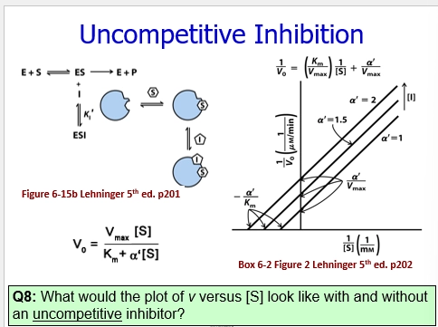
describe Mixed Inhibition in enzymes
where both Competitive & Uncompetitive enzyme inhibition, are both happening at the same time, relatively constantly
unlike Uncompetitive, the inhibitor doesnt require ES to bind. it can bind to E to form EI and inhibit this way. OR bind ES and trap them together
hard to know whats occurring mechanistically
ESP equation, we add the EI & ESI, but also a step betwen EI + S <=> ESI
decreases Vmax (rate decreases)
increases Km (binding affinity decreases)
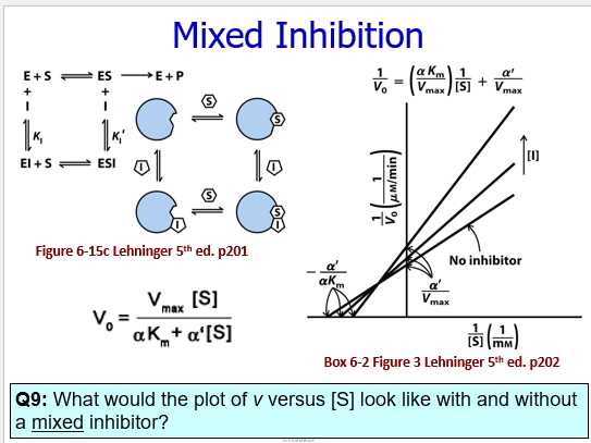
what are irreversible Enzyme inhibitors?
inhibitors that covalentyl bind to the E, typically the AS
one purpose is to make the E do their AS chemistry on the inhibitor, which utilises a side rxn to DESTROY the FG essential for enzyme activity
another means of inhibition is to form a stable, noncovalent, complex with the Enzyme - preventing catalysis as it is stable this way, and has no reason to free the AS

What are some beneficial uses of Synth Bio relating to metabolism?
Making medicine (eg bacteria producing insulin for diabetes treatment)
Improving food (eg yeast producing haeme for Impossible Burgers ‘meaty’ taste)
Making sustainable materials / fuels (eg making perfumes using chemicals for the smells, synthesised in the lab, rather than naturally derived involving mass farming)
Cleaning up waste / pollution (eg engineering metabolisms to break down waste metals etc, to remove from environmental harm)
describe these synthetic biology frameworks of thinking…
engineering framework
top down approach
bottum up approach
(engineering framework)
looking at pathways and enzymes as things we can engineer to change and do the things we want (solve our problem, achieve our end goal)
via creating a computer model => using to modify metabolisms / organisms / enzymes / pathways => analysing the outcomes of these via computer => refine etc
(top down approach)
using preexisting organisms (hosts), to insert your modified metabolisms / pathways / enzymes into - so we have this cellular system starting point
enables cost reduction, and fast production upscaling - when this is the purpose
hosts may be yeasts, bacteria, animal cells, plants
eg cultivate bacteria in bioreactors & maintain ideal growth & production conditions => with the tweaked gene of interest, we can get a great yield of the product of interest here (eg yeast & ethanol => Beer)
enables efficient conversion of raw materials, inton useful things. creating things (often waste productis) more efficiently than from previously industrialised
(bottom up approach)
building enzymatic networks / metabolic pathways, from their chemical elements, within the lab, within a test tube
in this test tube, aimes to create an artificial cellular environment - within which, new creations can be tested
eg purifying enzymes, putting them in the system to mimic other metabolic pathways, creating compartments to organise enzymes
(bottom up pros & cons)
beneficial to quickly check networks for tweaking, and to look at enzyme interaction
very modular, easy to combine enzymes & metabolites, in a controllable and visible way / ratios
easy to purify products / rxn components (in a test tube rather than in a cell / organism)
great potential applications for creating new enzymes, enzymatic pathways - particularly for small bespoke rxns (rather than industrial - maybe better for top down)
eg personalised medicine (tiny volumes of meds needed specifically for a specific person => eg via enzyme cascade)
HOWEVER - hard to scale up (this artificial cell environment, cells are small and organised)
to create larger systems, must purify every enzyme involved and put them in to make the artificial environment realistic
limited functionality in these synthetic systems, they are not evolving and adapting to changes you apply , they can only do a portion of cellular functions
THEY ARENT A CELL ITSELF. YOU LACK THIS FOUNDATION. CELLS ARE COMPLEX CHEMISTRY EVOLVED OVER BILLIONS OF YEARS

what is the CO2 Fixation example in Synth bio?
(premise)
Erb aimed to design a way to carry out this process more efficiently. expanding our ability to capture C, unique to biology, more efficient - to combat climate change etc
currently, 7 known CO2 fixation pathways in biology (eg Calvin Cycle via Rubisco Carboxylase activity)
(the CETCH cycle)
using a Bottom Up approach to mimic the cell environment in a test tube, adding chemicals & enzymes to create a new pathway, he designed the CETCH cycle
in this, CO2 is captured into GLycoxylate molecule, and cycled around. Fixes CO2, and is faster and generates more ATP than Calvin Cycle
shares 4 rxns with 3HP-4HB Archaea cycle
has mixed and matched enzymes from 9 organisms across the domains of life
= shows the modularity possible with this approach.

how is the idea of thinking as Enzymes & Metabolic pathways as parts, promising for future synth bio?
mixing and matching Metabolic Pahtways, Enzymes used in different ones - creates many new possibilities (novel enzymes, new chemistry, new rxns, new combinations & efficiencies)
applying to a situaton / problem of interest, creates great promise for advanced solutions (sustainable, efficient…)
thinking about the Pathway solution space, and the Enzyme solution space, on a graph’s x&y axis, we see the many combos and amount of variation possible in this system
existing pathways & enzymes exist in a tiny space in the total possibilities - copying, pasting, fine tuning, mixing & matching - the pathway variability has so much possibility to increase

in which ways may Enzymes be modified in Enzyme Engineering?
via Rational Design, Directed Evolution, De novo Design
often a workflow using a combination of these approaches, rather than in isolation
changing AA sequence, either randomly or rationally, to change a particular property, to make the enzyme better suited for your purpose
(specificity)
altering this, can favor new substrates (eg decrease specifity for old substrate, incr specificty for another possible pathway or a novel one)
can create new metabolic pathways
(stability)
can change how proteins are folded, to change their capability of existing at different conditions
eg folding in a way to resist denaturing in high T & pH
eg folding in a way to improve solvent or storage tolerability
gives them applications in new situations / cell environments / environmental conditions
(selectivity)
can change how selective they are in their substrate - eg favoring a certain stereoisomer over another
may gain efficiency in / change metabolic pathways. may make rxns more efficient, or more efficiently get the isomer you want
(enzymtic activity)
change AA sequence in such a way to incr catalytic rxn rate
OR turnover of the enzymes
regulate flux better (outflow of products, rate at which metabolites are produced in one enzyme / in a metabolic pathway)
how did the CETCH cycle development & design, use Rational Design EE methods?
the enzyme of target
their approach
use of rational design
their solution
used this method to improve one particular enzyme used as part of the pathway
in V1 of the cycle (1/5.4 for the final version), the rate limiting enzyme of CO2 fixation was Mcd (a type of Dehydrogenase)
this is due to the fact that Mcd catalytic ability was often coupled w an e transfer chain - which is difficult to recreate in a bottom-up (Test tube environment) approach
(their approach)
to recreate this e transport chain, it would be difficult to add a bunch of membrane proteins to solution - instead they looked at e acceptors
Ferrocenium waas first added, but this turned to Ferrocine upon reduction - and could not continuously cycle, and would coagulate enzymes at too high conc (too low conc, would be inefficient)
(rational design)
the Mcd AS structure was known, so Rational Design could be used
they targetted increasign the AS size, so it could accomodate the O2 molecule, to be the new e acceptor (more efficient! replicate this e transport chain, create their efficient CO2 fixation)
to figure out the specific mutations required to do so, enzymes with similar functions in other organisms, were looked at
eg Human Dehydrogenase - coordinates a cofactor (e donor) via pos charged AS pocket
eg Plant Oxidase - binds O2 with a large AS pocket
(solution)
they hypothesised, removing bulky AA residues in the Mcd AS, so O2 could come in and bind, to act as the e acceptor in the chain, to increase Mcd efficiency, as the limiting enzyme
=> designed a TRIPLE MUTANT via changing bulky Tyr residues => other AAs => O2 could bind (larger AS pocket, less negative residues)=> rxn rate increased
Mcd Dehydrogenase => Oxygenase

what is the De novo Design method of Enzyme Engineering?
a new and up and coming concept / method, in this space
(the approach)
using AI to guide the designing of proteins / enzymes from scratch / modifying existing ones, that are beneficial / more efficient for the target problem
the computer predicts mutations to make in the protein, that would make them work better (eg catalytic ability, specifity, selectivity) - via altering AA sequences / creating new ones
(pros and cons)
pros - novel folds & sequences available, unseen before in nature and science
cons - without experimental validation, these AI models may not be 100% correct. may be a beneficial aid, but not entirely accurate
(the origins)
Baker, Hassabis, Jumper - 2024 Nobel Chem prize
Baker - passionate about computer folds, came up with a protein structure folding game
developed Rosettafold => using computers for designing folds (difficult, not really matching experimental results)
developed Alphafold => 2020 AI boom lent to this development, helping our understanding, and allowing computers to predict novel enzyme structures & metabolic pathways & mprove pathways
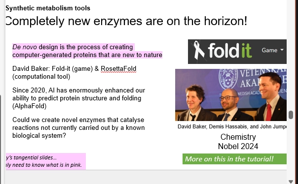
what is one way that metabolic flux can be shifted synthetically?
give an example of this in practice
colocalising enzymes, in the same space in the cell, to incr the flowthrough of metabolites through their catalysis (= incr flux)
via Synthetic Condensates - organisation of metabolites & machinery, as a form of control
(Synthetic Condensates)
design a condensate (cellular & metabolic engineering) => assemle => recruit cargo (eg enzymes & substrates)
=> reactions (enzymes & substrates recruited) are colocalised, so will happen with greater flux (better flow of metabolites, greater efficiency)
can either be selective of the cargo they contain, excluding certain molecules (exclusion), or targetting certain molecules (enrichment)
(Eg - Light Sensitive Proteins)
synthetic condensates & light sensitive proteins, to trigger disassembly with certain stimuli (light)
assembling a condensate containing loght senstiive proteins => when shining a light, they fall apart, as binding is dissociates => the droplet disassembles => metabolites & enzymes are released
therefore in the dark, these condensates assembled (colalisation control occurring. controlling flux by compartmentalising enzymes synthetically)
and in the light, these condesnates disassembled (Resetting colocalisation & metabolic control, back to cell default)
in Yeast, they enriched enzymes of interest into this droplet (targetting certain molecules to recruit as their cargo)
this enabled targetting a certain metabolite, and increasing its flux. with the various enzymes associated with a metabolic pathway branch point (where a substrate could go on to form different products depending on the enzyme used)
to favor their product of interest, the enzymes in the pathways of interest were colaclised in this droplet
resultied in a 6-fold product formation increase, in dark exposure.

define metabolic flux
the rate that a metabolite moves through a metabolic pathway
how fast it flows through. turned from substrate to product. then moving onto the next pathway
this enables cells to tweak metabolic rxn rates, based on how metabolites flow through patwhays, which influences the amount available for the next pathway (and therefore its own rxn spped & efficiency)
what is a main way that cells can regulate metabolism / metabolic flux?
what are the 6 types of this
via changing enzyme activity - directly or indirectly!
(the types)
substrate levels (less substrate available, reduces enzyme rxn rate)
cooperativity
allosteric effectors (may require activation by an external molecule)
substrate cycle
covalent modification (eg phosphorylation, may increase or decrease rxn rate, or prevent / activate catalysis)
changing enzyme concentration (less enzyme present, lowers catalysis overall - may be due to gene expression etc)

how does changing substrate levels, affect enzyme activity (& therefore regulate metabolism / metabolic flux)
how
versatility of this regulation
what is a specific enzyme example
based on the MM equation, changing [S] changes Vmax & Km (maximum catalysis rate & [S] req to fill ½ AS)
only specific enzymes are suitable to be modelled by this equation
(changing [S] effects on MM)
changing [S] at levels close to Km ([S] to fill ½ AS) has larger effects on activity rate (line slope)
less sensitive response for changes to [S] further away from Km
(versatility as a regulator)
only certain metabolic pathways, with certain [S] (close to Km), for certain Enzymes (suitable for MM modelling) - can be regulated by this method
most enzymes exist at [S] levels much higher than Km - so regulation is inefficient by this means (changing [S] will only have small changes)
(specific example)
Hexokinase IV (Glucokinase)
this E catalyses a first step in Glycolysis (different isoforms (slightly different versions) of Hexokinases carry out this rxn in the same way). Redundancy
this E is very sensitive to [S] changes (Glucose) - existing at [Glucose] ~ Km (has a high Km), so its activity is sensitive to [Glucose] changes
exists in Pancreatic Beta Cells, so activity is sensitive to Blood [Glucose] changes, particularly important after eating food
this enables them to carry out their function - [Glucose] increase causes increased activity, to increase production of Insulin (metabolites) to be secreted (to regulate blood [Glucose])
a ‘Glucose Sensor’. additionally, their membranes are higly Glucose permeable (so quite sensitive even additionally) - insulin dependent GLUT2 transporters
compared to Hexokinase I, that isnt specific to this function, which has a much lower Km than [Glucose] it exists in, so it isnt able to be regulated by these means
![<ul><li><p>based on the MM equation, changing [S] changes Vmax & Km (maximum catalysis rate & [S] req to fill ½ AS)</p></li><li><p>only specific enzymes are suitable to be modelled by this equation </p></li></ul><p>(changing [S] effects on MM)</p><ul><li><p>changing [S] at levels close to Km ([S] to fill ½ AS) has larger effects on activity rate (line slope)</p></li><li><p>less sensitive response for changes to [S] further away from Km</p></li></ul><p>(versatility as a regulator)</p><ul><li><p>only certain metabolic pathways, with certain [S] (close to Km), for certain Enzymes (suitable for MM modelling) - can be regulated by this method</p></li><li><p>most enzymes exist at [S] levels much higher than Km - so regulation is inefficient by this means (changing [S] will only have small changes)</p></li></ul><p>(specific example)</p><ul><li><p>Hexokinase IV (Glucokinase)</p></li><li><p>this E catalyses a first step in Glycolysis (different isoforms (slightly different versions) of Hexokinases carry out this rxn in the same way). Redundancy</p></li><li><p>this E is very sensitive to [S] changes (Glucose) - existing at [Glucose] ~ Km (has a high Km), so its activity is sensitive to [Glucose] changes</p></li><li><p>exists in Pancreatic Beta Cells, so activity is sensitive to Blood [Glucose] changes, particularly important after eating food</p></li><li><p>this enables them to carry out their function - [Glucose] increase causes increased activity, to increase production of Insulin (metabolites) to be secreted (to regulate blood [Glucose])</p></li><li><p>a ‘Glucose Sensor’. additionally, their membranes are higly Glucose permeable (so quite sensitive even additionally) - insulin dependent GLUT2 transporters</p></li><li><p>compared to Hexokinase I, that isnt specific to this function, which has a much lower Km than [Glucose] it exists in, so it isnt able to be regulated by these means </p></li></ul><p></p>](https://assets.knowt.com/user-attachments/cddfedf5-b267-483b-977a-63a99edab7b9.png)
how can cells regulate enzyme activity (& regulate metabolism / metabolic flux), by directly changing enzyme conc in the cell?
give a specific enzyme example
inuitively, increasing [E], would increase metabolic flux, as more catalysis is being carried out, to produce more metabolites, from more substrates
(how)
via changing mRNA txn of these enzymes => changes amt of enzyme translated => changes amt of enzyme available in the cell
= txnal activators (various types of molecules that activate TFs. eg signalling molecules activating TFs downstream, TFs themselves activated)
slower, long term, environmental response, changes in regulation (hours-days) VS quicker changes in regulation from stimuli in the other regulation methods (seconds)
proteins stay around for ~1 day, then are degraded, so has a while to take effect from the previous state (so not utilised in a rapid response)
(specific example)
High Carb Diet
induces txn of genes that code for enzymes involved in glycolysis, to breakdown the incr amount of glucose
via Glucose metabolites produced as it reacts - that therefore signal HOW MUCH glucose is present in the cell
specifically Xylulose-5-Phosphate => activates Phosphatase => removes P from ChREBP (Carbohydrate Response Element) TF => enables it to transport into the nuc => can bind onto DNA promotor region for genes of interest
genes related to Fatty Acid Synthesis & Glycolysis (response to lots of glucose = store more, break more down!)
mRNA txn incr => enzymes produced incr => we change the enzyme conc available for rxn => regulating the flux of Glucose to controlled pathways and action
![<ul><li><p>inuitively, increasing [E], would increase metabolic flux, as more catalysis is being carried out, to produce more metabolites, from more substrates</p></li></ul><p>(how)</p><ul><li><p>via changing mRNA txn of these enzymes => changes amt of enzyme translated => changes amt of enzyme available in the cell</p></li><li><p>= txnal activators (various types of molecules that activate TFs. eg signalling molecules activating TFs downstream, TFs themselves activated)</p></li><li><p>slower, long term, environmental response, changes in regulation (hours-days) VS quicker changes in regulation from stimuli in the other regulation methods (seconds)</p></li><li><p>proteins stay around for ~1 day, then are degraded, so has a while to take effect from the previous state (so not utilised in a rapid response)</p></li></ul><p>(specific example)</p><ul><li><p>High Carb Diet </p></li><li><p>induces txn of genes that code for enzymes involved in glycolysis, to breakdown the incr amount of glucose</p></li><li><p>via Glucose metabolites produced as it reacts - that therefore signal HOW MUCH glucose is present in the cell</p></li><li><p>specifically Xylulose-5-Phosphate => activates Phosphatase => removes P from ChREBP (Carbohydrate Response Element) TF => enables it to transport into the nuc => can bind onto DNA promotor region for genes of interest</p></li><li><p>genes related to Fatty Acid Synthesis & Glycolysis (response to lots of glucose = store more, break more down!)</p></li><li><p>mRNA txn incr => enzymes produced incr => we change the enzyme conc available for rxn => regulating the flux of Glucose to controlled pathways and action </p></li></ul><p></p>](https://assets.knowt.com/user-attachments/5c0e07f0-af57-402f-978d-a87b4d458a0a.png)
how does cooperativity affect enzyme activity (& therefore regulate metabolsim / metabolic flux?)
what is this
what are the two models
give an example
(cooperativity)
Enzymes are dynamic, as AA chains move around, closing and opening AS & pockets - changing how Substrates can bind
ligand binding therefore, can change how the AS binds onto Substrate / how available it is / how many products are made (regulate Metabol)
the substrate itself, binds to enzyme AS, which changes its activity (S itself regulating enz activity & flux) = > via changing the stability of the OTHER subunits of the quaternary enzyme
cooperation between the ligand, and the enzyme, to change how much Substrate is catalysed (Regulate rxn)
may change confo to open a new AS, close an AS,
two different models, hard to tell the difference between as enzymse move super quick, may be a combo of both or differ between enzymes
(concerted model)
in a Quaternary complex in a less reactive state, ligand binding can shift eq to a more reactive state
incr Substrate binding => incr Enzyme Activity
this increases more so, as more ligands bind
(sequential model)
in a Quaternary complex in a less reactive state, ligand binding happens sequentially
one ligand binds onto one protein => may readily dissociate but slightly shifts eq
as more ligands accumulate => eq shifts to a more reactive state => ligands bind for longer => complex becomes more reactive => more Substrate Enzyme Catalysis
typically, the ligand being realted to the substrate (Eg a metabolite in the pathway, the substrate itself), so higher conc signalling a higher substrate conc (so more Enzyme activity is required)
(example)
Globins (O2 binding enzymes) monomers / subunits make Haemoglobin Quaternary structures (2 of each type per complex, alpha & beta)
Globins have Fe coordinated by AA side chains (histodine w O groups) to create their AS (binds O2)
Deoxyhaemoglobin w/o O2 bound. a stable state (tense state) as more ion AAs are paired to bind subunits. high O2 affinity (ensure it remains bound)
purpose in the high O2 conc lungs (bind up lots)
Oxygenated haemoglobin w/ O2 bound. ion pairs are broken so is less stable (more relaxed), due to ligand binding (O2), which means at arrival to the tissue, the bound O2 can be delivered (move from high => low conc, not stuck on the enzyme in a stable arrangement)
purpose in the low O2 tissues (release bound)
= enzyme activity changing based on O2 binding (cooperativity with O2 as the ligand)

how is enzyme activity regulated by Allostery (and therefore metabolism / metabolic flux?)
what is this
difference from cooperativity
give an example
(what)
when a ligand binds to the enzyme / complex away from the AS / substrate binding sites - on a seperate allosteric site
here it modulates the affinity of the AS for the S & TS (-based on changing confo, changing charge attractions and repulsions, etc)
it may increase S binding affinity (eg change AS shape to be less complementary) or increase S binding affinity
in turn, increasing or decreasing enzyme activity - based on affecting S binding => affecting the metabolic flux (as more / less substrate flow thru)
(difference)
cooperativity involves the substrate binding to the AS of one quaternary complex subunit, which increases binding affinity for substrates onto the other subunits
so as more S joins and is in conc and is catalysed, the more activity the enzyme complex is regulated to carry out
VS allosteric, a seperate effector (typically not the substrate) binds to a different site on the enzyme, which in turn changes binding to the AS (and thus regulates E activity)
HOWEVER - they often show cooperativity AND allosteric effectors together for regulated enzymes
(example)
Phosphofructokinase I (Glycolysis enzyme, involved in Phosphorylating Fructose-6-phosphate => Fructose-1,6-Bisphosphate)
in high ATP conditions, we dont need to do this step to make more ATP and carry out Glycolysis (rather use the gluc for something else)
PFK1 is allosterically inhibitited by ATP. ATP binds to allosteric site => confo change of AS => decreases binding affinity for AS to substrate (rxn rate lowered, less catalysis)
in low ATP conditions, we want to incr GLycolysis rate via this enzyme (to make ATP)
allosterically activsted by AMP (in high conc with low ATP). AMP binds to allosteric site => confo change of AS => incr S binding affinity (rxn rate incr, more catalysis)
Pyruvate kinase (Glycolysis enzyme, involved in Phosphorylation of ATP from removing Phosphate from Phosphoenol Pyruvate => Pyruvate)
an allosteric effector is Fructose-1-6-Bisphosphate!
binds => incr S binding affinity => aids metabolic flux by upregulating feeding S into the next part of Glycolysis (more F16BP means do more of this final step!)
smoothens the flow thru (metabolic flux)

how do substrate cycles regulate enzyme activity (& regulate metabolism / metabolic flux therefore?)
describe
example
(substrate cycles)
using small molecules, generated as secondary in metabolic pathways (eg not the main focus of the pathway), for a purpose alike signalling molecules
they amplify a regulaatory response, by binding to enzymes, and altering their activity (incr activity, dcr activity, affecting metabolism & metabolic flux)
they help link metabolic pathways - altering enzyme activity in one, can change others, and these small molecule signals are the messengers between
(example)
AMP! a key indicator of cellular enegry status, as Adenylate kinase converts 2ADP=> AMP + ATP, at eq
so as cells use up ATP, AMP is produced = low cell energy. per 2ADP used, 1AMP forms (so a very sensitive indicator)
PFK1 & FBPase-1 (Reverse enzymes of the Phospohorylation of Fructose-6-Phosphate) are regulated by AMP conc
incr AMP = incr PFK activity & decr FBPase activity = huge fold changes in flux (via changing enzyme activity) = incr ATP production

how does substrate cycle enzyme regulation, relate to allosteric enzyme regulation ?
substrate cycles is the side-occurring cycling of substrates into other molecules (typically small ones prodced as a side product) that then go on to bind to enzymes allosterically, and change AS confo / confo, to incr or decr binding affinity of the substrate
therefore incr or decr enzyme activity => incr or decr metabolic flux
based on how much conc of the substrate, indicated by the sideline cycling of it that is also going on
eg 2ADP => ATP + AMP occuring via Adenyl kinase, therefore AMP conc indicates ATP conc - without having to use ATP as the direct effector (AMP smaller, more diffuse, better signaller)
therefore these means of regulation are closely related. substrate cycling can lead to production of allosteric effectors, to regulate enzyme activity based on the conc of the substrate indirectly.
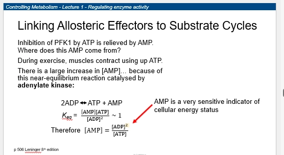
how can covalent modifications regulate enzyme activity (and therefore incr or dec metabolic flux / regulate metabolism?)
what
example
how does this link to other means of enzyme regulation?
(what)
covalently linking components to AAs of proteins, post translation - to change how they interact with molecules within the cell / fold
thus directly affecting enzyme activity (changes how they can interact with S, what rxns they can do, how efficient they are)
eg phosphorylations, methylation, adenylation, ubiquitation
(example)
the key one - Phosphorylation! (addition of P to Tyr, Ser, Thr, His, residues via Kinase, removed by Phosphatases - the reverse)
Phosphorylation is carried out by an Enzyme (Kinase) - which itself can be regulated by all other means, to regulate Phosphorylation of other Enzymes, which regulates their activity (=large effect on metabolic flux)
eg AMP allosterically activates AMP Kinase, which Phosphorylates key proteins to monitor cellular energy status (based on AMP conc outweighing ATP conc, hence high ADP low ATP)
AMP binds => protein subunits associate closer => rxn rate efficient. AMP dissociates => protein subunits dissociate a bit => AS structure not efficient (Inactive)
activated AMP Kinase, can then go around and Phosphorylate (Covalently modify) various other enzymes related to cellular energy levels (= tagging enzymes via phosphorylation. signal amplification, cascade, activations. they can then go onto do other stuff)
various isoforms of AMP kinase (differences in subunits, different tissues) attuned to the needs of the tissue (eg Muscle vs Liver, use vs store energ)

what is another key way of regulating metabolism & metabolic flux - in terms of location in the cell?
how do euks do this?
how do proks do this?
compartmentalisation!
controlling WHERE a metabolic pathway is carried out, can incr or decr its flux
controlling where the S is vs E, local S conc, spatially relating certain metabolic pathways that flow on from eachother - can all incr or decr flux
(euks)
via organelles
membrane-bound cellular compartments, that each have specific functions - so to be efficient they concentrate enzymes & substrates for their respective niche, within these compartments
can have varying conditions to the rest of the cell
can concentrate parts of metabolic pathways (accelerate), can localise various metabolic pathways so they flow onto eachother (incr flux)
eg lipid metabolism
(proks)
they compartmentalise in the cell too - just not with defined membrane-bound parcels
still have rxns & enzyme & susbtrate conc & pathways happening in certain areas of the cell
still largely unknown in science
why is it tricky to measure local metabolite concentrations within cells?
why is this info helpful to know
(helpful?)
we can investigate metabolic pathways, and see how [substrate] may affect their flux
from this, we can figure out how we can manipulate this flux (based on how its reglated) = how we can control these metabolic pathways.
(tricky?)
we can lyse cells and measure [metabolite], but this just gives the average within the whole cell (Rather than in compartments)
we want to know - where was the metabolite bound, where was it free, if it was bound to an organelle, or if bound to an enzyme
eg brain cells [ADP free] < < [ADP total], as it is largely bound in the mitochondria ready to be fed into ATP generation
eg muscle cell [ADP free] < < [ADP total], bound to actin ready for contraction
![<p>(helpful?)</p><ul><li><p>we can investigate metabolic pathways, and see how [substrate] may affect their flux</p></li><li><p>from this, we can figure out how we can manipulate this flux (based on how its reglated) = how we can control these metabolic pathways.</p></li></ul><p>(tricky?)</p><ul><li><p>we can lyse cells and measure [metabolite], but this just gives the average within the whole cell (Rather than in compartments)</p></li><li><p>we want to know - where was the metabolite bound, where was it free, if it was bound to an organelle, or if bound to an enzyme </p></li><li><p>eg brain cells [ADP free] < < [ADP total], as it is largely bound in the mitochondria ready to be fed into ATP generation</p></li><li><p>eg muscle cell [ADP free] < < [ADP total], bound to actin ready for contraction </p></li></ul><p></p>](https://assets.knowt.com/user-attachments/40a66dcf-d4aa-468f-88a2-e606e4f63a37.png)
how can we actually measure local cellular metabolite conc in the muscle cells as an example?
to measure ATP / ADP conc, theres a method using Creatine Kinase & MRI (a special type of NMR)
identifying the metabolites present, by looking at chemical shifts of molecules
can be done in real time with muscle contraction (MRI)
(the premise)
Phosphocreatine (Phosphorylated Creatine) acts as a Phosphate ‘storage’ for Creatine Kinase to chop Phosphates from => enables conversion of ADP => ATP in other metabolic pathways
Creatine Kinase also catalyses the reverse rxn, Phosphorylating Creatine from ATP Phosphate
lots of ATP is required for muscle contraction in these cells (for microfilament motor proteins)
(figuring out metabolite conc)
we can rearrange the equilibrium equation of this conversion, and put [ADP] as the subject, based on [ATP], [Creatine], Keq, [Phosphocreatine2-] & [H+]
we can figure out these other parameters with MRI (a special type of NMR), which non-invasively measure [P31 Phosphorus Isotope]
[Phosphocreatine] is simply provided by chemical shifts - it drops and increases noticeably, and buffers [ATP] (to enable constant contraction)
this makes sense considering the eq eqatuon
to figure out [ATP], measured from backk-calculating detectable signals from the other components chemical shifts (as ATP produces quite a variety that cannot be directly measured)
![<ul><li><p>to measure ATP / ADP conc, theres a method using Creatine Kinase & MRI (a special type of NMR)</p></li><li><p>identifying the metabolites present, by looking at chemical shifts of molecules </p></li><li><p>can be done in real time with muscle contraction (MRI)</p></li></ul><p>(the premise)</p><ul><li><p>Phosphocreatine (Phosphorylated Creatine) acts as a Phosphate ‘storage’ for Creatine Kinase to chop Phosphates from => enables conversion of ADP => ATP in other metabolic pathways</p></li><li><p>Creatine Kinase also catalyses the reverse rxn, Phosphorylating Creatine from ATP Phosphate </p></li><li><p>lots of ATP is required for muscle contraction in these cells (for microfilament motor proteins)</p></li></ul><p>(figuring out metabolite conc)</p><ul><li><p>we can rearrange the equilibrium equation of this conversion, and put [ADP] as the subject, based on [ATP], [Creatine], Keq, [Phosphocreatine2-] & [H+]</p></li><li><p>we can figure out these other parameters with MRI (a special type of NMR), which non-invasively measure [P31 Phosphorus Isotope]</p></li><li><p>[Phosphocreatine] is simply provided by chemical shifts - it drops and increases noticeably, and buffers [ATP] (to enable constant contraction)</p></li><li><p>this makes sense considering the eq eqatuon</p></li><li><p>to figure out [ATP], measured from backk-calculating detectable signals from the other components chemical shifts (as ATP produces quite a variety that cannot be directly measured)</p></li><li><p></p></li></ul><p></p>](https://assets.knowt.com/user-attachments/6477df0d-fdd1-432b-96c8-2c39bc96d0cc.png)
how can we measure local metabolite concentration in a cell, using NMR?
Nuclear Magnetic Resonance - experimental setup that gives spatial insight (rather than real time info like MRI subset - as we req hours of prep)
(setup)
use NMR-labelled metabolic precursors (eg C13 & N15 containing) incorporated into metabolites (eg Glucose with C13) to label the metabolites so we can see how they are converted through pathways
then take cell samples from tissues of interest
then purify the organelle / cellular compartment of interest (various methodologies - eg homogenisation & centrifuge)
to remove the metabolites of interest, use solvents
create spectra of these metabolites => analyse to see if they contain the isotopically labelled elements
from tracing these labelled, taking samples at differnet times, with different profiles if we want to test a treatment, we can see how specific metabolites move throughout the cell spatially
we do suffer with the lack of real time info, as metabolism & changes & enzyme activity can occur fast

how can we measure local metabolite concentration by using metabolic gradients?
(the premise)
metabolites create gradients across the cell / compartment, between their source (Created, high conc) and sink (consumed, low conc)
so to look at local metabolite conc, you can express this in terms of metabolic gradients - a more subtle form of compartmentalisation
defining the source and sink locations, gives insight into metabolic pathways & how metabolites are regulated
( the solution)
needs something in real time, rather than NMR- as it happens quickly
we need cellular imaging - which requires a reporter enzyme (something we can visualise in imaging, to mark the location of other molecules
eeg for ATP, use Luciferase ATP Reporter Enzyme, which converts chemical E (ATP) into light (=bioluminessence in fireflies & glowworms!)
this can be observed when imaging the cell, to indicate ATP conc (& gradients therefore! btwn source & sink areras)
also can indicate WHERE organelles are located spatially compared to others (based on similar ATP / Metabolite concs = closer, larger gradient = Further apart)
how does Luciferase work?
how can we use Luciferase to report ATP location in the cell / compartment?
give an example & what it helped us find out
(Luciferase)
Luciferin molecules are produced in the cell
Luciferin + ATP intermediate (Luciferyl adenylate), acts as a Substrate for Luciferase => forms Oxyluciferin + Photon (= light!)
(Reporting ATP location)
even picomoles of ATP can be detected with Luciferase, as they will react with Luciferin and cycle through to Oxyluciferin + Light, which can easily be detected when imaged (light vs no other light in the cell - good signal, low bckgrnd noise)
if we tag Luciferase to certain cellular locations, inserted into membranes (via compartment specific membrane proteins), we localise it to an organelle of interest / cellular area
here, it’ll provide a good idea of ATP concentration - in real time as it reacts with Luciferin
(Pancreatic B-Cells)
anchor 1mM Luciferase, to different cellular membranes (different cellular locations), and graph together based on Photons per area (ATP per area)
adding a treatment (GLucose), Cytoplasmic [ATP] increased then decreased, while Mitochondria & Plasma Membrane increased and stayed high
firstly - indicating mitochondira are located near the outer PM in a B-cell type, where they provide a localised pool of ATP near the membrane
why - they are responsibe for insulin secretion!
high [glucose] bloodstreatm => taken into cells at membrane => enter mitochondria for respiration => produce an ATP pool near the Plasma Membrane
at high enough [ATP], membrane potential changes cause Insulin secretion into the bloodstream
high [ATP] => K ion channels close => membrane depol => Ca channels open => Ca influx into the cell => Insulin from granule compartments triggered release => Exocytosed
mutations preventing the ATP pool creates Insulin release defects (EG DIABETES)
Ca also activates the TCA cycle to use up bloodstream glucose to form ATP (a positive feedback loop to use the lots of Glucose, also helps to sustain the ATP pool)
![<p>(Luciferase)</p><ul><li><p>Luciferin molecules are produced in the cell</p></li><li><p>Luciferin + ATP intermediate (Luciferyl adenylate), acts as a Substrate for Luciferase => forms Oxyluciferin + Photon (= light!)</p></li></ul><p>(Reporting ATP location)</p><ul><li><p>even picomoles of ATP can be detected with Luciferase, as they will react with Luciferin and cycle through to Oxyluciferin + Light, which can easily be detected when imaged (light vs no other light in the cell - good signal, low bckgrnd noise)</p></li><li><p>if we tag Luciferase to certain cellular locations, inserted into membranes (via compartment specific membrane proteins), we localise it to an organelle of interest / cellular area</p></li><li><p>here, it’ll provide a good idea of ATP concentration - in real time as it reacts with Luciferin </p></li></ul><p>(Pancreatic B-Cells)</p><ul><li><p>anchor 1mM Luciferase, to different cellular membranes (different cellular locations), and graph together based on Photons per area (ATP per area)</p></li><li><p>adding a treatment (GLucose), Cytoplasmic [ATP] increased then decreased, while Mitochondria & Plasma Membrane increased and stayed high</p></li><li><p>firstly - indicating mitochondira are located near the outer PM in a B-cell type, where they provide a localised pool of ATP near the membrane</p></li><li><p>why - they are responsibe for insulin secretion! </p></li><li><p>high [glucose] bloodstreatm => taken into cells at membrane => enter mitochondria for respiration => produce an ATP pool near the Plasma Membrane</p></li><li><p>at high enough [ATP], membrane potential changes cause Insulin secretion into the bloodstream </p></li><li><p>high [ATP] => K ion channels close => membrane depol => Ca channels open => Ca influx into the cell => Insulin from granule compartments triggered release => Exocytosed</p></li><li><p>mutations preventing the ATP pool creates Insulin release defects (EG DIABETES)</p></li><li><p>Ca also activates the TCA cycle to use up bloodstream glucose to form ATP (a positive feedback loop to use the lots of Glucose, also helps to sustain the ATP pool)</p></li></ul><p></p>](https://assets.knowt.com/user-attachments/8390ddf2-e1fa-43a3-a65a-37be96d8c2f1.png)
what is substrate channelling?
how does this help regulate metabolism?
(substrate chanelling)
spatially controlling enzymes in crowded cellular environments, to control flux
cells are densly packed with proteins / enzymes / DNA / organelles / cytoskeleton / polymers
this promotes formation of multi-enzyme complexes, as they are likelier to form transient (non-covalent) interactiions with eachother
due to solvent exclusion effects - increasing the effective enzyme conc (more enzymes in a crowded space, occupy a smaller volume each, as solvent is excluded)
essentially MORE CONCENTRATED with enzymes (vs a non-crowded space, larger) => likelier to interact in random movements => likelier to form multienzyme complexes
EXCEPTIONS - enzymes with lots of confo changes (eg active & inactive) with less space to move around (disfavors rxn)
DIFFICULT TO STUDY - crowded environment is characteristic of the cell, and hard to mimic in the test tube
(regulating metabolism)
multienzyme complexes, hold enzymes closer together transiently, so substrates can be readily CHANNELLED from one AS pocket enzyme, to the next - so flux increases
avoids substrates diffusing out of a pathway for use in something else
prevents the intermediate from going down competing rxn pathways
protects toxic & unstable intermediates from damaging the cell, if they are quickly shuffled
pulls eq forwards towards complete rxn, due to these effects

Name and describe the 5 modes of Substrate Chanelling
how are substrates directly passed between enzymes in multienzyme complexes, to increase flux by directly shuttling them?
(Molecular tunnel)
AA pocket specifically for chanelling S from E1 => E2
seen particularly in AA Biosynthetic pathways, which are important so require tight control to directly form the product from each metabolite
eg Tryptophan Synthase. Indole AA tunell => channels metabolite from A subunit => B sunit => via AA residues guiding the intermedaite through (Favorable, stabilising, interactions)
(Electrostatic surface / bridge)
enzymes with AA charged surfaces, with grooves, to pass metabolites along by forming favorable interactions (Stabilising the intermediates etc) => ASs of other enzymes in the complex to continue metabolism
surface AA residues are specific to stabilising the metabolic intermediate, typically charged + (mutating to - or neutral would decrease rate)
eg TCA cycle Malate Dehydrogenase + Citrate Synthase + Aconitase (positive electrostatic surface)
(Molecular tether / Swinging arm)
localising enzymes that facilitate direct movement of a metabolite through their AS for the rxn pathway, via ‘grabbing’ the metabolite with a ‘swinging arm’ enzyme AA module
eg Pyruvate dehydrogenase complex, see in cryo EM. 3 enzyme complex, with E2 having the swinging arm (Lipoate domain. long chains)
localises 3 enzymes all part of a metabolic pathway, uses the swinging arm to move susbtarte through efficiently and controlled, to complete rxn
Pyruvate => remove CO2 (E1) => E2 swings it over to itself => add Acetyl (E2) => Acetyl CoA (E3 recycles electron carriers to recycle the Lipoate arm)
(Molecular cage)
positions enzyme domains of a complex, in such a way that props them together to form a ‘cage’ / enclosed space where the intermediate is produced
eg Propionyl CoA Synthase, is part of a metabolic pathway that produces a toxic intermediate along the way (Acrylyl-CoA)
producing it enclosed in this cage, prevents damage to other parts of the cell
(Biomolecular condensates)
localising enzymes & substrates by forming membrane-less cellular compartments, to incr metabolic flux (substrate and enzymes kept together. multiple enzymes of a pathway, would be handy)
different types can coexist in the cell, various functions and uses, for both proks & euks
spontaneously form droplets of liquid-liquid phase seperations in the cell, and based on the noncovalent transient interactions of enzymes, metabolism is localised in this way to iincr flux
can occur also with transient intearctions between DNA & RNA (nucleic acids)
can also form in response to stress & signals (an easy means of regulation)
eg Purinosome in Euks, for Purine biosynthesis (Nucleic acids), involing 6 enzymes that localise in droplets, to make this pathway efficiently flux regulated
7-fold synthesis incr compared to freely floating
doesnt even require an organelle.
thought to be energy intensive -they localise close to the mitochondria (ATP)