Hip Special Tests & Midterm Imaging
1/53
There's no tags or description
Looks like no tags are added yet.
Name | Mastery | Learn | Test | Matching | Spaced |
|---|
No study sessions yet.
54 Terms
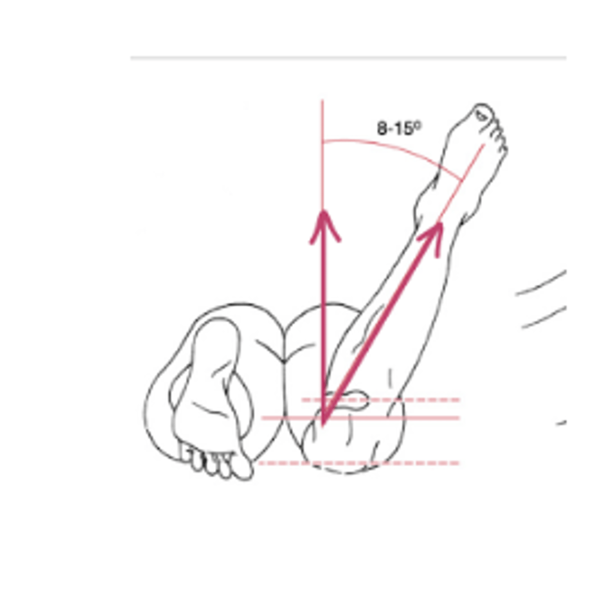
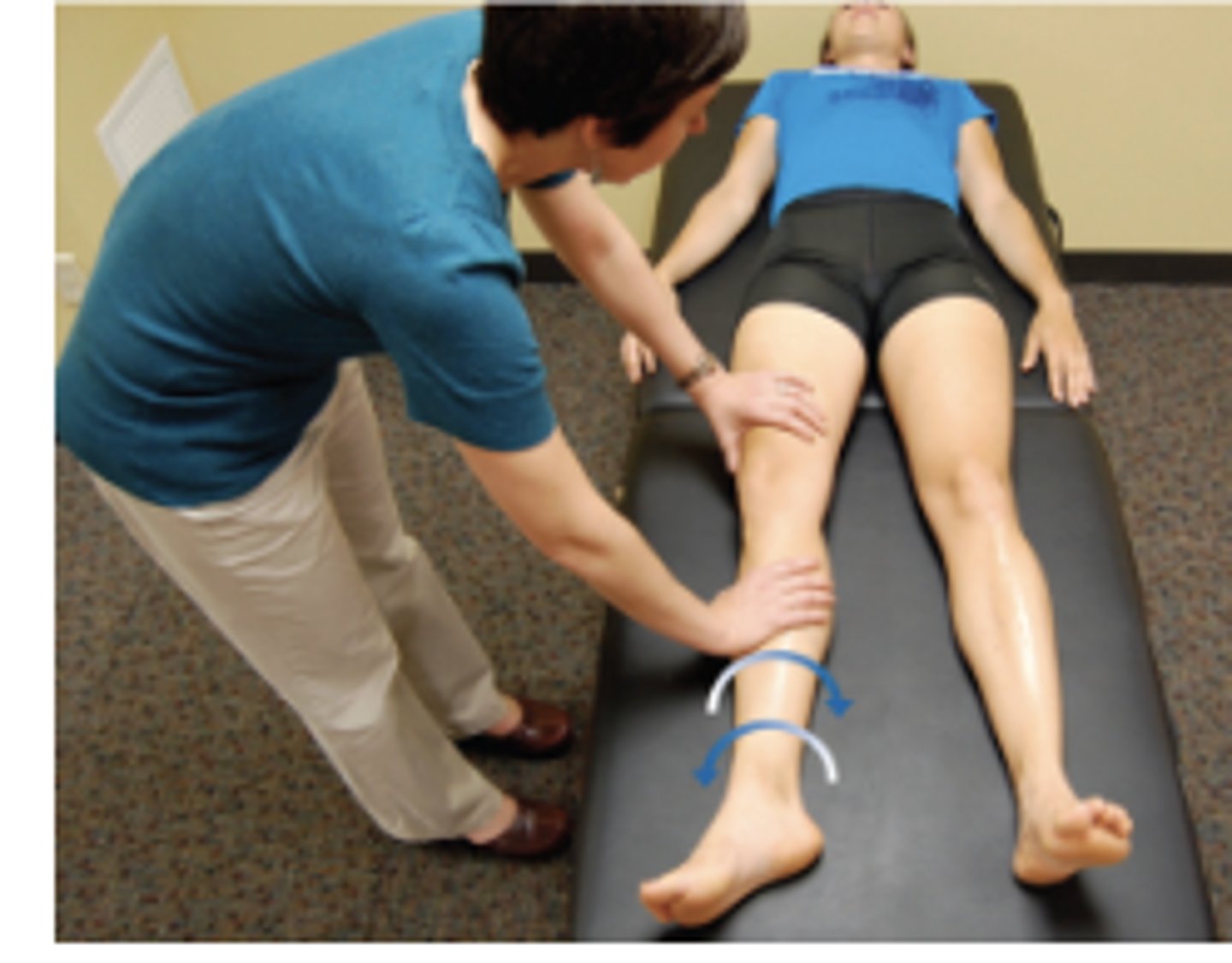
Craig Test
Normal males: 8-12 degrees
Normal females: 12-18
Put hand on greater trochanter and IR/ER femur
Tests for femoral anteversion → if lots of IR you have anteversion
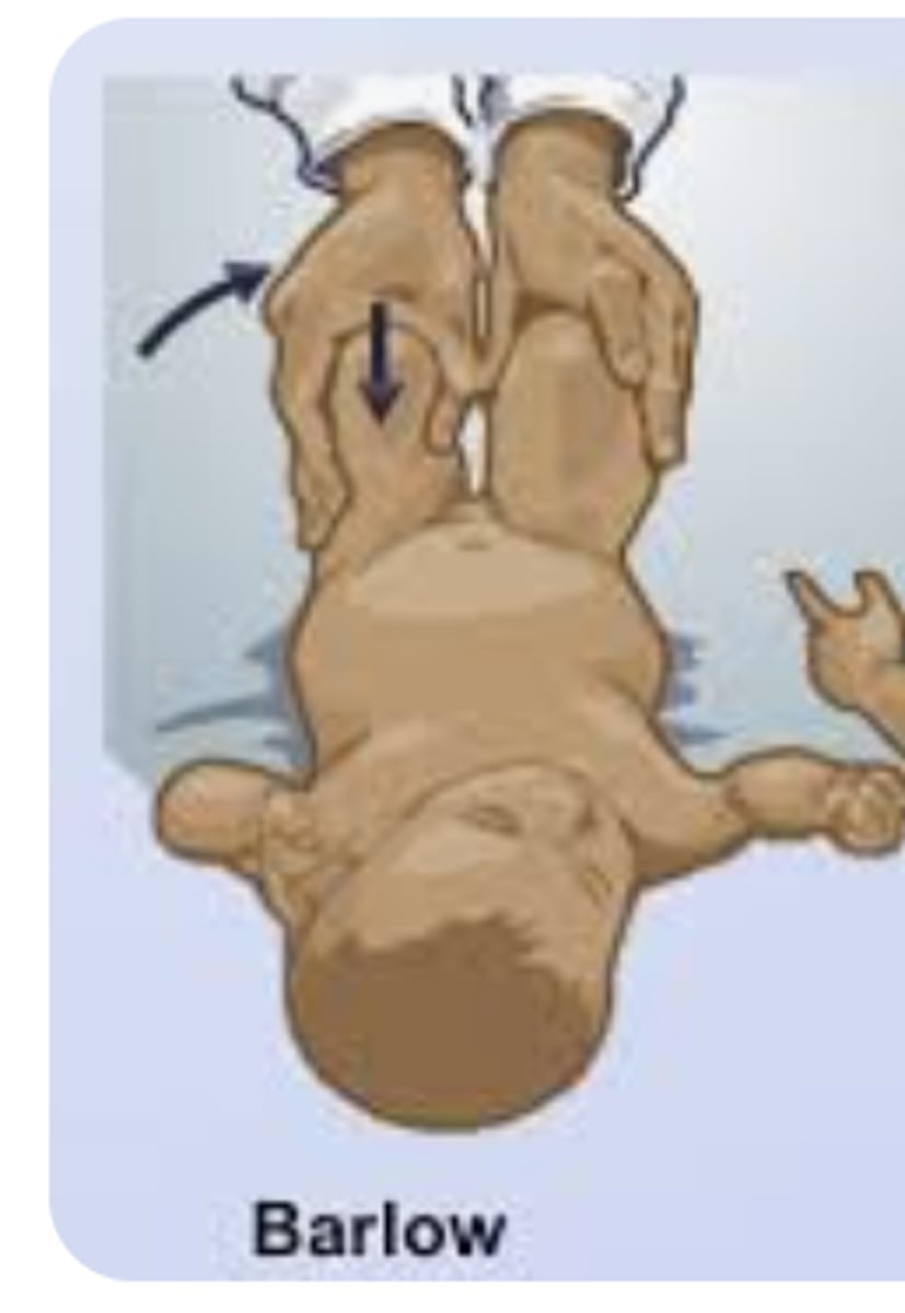
Barlow's Test
Flex + Adduct Hip
Apply posterior force
(+) = dislocation / sublux
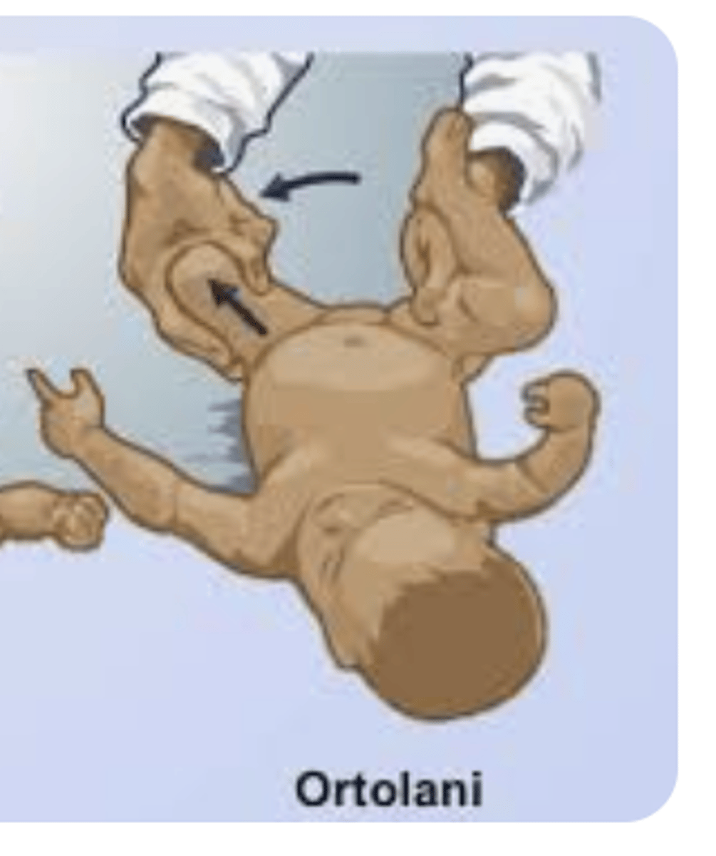
Ortolani's Test
Flex Hips/Knees + Abduct
(+) = relocation
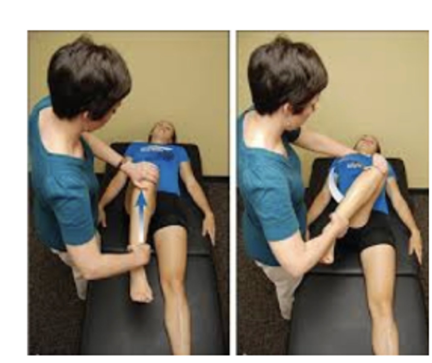
FADDIR
just flex to 90
IR and adduct
Tests for: FAI, hip OA
FABER
Tests for: FAI, hip OA
Quadrant
NO axial load
flex/add/IR quad
flex/abd/ER quad
Tests for: FAI, Labral injury
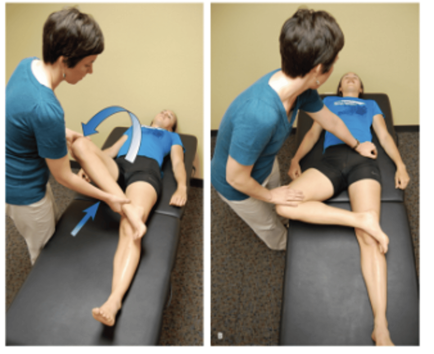
Hip Scour Test
WITH an axial load
flex/add/IR quad
flex/abd/ER quad
Tests for: FAI, Labral injury, OA
Log Roll
Tests for: FAI, labral injury, generalized laxity
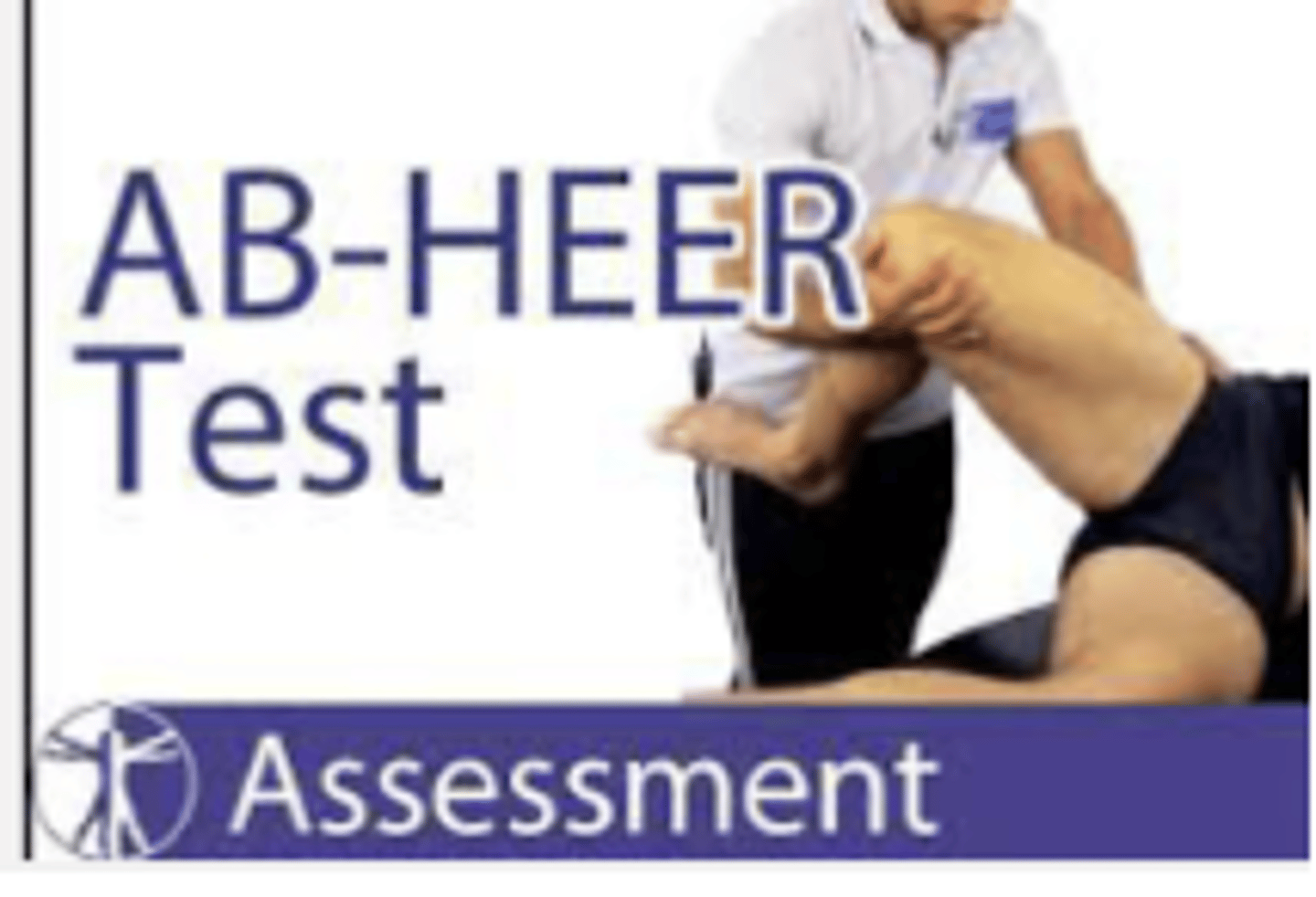
AB-HEER
positive= pain provocation of anterior pain or feeling of instability or apprehension
Tests for: microinstability
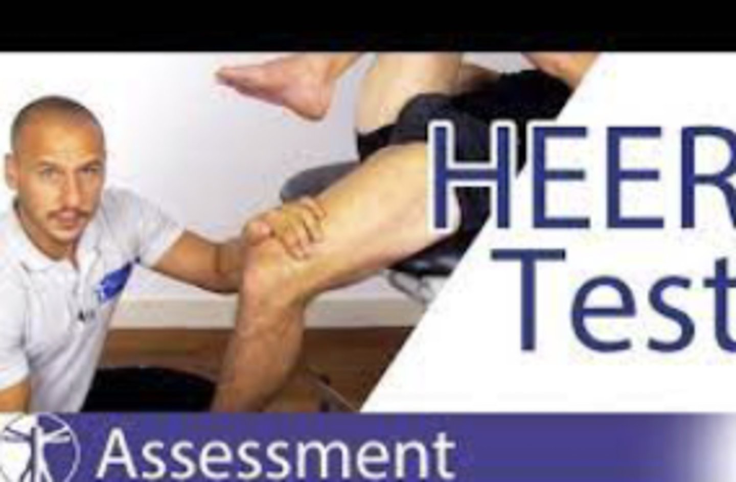
HEER
We are just looking for pain or apprehension here
Be at very edge of table, let them grasp the opposite knee
Push into more extension and ER
Tests for: microinstability
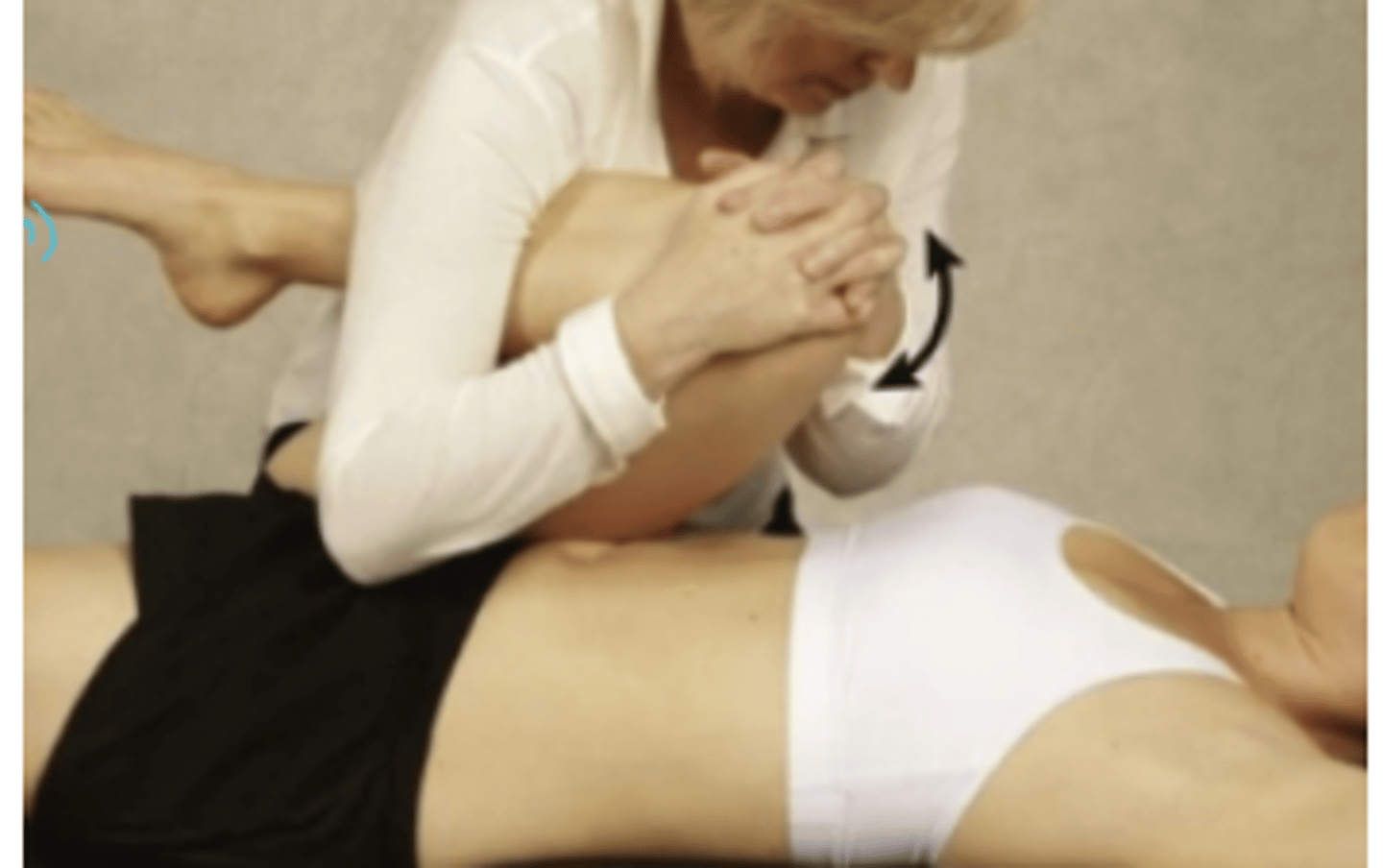
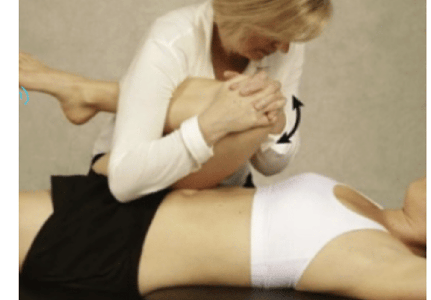
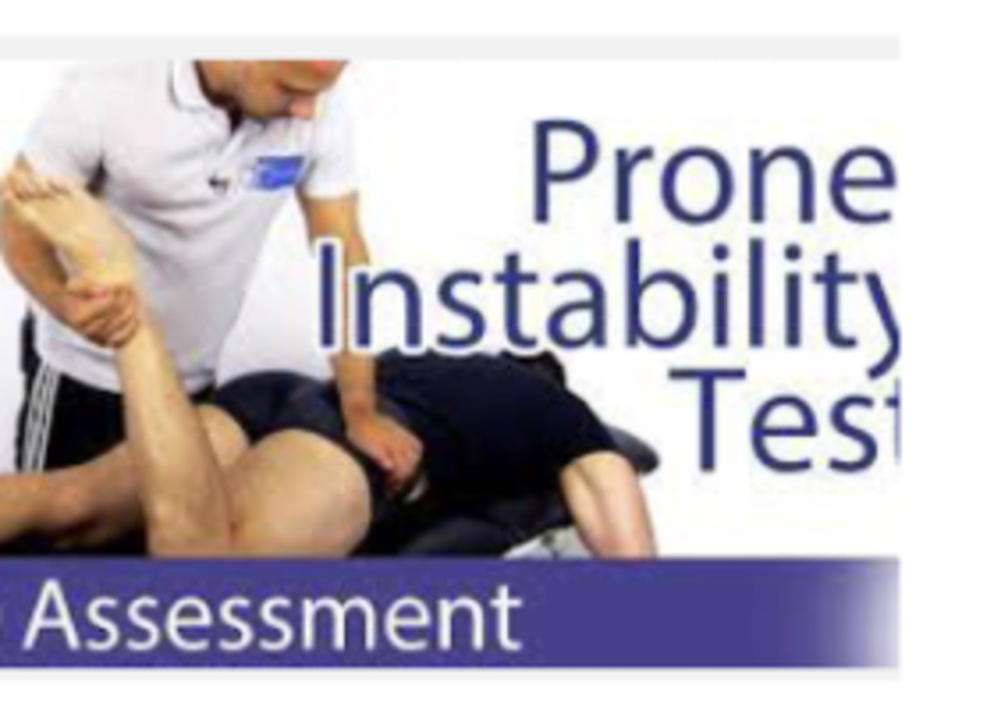
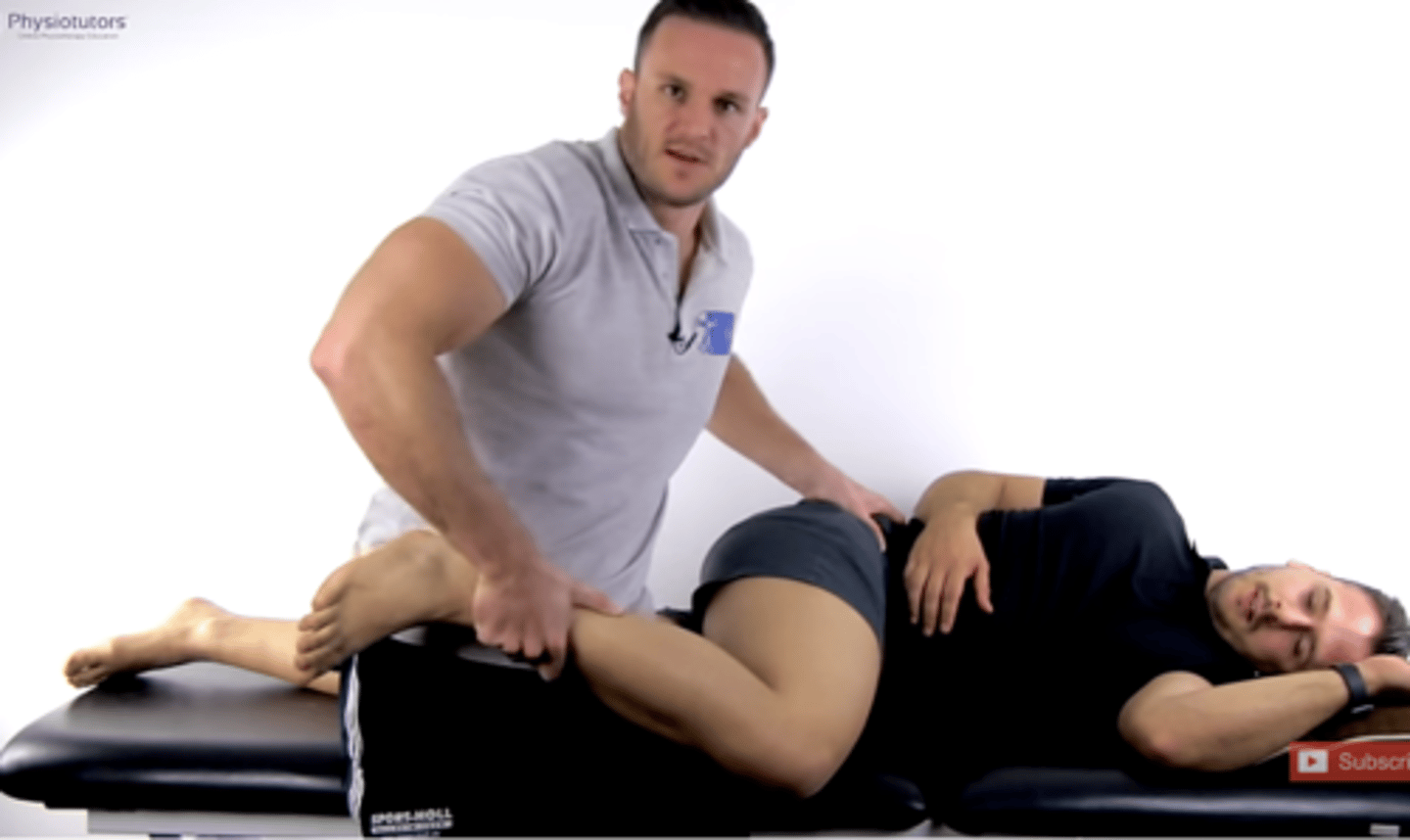
Prone Instability
Prone, flex to 90, ER the femur
Stretching anterior structures
Posterior to anterior glide of femoral head
Move butt cheek out of the way
**make sure your elbow is extended
Tests for: microinstability
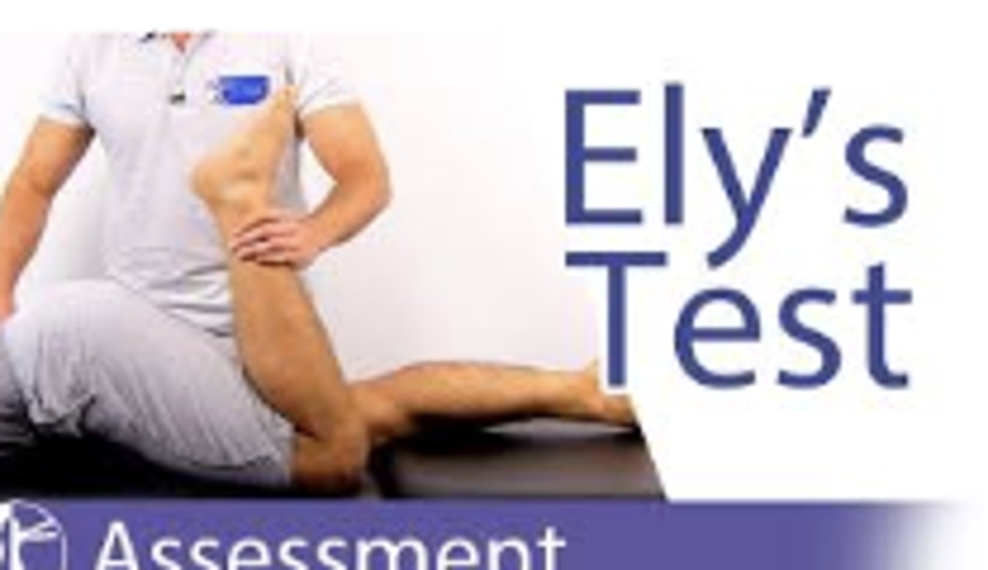
Ely's Test
(+) potential tightness of the rectus femoris muscle (and length) by pelvis lifting off of the table. *** if you have lengthened or weakened abs, that can give you a false positive
Tests for: Internal Coxa Saltans
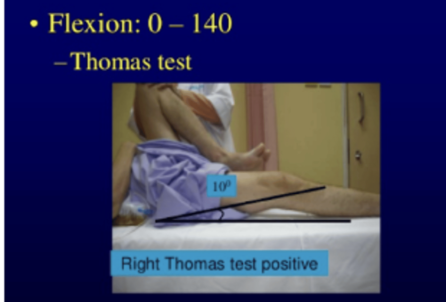
Thomas Test
positive= femur lifted 10 degrees off of the bed
Tests for: Internal Coxa Saltans, tight hip flexors/contracture
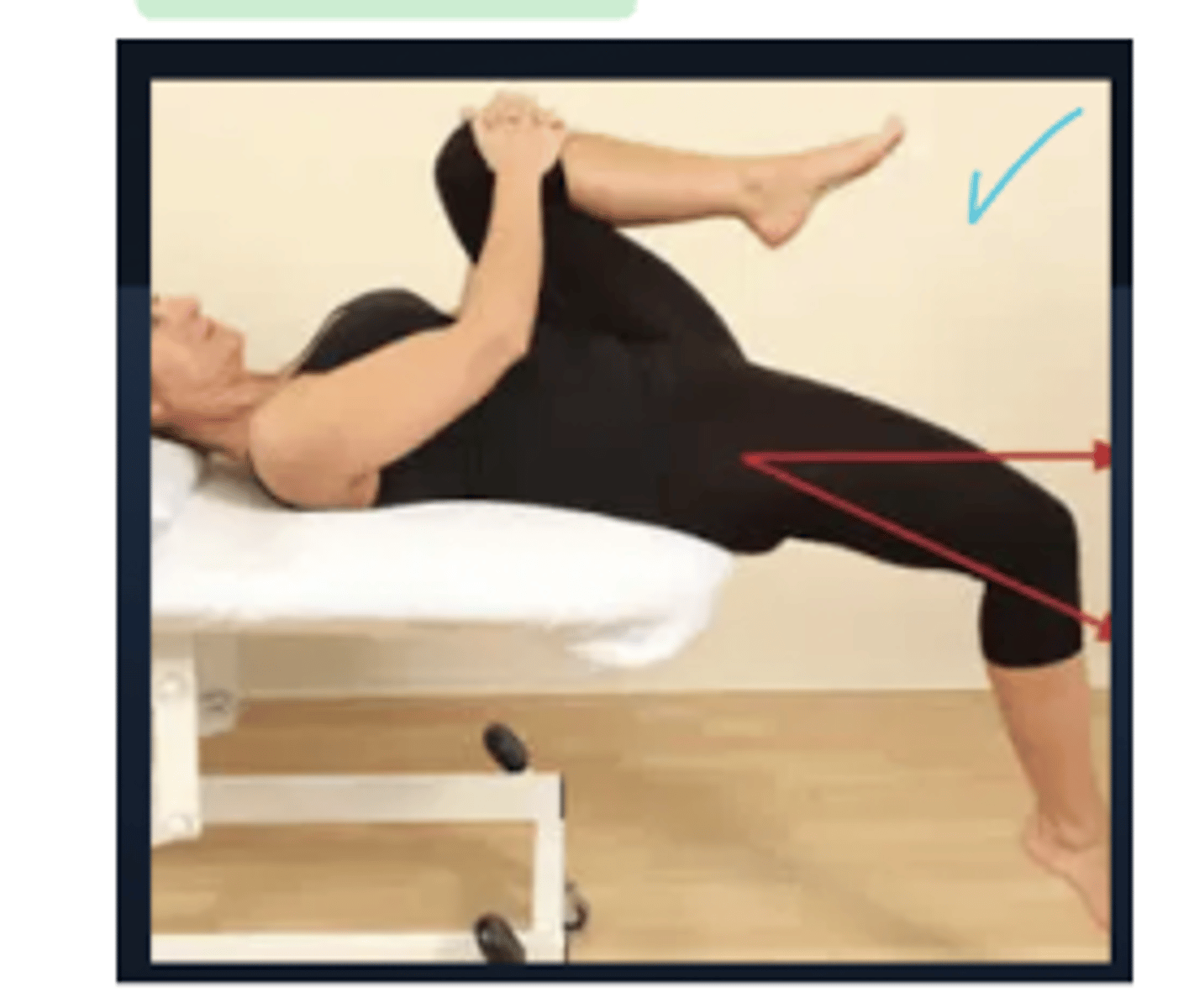
Modified Thomas Test
Iliopsoas - 1 joint muscle → if high is parallel or below the table this is good
Rectus femoris - 2 joint muscle → amount of knee flexion, if its a large angle its probably tight
TFL → IR means tightness
Sartorius → ER means tightness
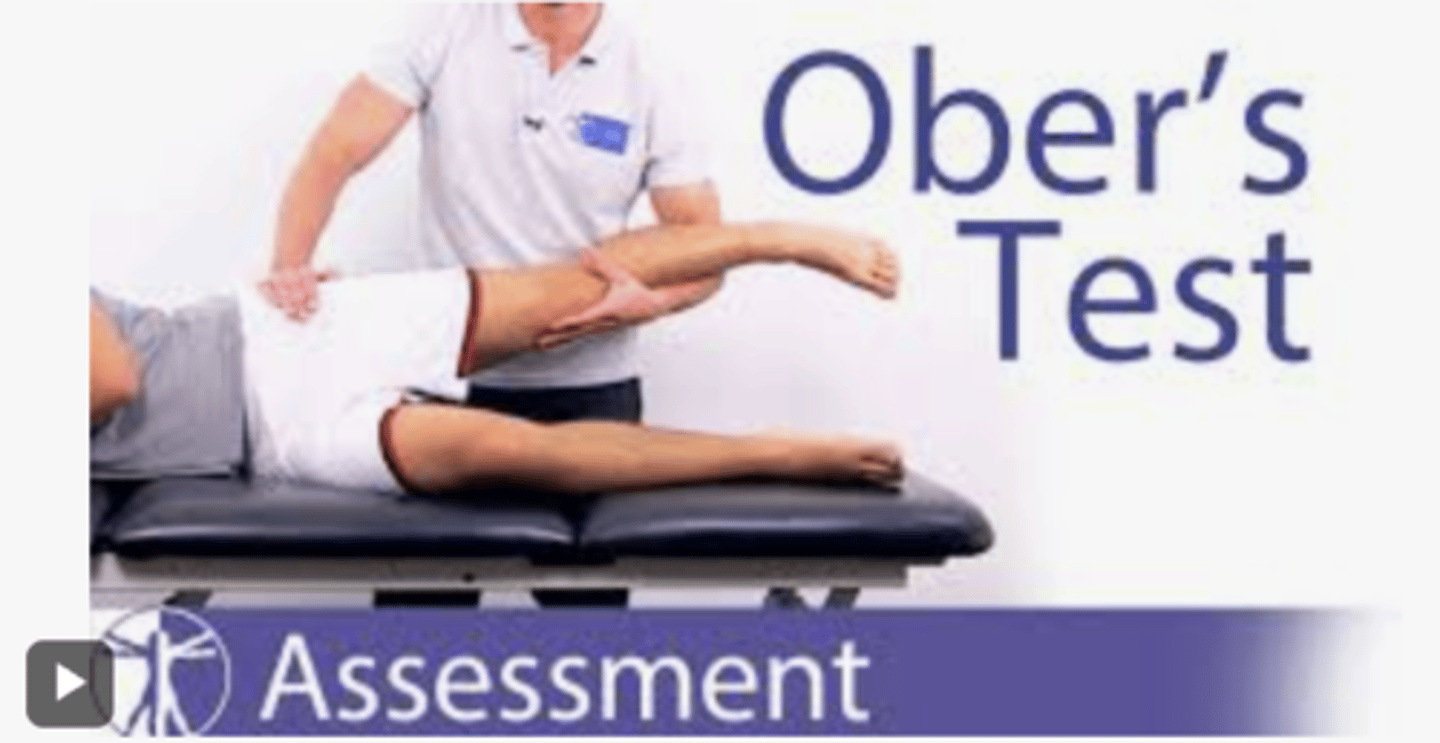
Ober's Test
Abd and extend hip, if the leg does not fully adduct or drops back down to the table, it indicates tightness or restriction in the ITB and TFL. Bottom leg is bent, top leg is bent to 90, closest to pt will stabilize pelvis. *keep in ext the whole time
GOLD STANDARD
Tests for: External Coxa Saltans , IT Band Syndrome or TFL tightness/superior hip structures
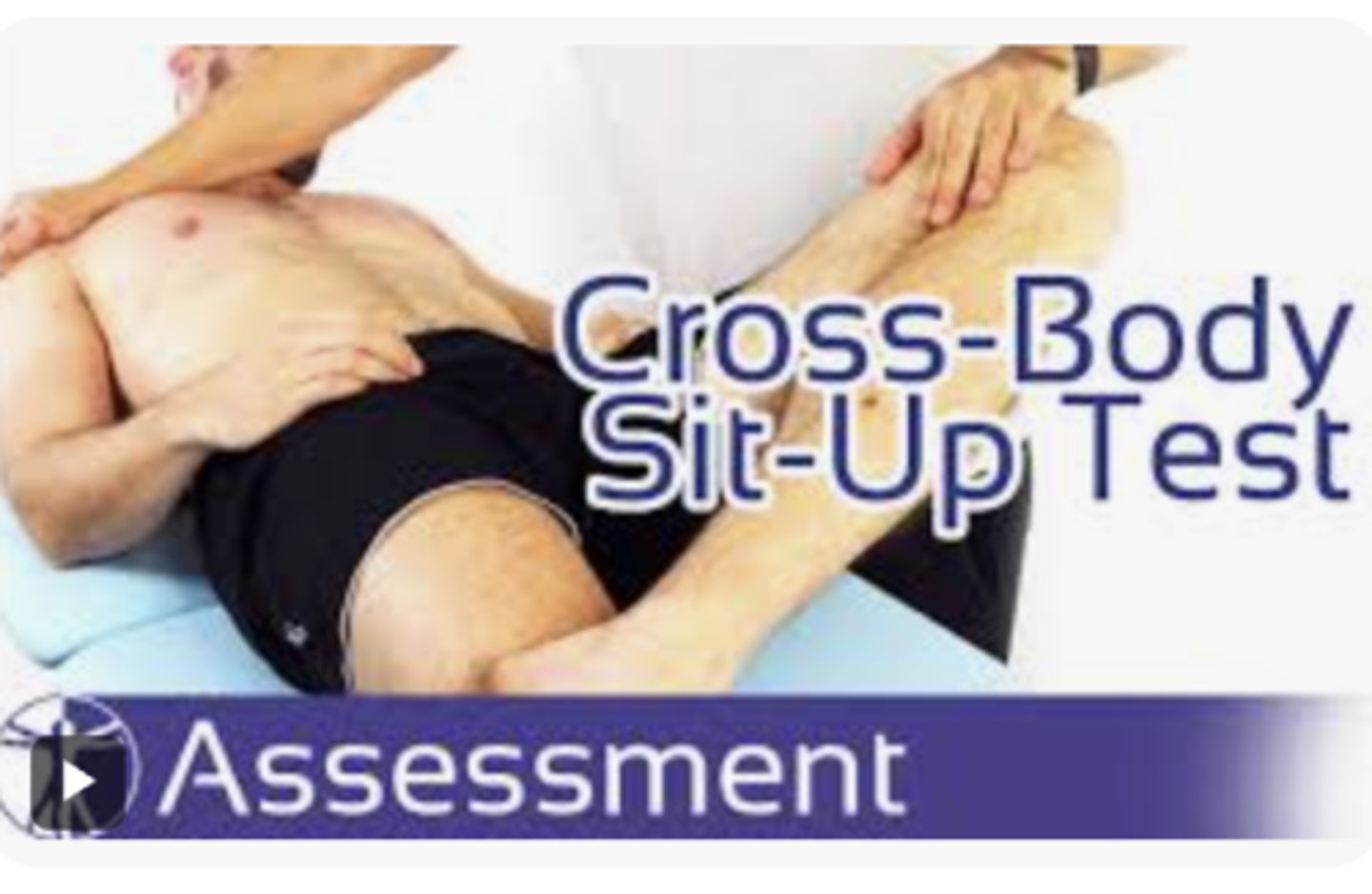
Resisted Cross Body Sit-up Test
Tests for: athletic pubalgia
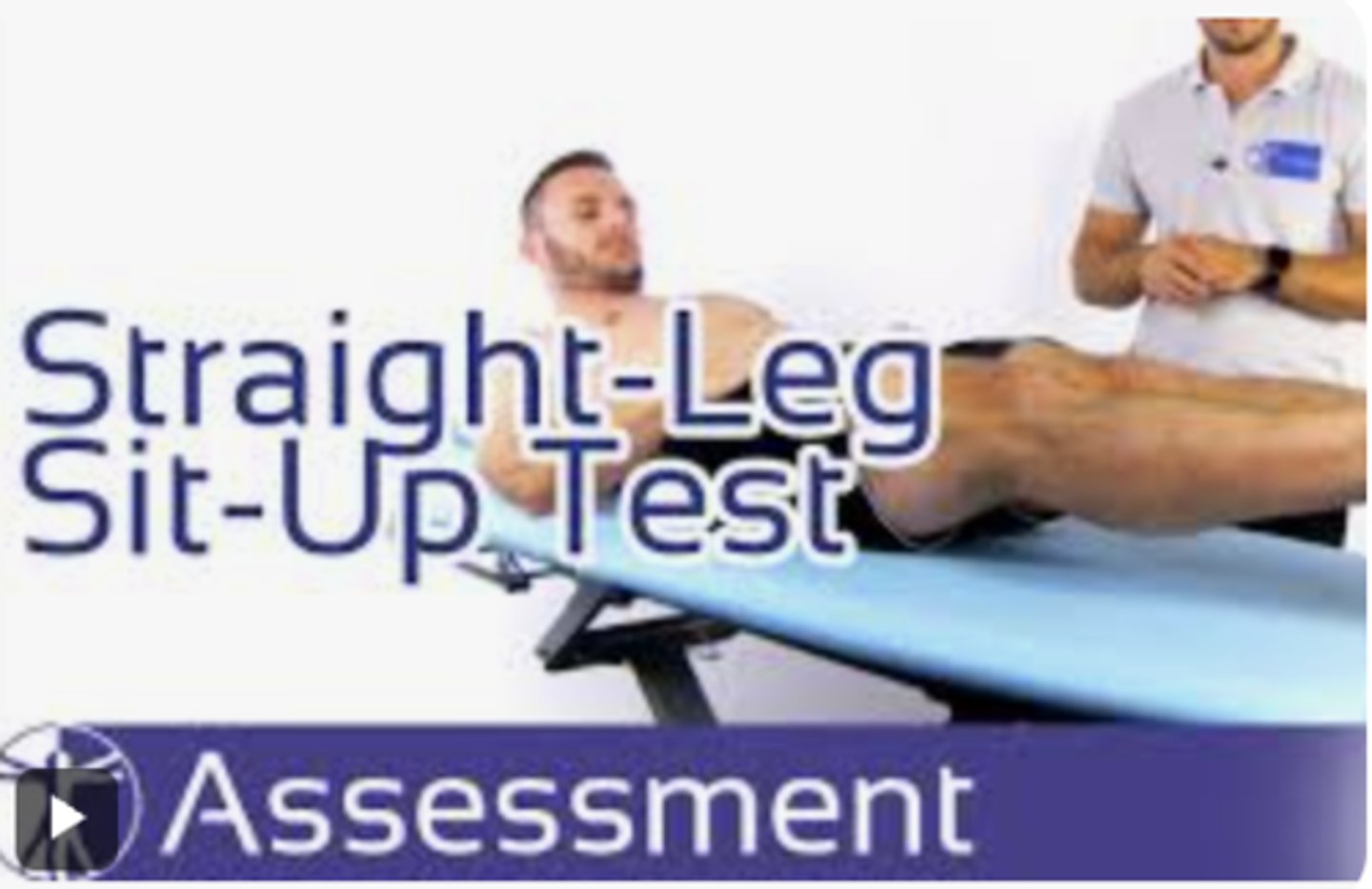
Straight leg Sit-up Test
Test for: athletic pubalgia
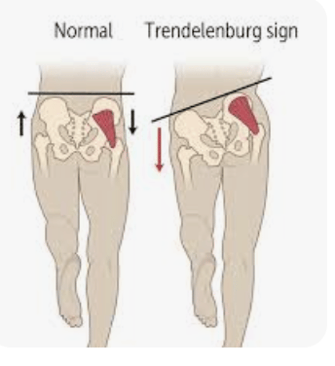
Trendeleburg Sign
Gluteus medius endurance weakness
C/L hip drop
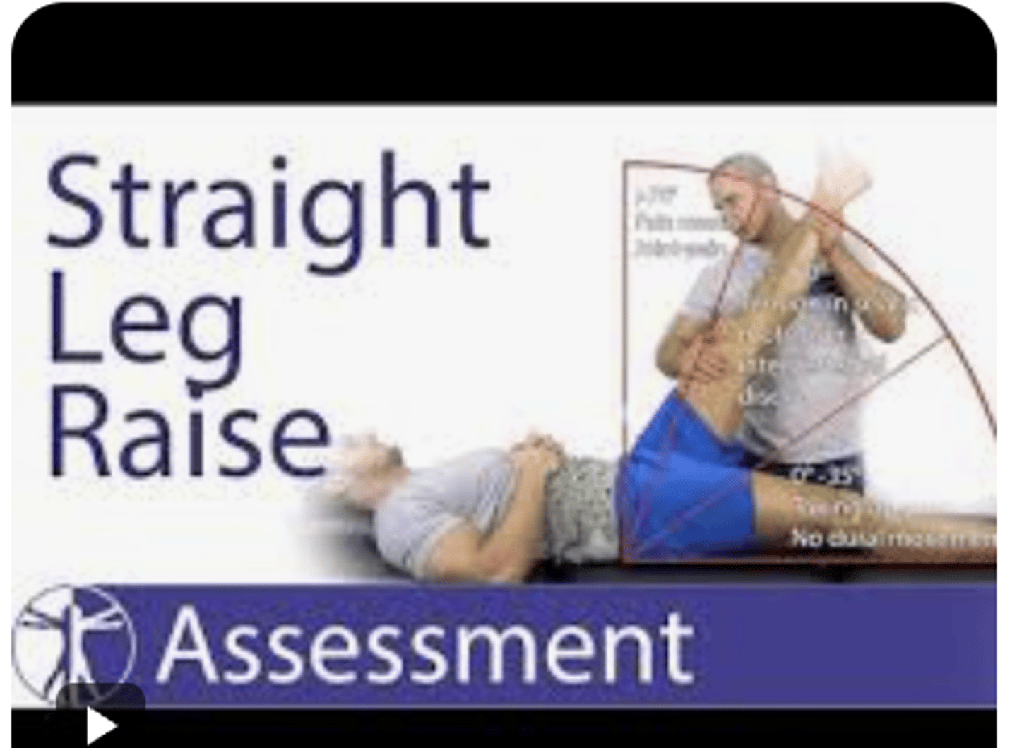
SLR
Hamstring length
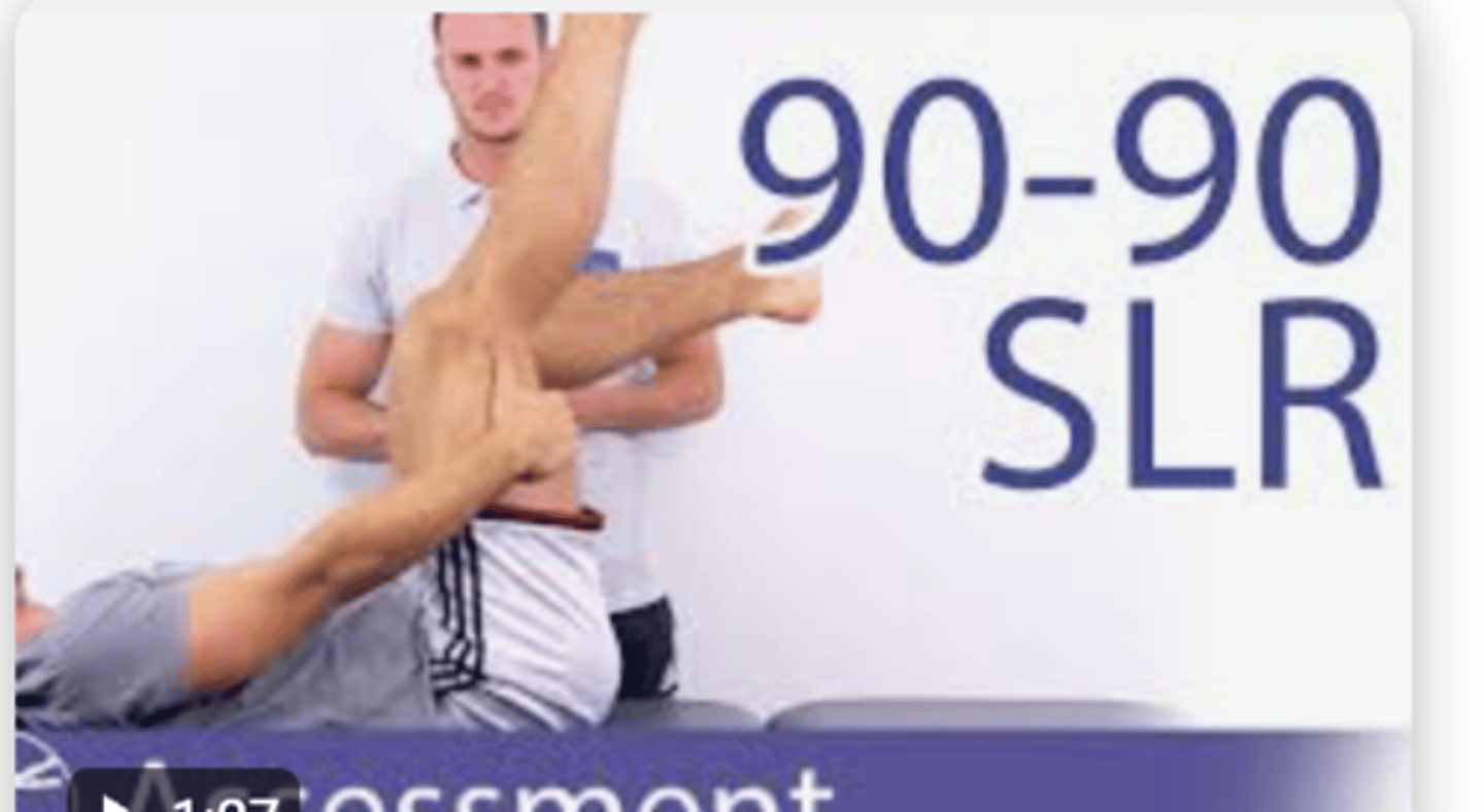
90/90
Hamstring Length
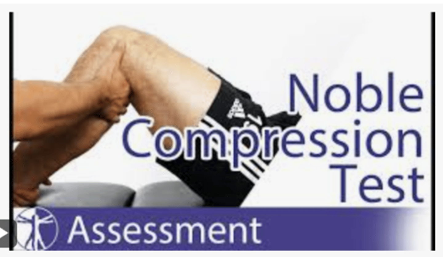
Noble's Compression Test
It involves applying pressure over the lateral epicondyle while the patient passively extends the knee from 90 degrees of flexion. A positive test is indicated by pain or snapping felt at 30 degrees of knee flexion, suggesting irritation or friction of the iliotibial band.
Tests for: IT band syndrome
Femoral N. Tension Test
The femoral nerve tension test is not specific for hip osteoarthritis (OA), but it can be used to differentiate hip pain from lumbar radiculopathy or nerve root involvement... a negative test supports hip OA
Tests for: Hip OA
SI Joint Test
?
Tests for: Hip OA
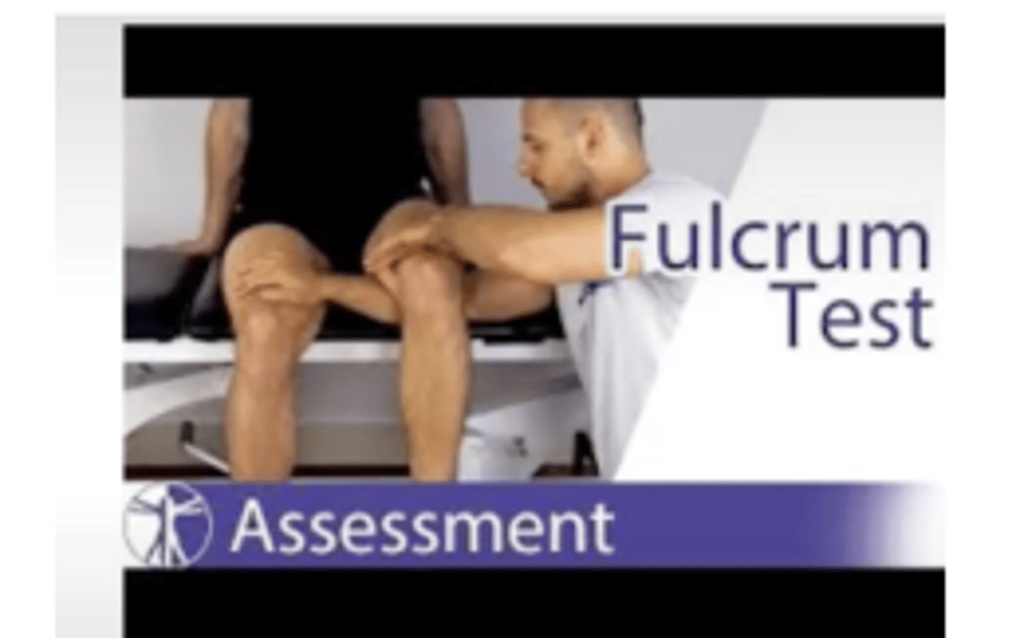
Fulcrum Test
Tests for: Femur stress fx
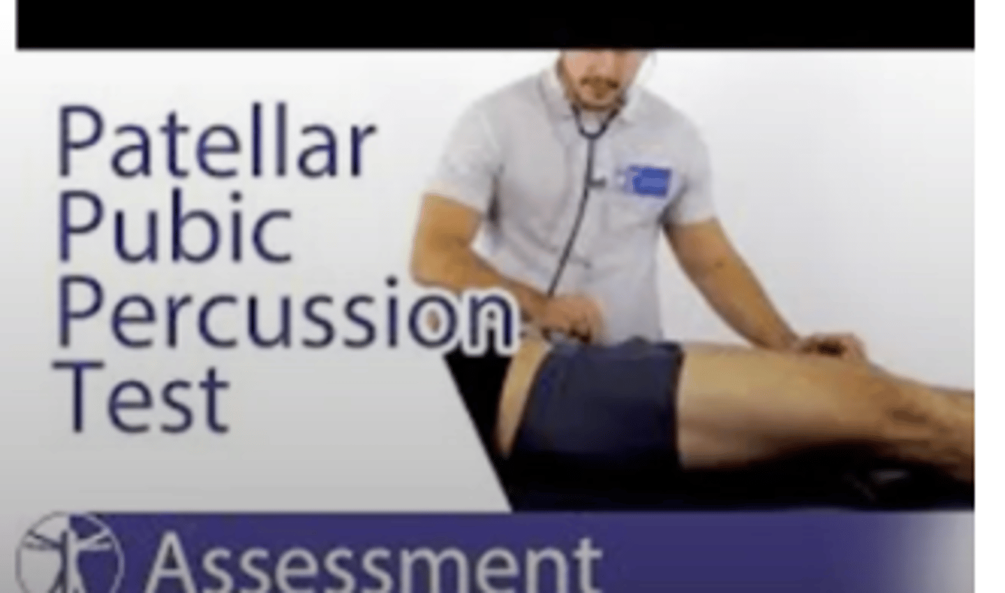
Patellar Pubic Percussion Test
Not pain provocation, if there is a stress fracture it will decrease the conductance of vibration/sound through the bone. let the patient find their pubic bone. Middle finger on belly button and heel of hand on pubic bone
(+)= muffled sound on affected side
Tests for: femur stress fx
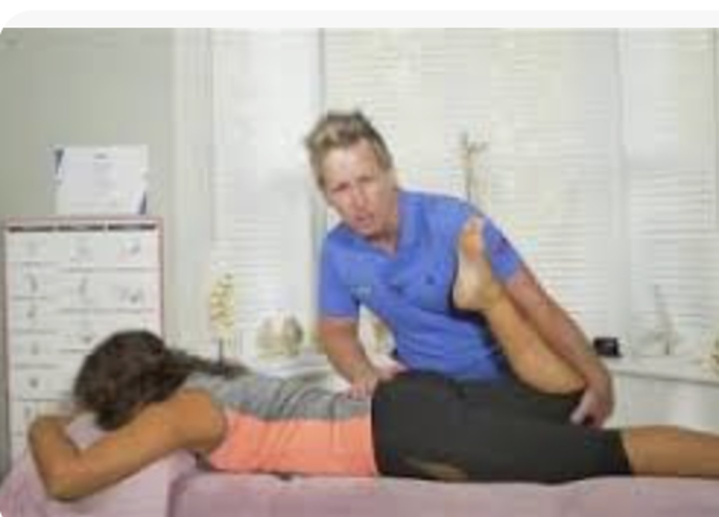
Piriformis Test (FAIR Test)
-Patient sidelying with top hip flexed to 60 degrees with knee flexed
-PT stabilizes hip with one hand and applies downward pressure to knee
-Positive = pain during pressure
Sign of the Buttock
take them info SLR first, if you bend the knee if its hamstring tightness you should be able to flex hip farther. Positive would be no change in hip flexion from SLR to knee bent position
(+)= NOT a good sign! a space occupying a lesion in the gluteal region like neoplastic tumor/ tumor. Positive if they CANNOT get more motion in SLR as knee at 90 and hip flexion
*non-musculoskeletal = you can't reproduce the symptoms that brought them in --> you need to refer OUT!
Modified Ober's Test
Top leg is now extended. If they have IT band syndrome at distal lateral knee you extend the leg to decrease the pain
The limb should be able to drop into parallel.
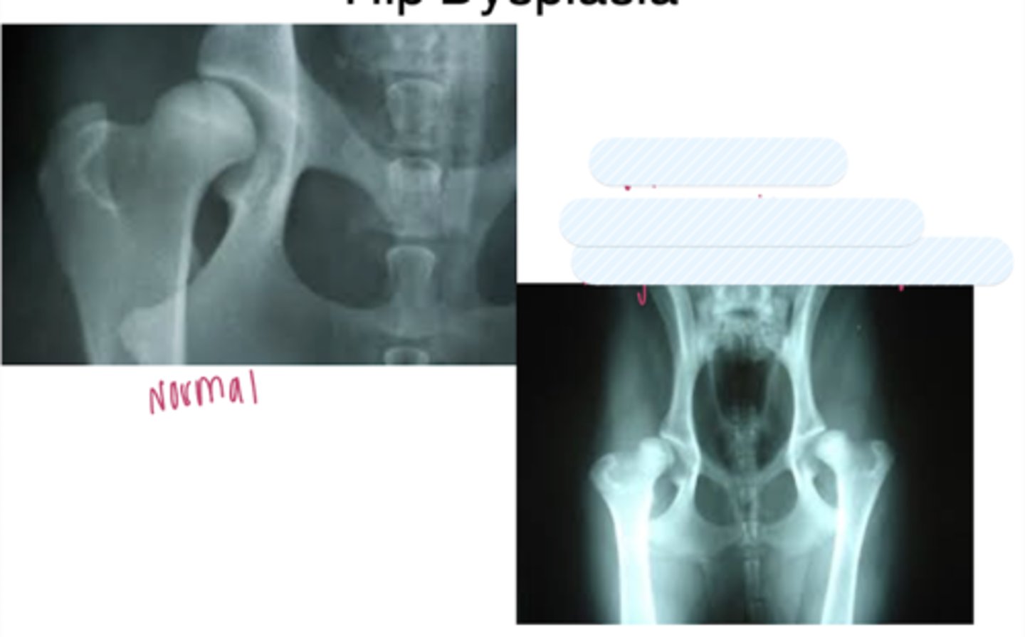
hip dysplasia on the Left photo
the L hip is worse than the R hip
very acute center edge angle
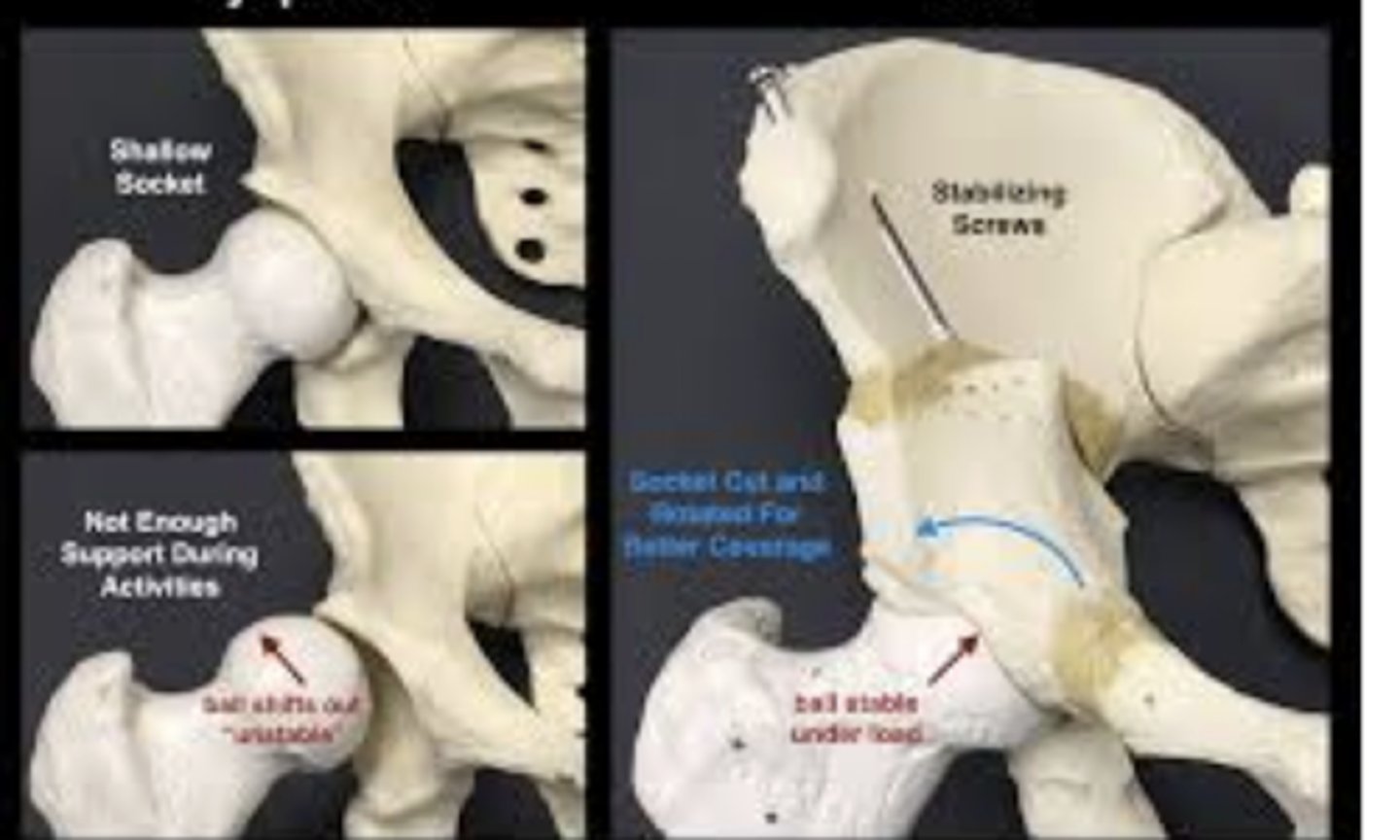
periacetabular osteotomy
used for hip dysplasia. break femur and pelvis, rotate them to provide greater joint angle
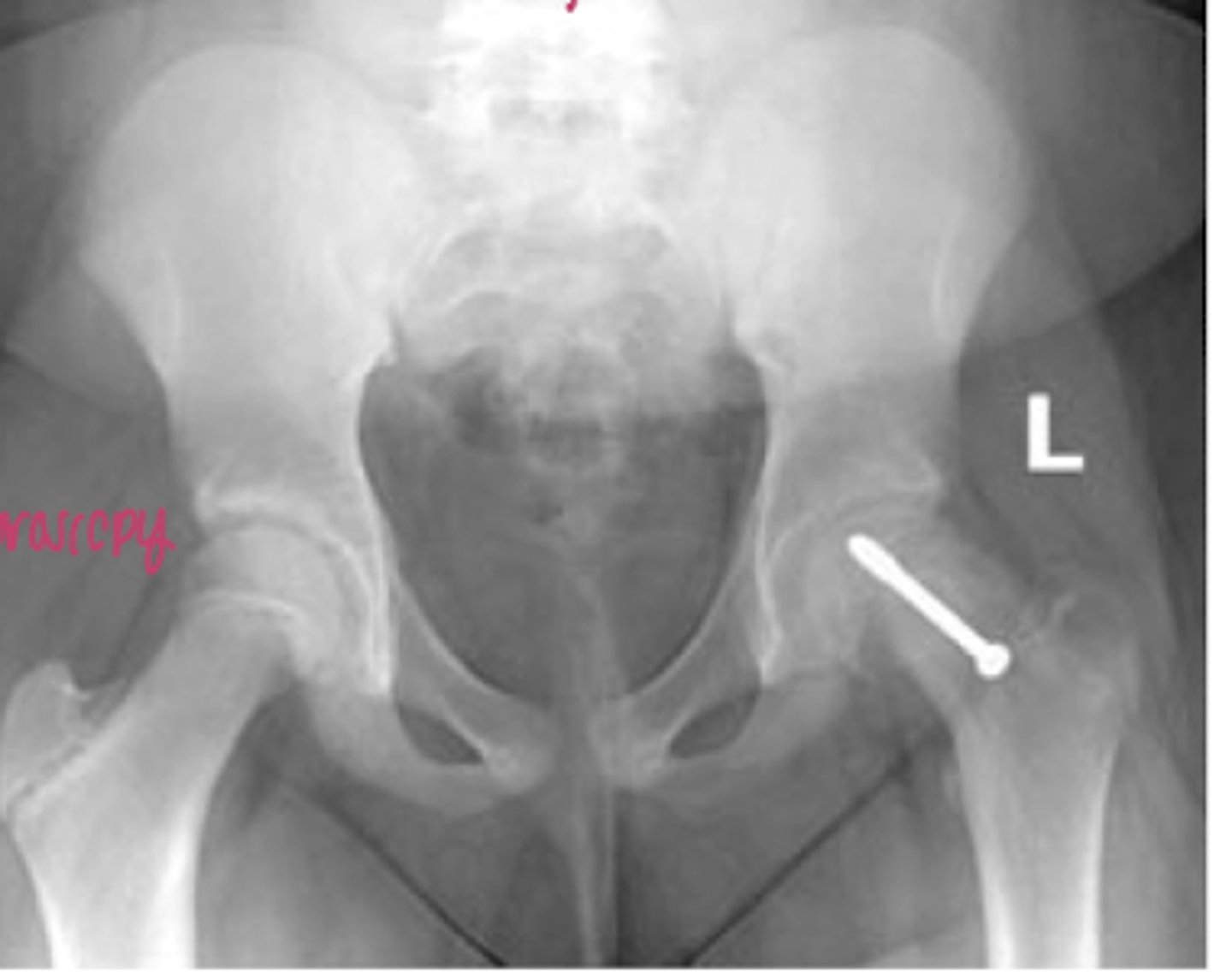
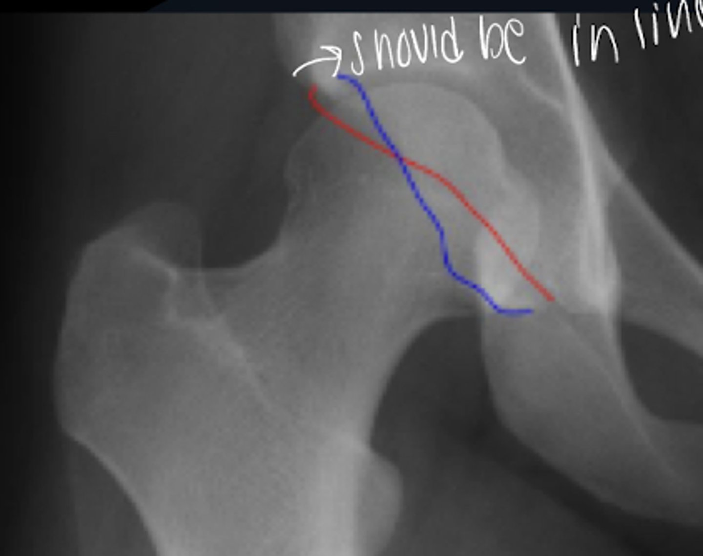
SCFE
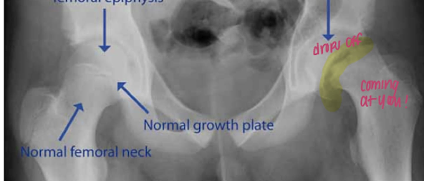
percutaneous pinning for SCFE
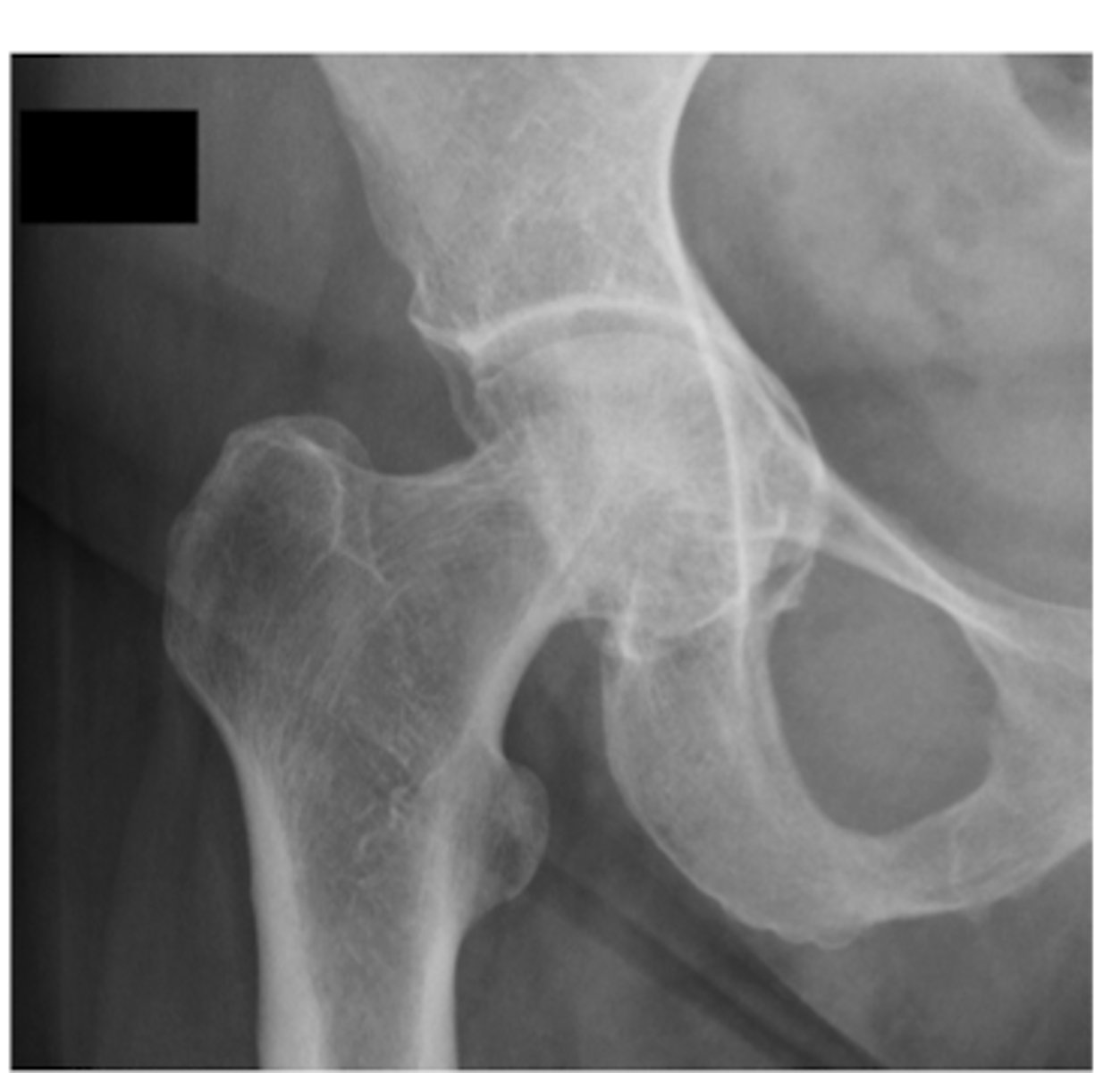
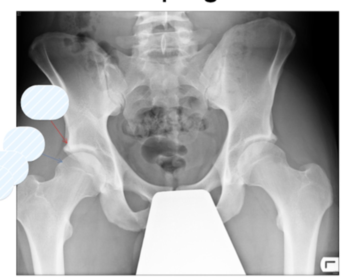
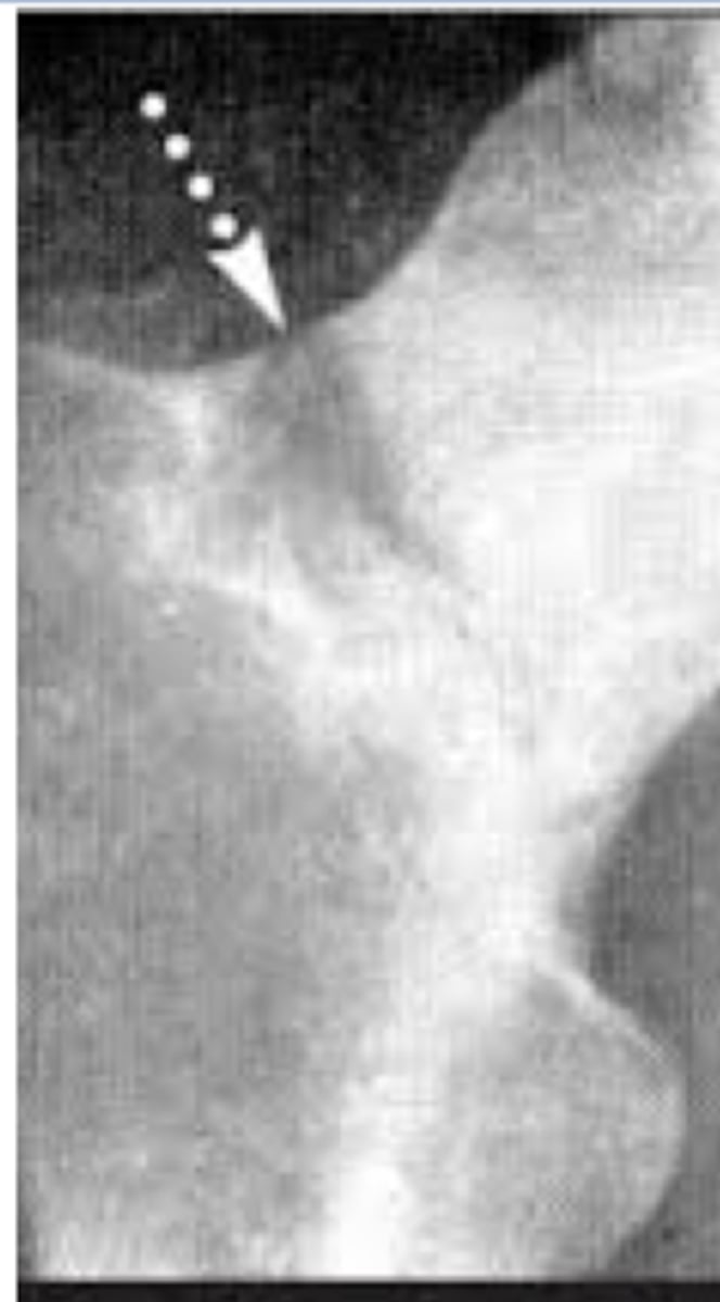
crossover sign
(+) means acetabular retroversion and a PINCER
posterior acetabular rim is medial to the center of the femoral head
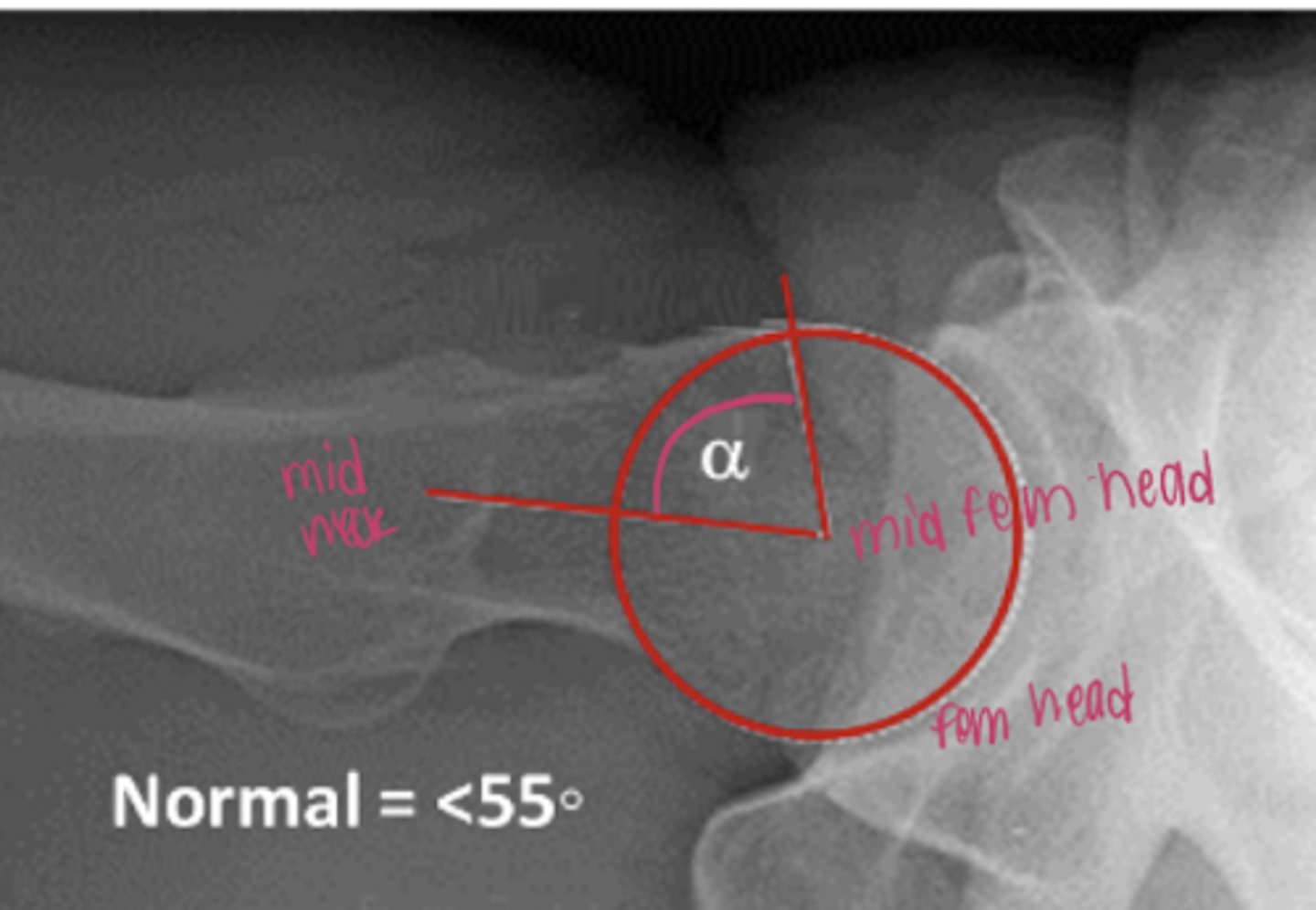
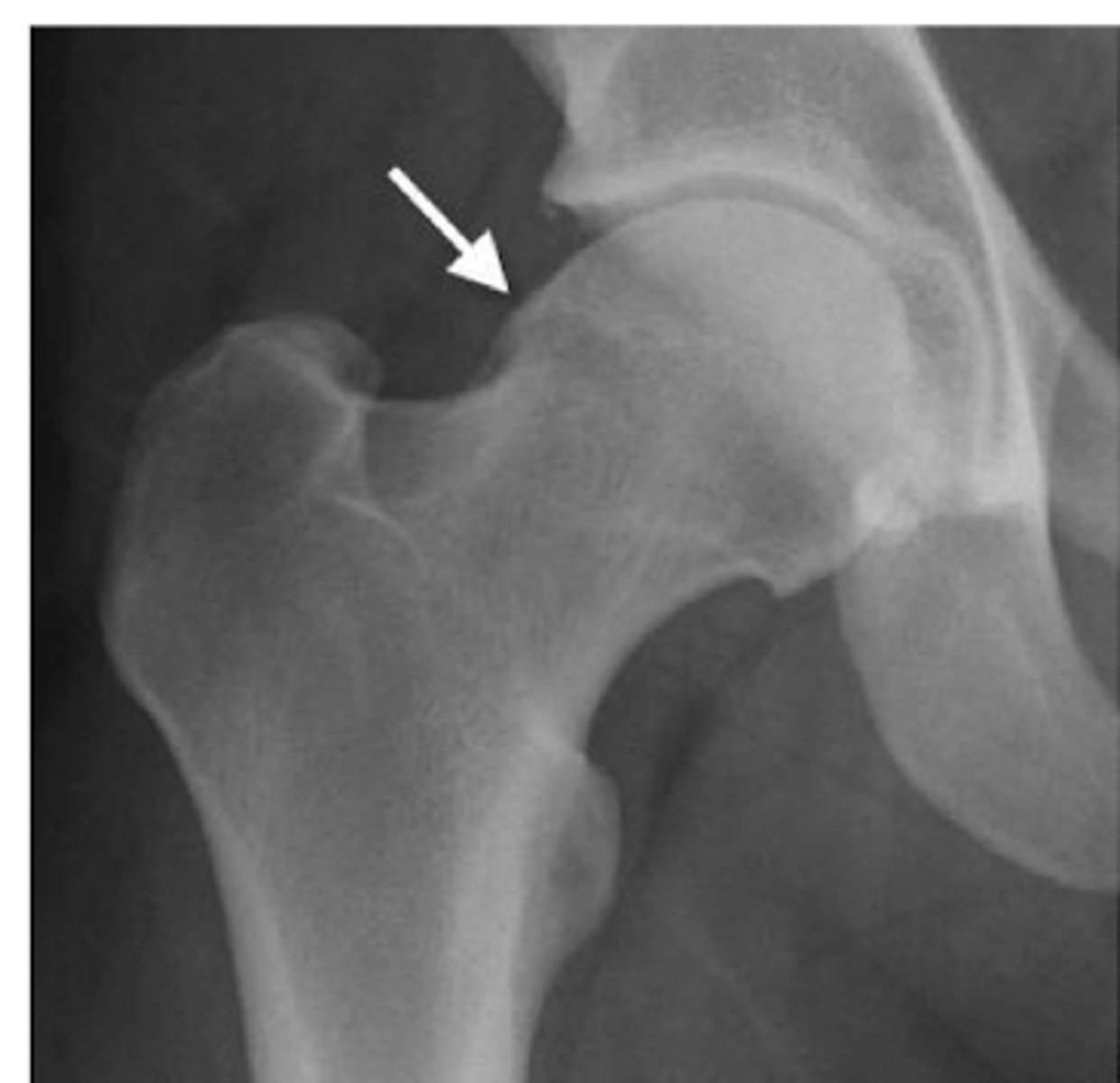
alpha angle
used to diagnose FAI CAM (large femoral head)
Pincer
CAM
mixed impingement
CAM + Pincer
alpha angle > 55
angle of wiberg > 35
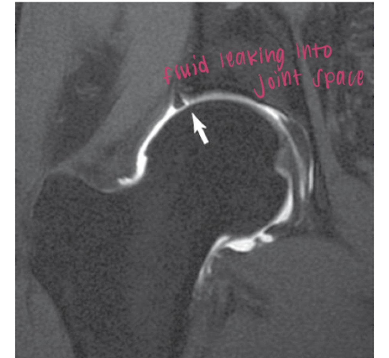
MR arthrogram of labral tear
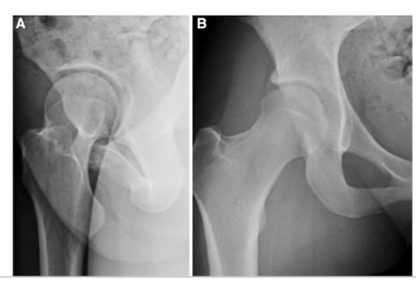
FAI arthroscopy
A pre op
B post op
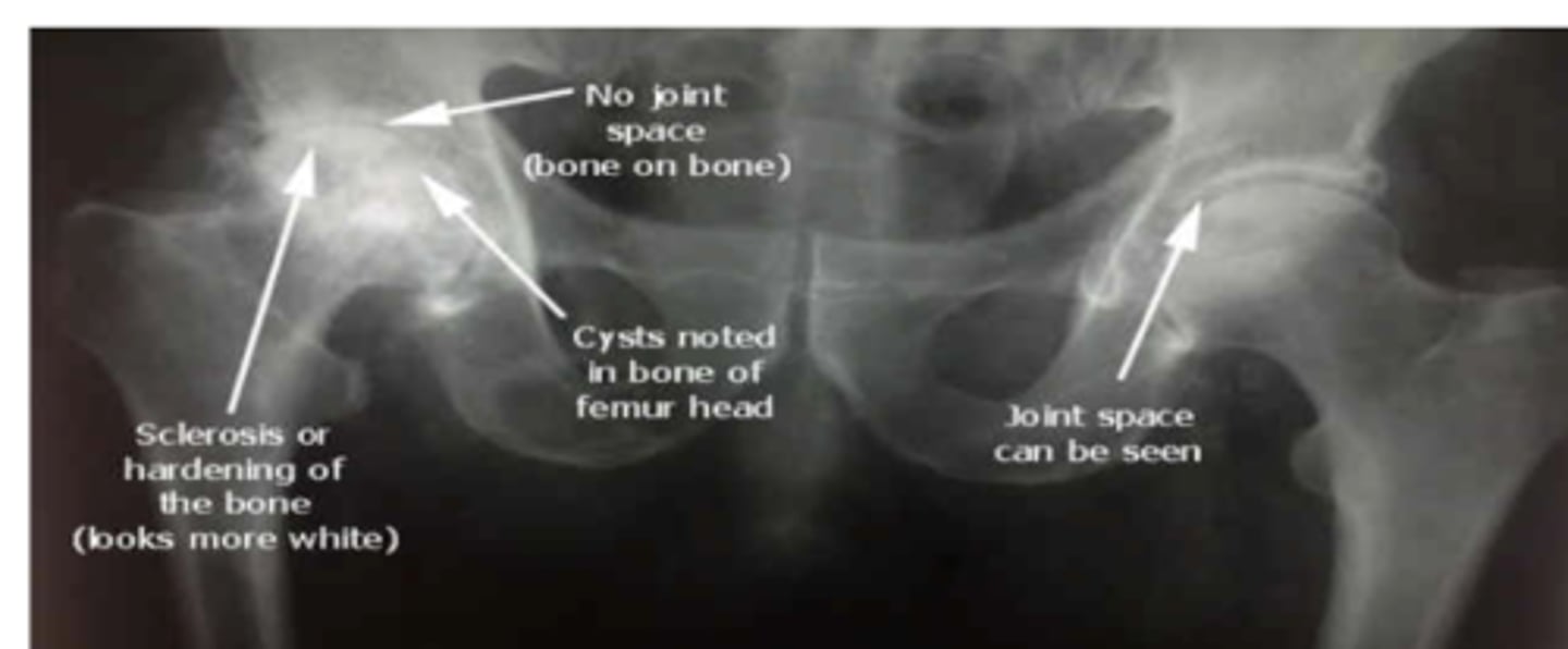
Hip OA
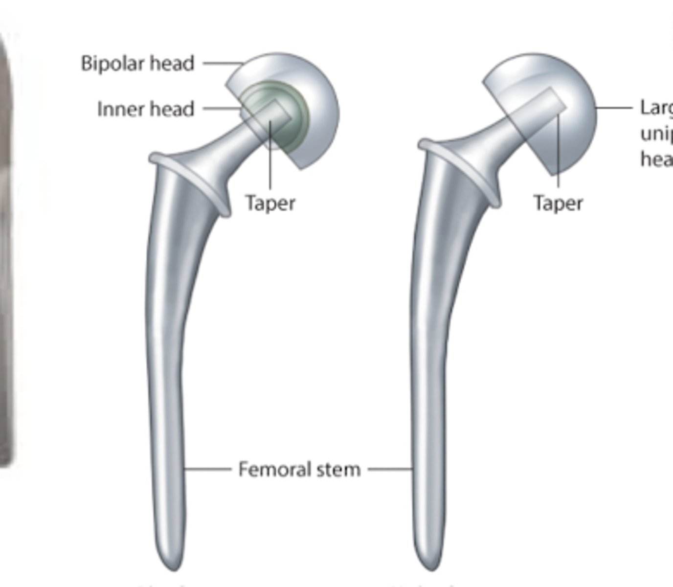
L: bipolar hemiprosthesis
R: unipolar hemiprosthesis
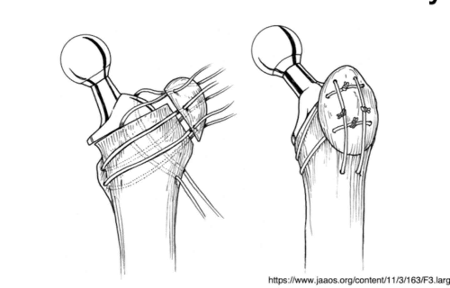
trochanteric osteotomy
used for THA revisions

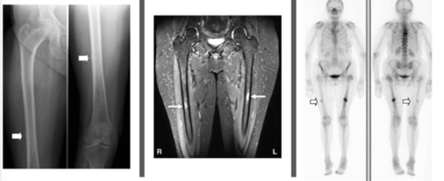
femoral stress fracture
femoral stress fracture
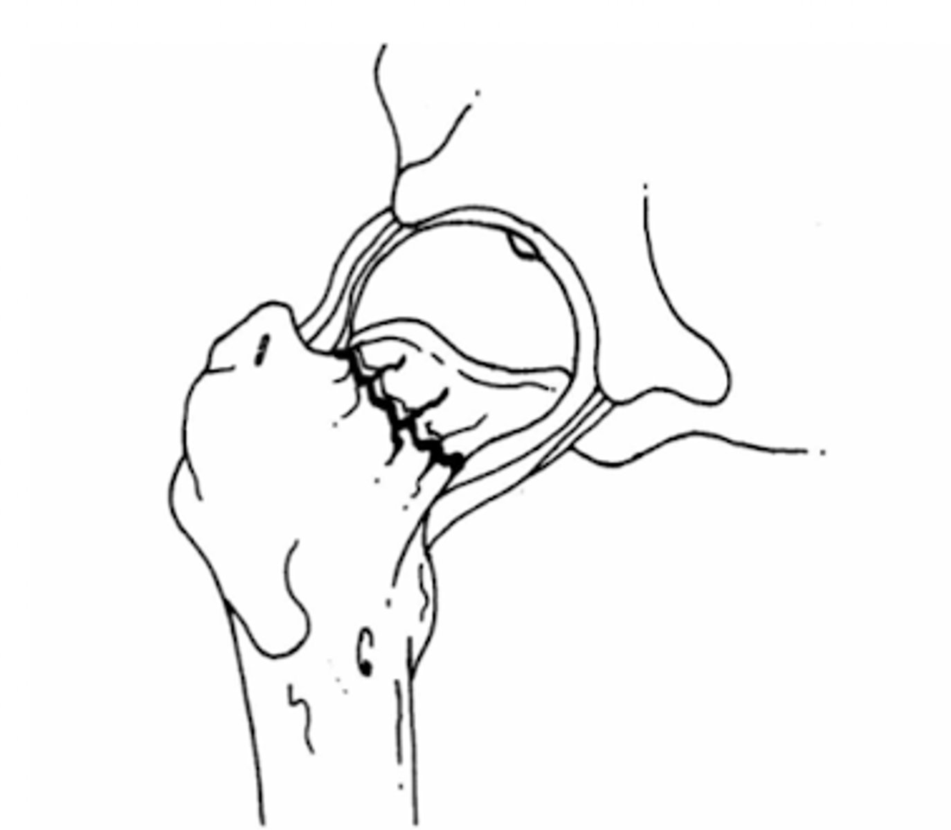
gardens I fx
gardens II fx
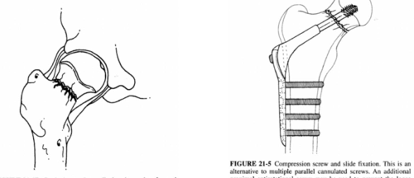
gardens III fx
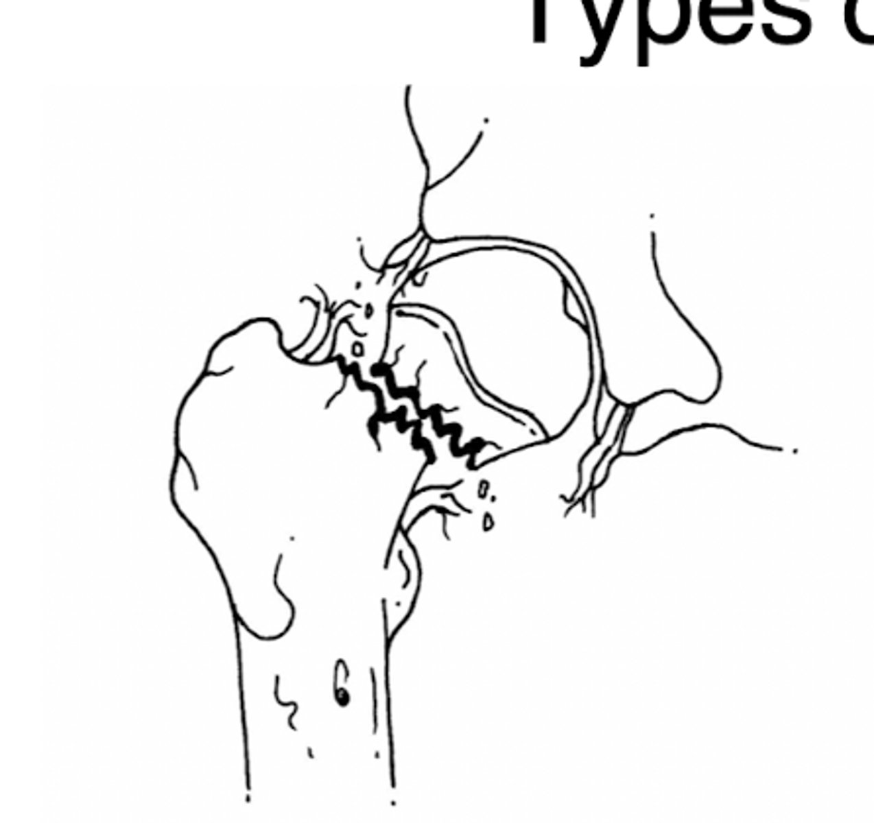
gardens IV fx
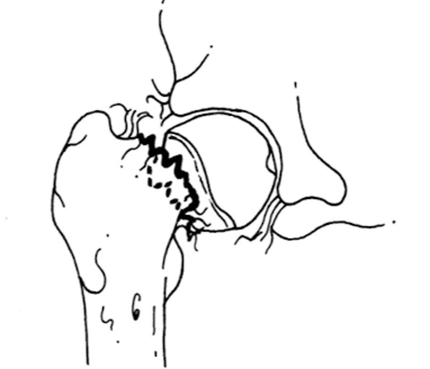
intercapsular fx
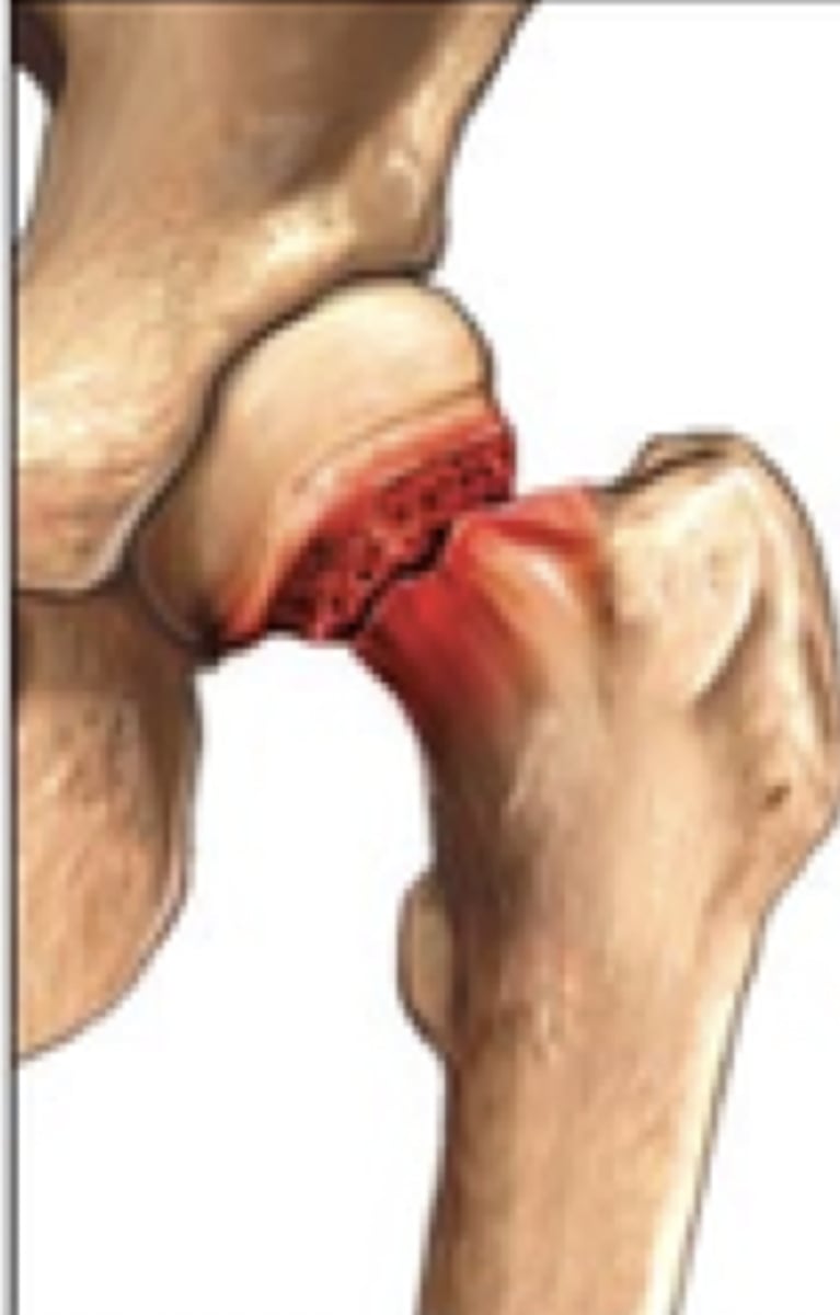
intertrochanteric fx
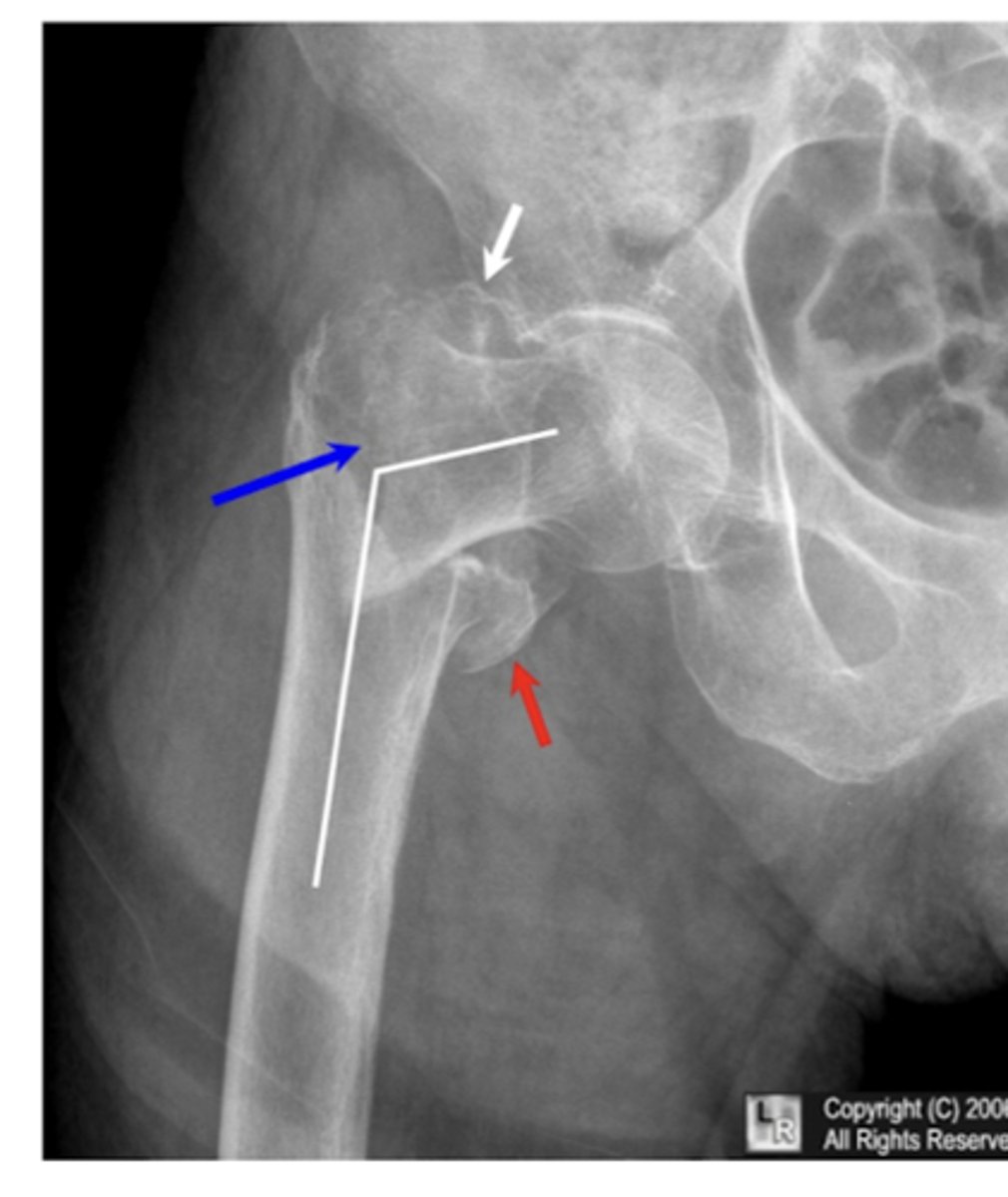
intertrochanteric fx
Comminuted intertrochanteric fracture. There is a fracture from the greater to the lesser trochanter (blue arrow). There are separate fragments of the greater trochanter (white arrow) and lesser trochanter (red arrow). There is varus deformity (white line) of the femoral shaft.
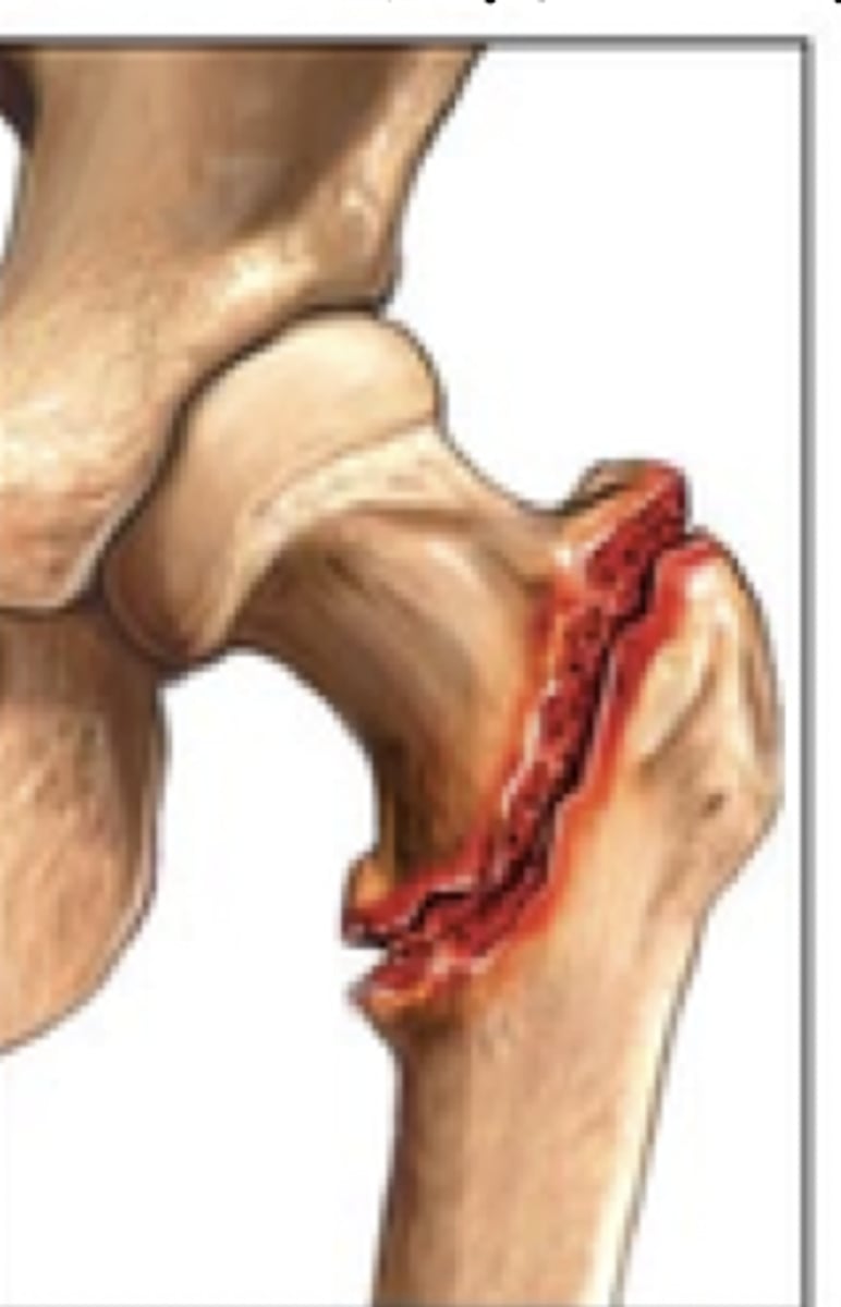
subtrochanteric fx
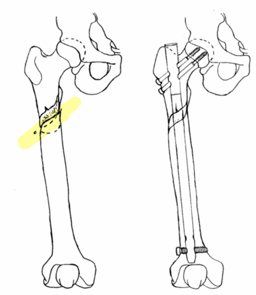
subtrochanteric fx
