Feline Neuro ICVA Diseases
 Call Kai
 Learn
Learn Practice Test
 Spaced Repetition
 Match
Match Flashcards
 Knowt Play
1/22

There's no tags or description
Looks like no tags are added yet.
Name | Mastery | Learn | Test | Matching | Spaced | Call with Kai | Chat |
|---|
No analytics yet
Send a link to your students to track their progress
23 Terms
Cerebellar Hypoplasia
Caused by panleukopenia
Diabetic Polyneuropathy/Peripheral Neuropathy
Feline diabetic polyneuropathy occurs as a result of prolonged hyperglycemia, which leads to a series of metabolic and vascular changes that ultimately damage peripheral nerves. The exact pathophysiology involves several mechanisms:
Sorbitol Pathway Activation: Chronic hyperglycemia activates the polyol pathway, where glucose is converted to sorbitol by the enzyme aldose reductase. Sorbitol accumulation within neurons increases osmotic stress, leading to cellular damage.
Oxidative Stress: Hyperglycemia also contributes to increased production of reactive oxygen species (ROS), causing oxidative damage to nerve cells and impairing their function.
Advanced Glycation End-products (AGEs): Hyperglycemia promotes the formation of AGEs, which alter the structure and function of proteins within nerve cells, further contributing to neuropathy.
Vascular Changes: Chronic hyperglycemia can cause endothelial dysfunction and microvascular damage, reducing blood supply to peripheral nerves and exacerbating neuronal damage.
Nerve Demyelination: The combination of metabolic and ischemic damage leads to demyelination and axonal degeneration, which are key pathological features of diabetic polyneuropathy.
Diabetic Polyneuropathy/Peripheral Neuropathy:C.S
Clinical Signs
The clinical presentation of FDP varies depending on the severity and extent of nerve involvement. Common clinical signs include:
Plantigrade Stance: One of the hallmark signs of FDP is a plantigrade stance, where the cat walks with its hocks touching the ground. This is due to weakness in the distal muscles of the hind limbs.
Muscle Atrophy: Prolonged neuropathy leads to muscle wasting, particularly in the hind limbs, resulting in visible atrophy.
Paresis and Ataxia: Affected cats may show mild to moderate paresis (weakness) and ataxia (incoordination), especially in the hind limbs.
Reduced Reflexes: Neurological examination may reveal reduced or absent spinal reflexes, such as the patellar reflex, due to impaired nerve function.
Sensory Deficits: Although less commonly reported in cats than in dogs, some cats with FDP may exhibit sensory deficits, including decreased pain perception and proprioception.
Hyperesthesia and Pain: In some cases, cats may develop hyperesthesia (increased sensitivity) and neuropathic pain, characterized by discomfort or pain upon light touch or movement.
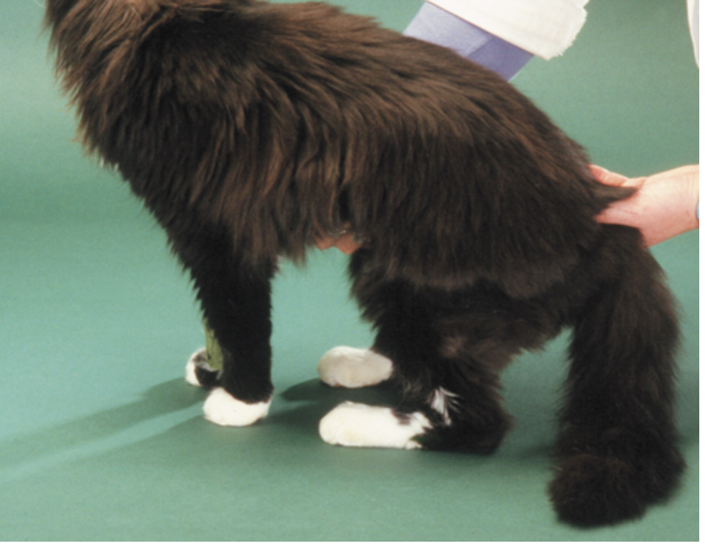
Plantigrade stance
Diabetic Polyneuropathy/Peripheral Neuropathy:D.X
Diagnostics
Diagnosis of FDP involves a combination of clinical evaluation, laboratory testing, and advanced diagnostics:
Clinical Evaluation: A thorough neurological examination is essential for identifying the characteristic signs of FDP, such as a plantigrade stance and muscle atrophy.
Blood Glucose Monitoring: Persistent hyperglycemia is a key indicator of poorly managed diabetes. Serial blood glucose measurements and fructosamine levels help assess long-term glucose control.
Electromyography (EMG): EMG can detect abnormalities in nerve conduction and muscle activity, revealing evidence of peripheral nerve dysfunction consistent with polyneuropathy.
Nerve Conduction Velocity (NCV) Studies: NCV studies are useful for quantifying the extent of nerve damage. Cats with FDP typically show decreased nerve conduction velocities due to demyelination and axonal loss.
Biopsy and Histopathology: In rare cases, a nerve biopsy may be performed to confirm the diagnosis, revealing characteristic features such as myelin loss, axonal degeneration, and endoneurial fibrosis.
Differential Diagnosis: It is important to rule out other conditions that may mimic FDP, such as spinal cord injury, myasthenia gravis, hypokalemic polymyopathy, and metabolic myopathies.
Diabetic Polyneuropathy/Peripheral Neuropathy:T.X
Treatment
The primary goal of treatment for FDP is to restore glycemic control and manage neuropathic symptoms. Key components of treatment include:
Tight Glycemic Control:
Insulin Therapy: Ensuring that the cat's diabetes is well-controlled through appropriate insulin therapy is the cornerstone of managing FDP. Regular monitoring of blood glucose levels and adjusting the insulin dose as needed are critical.
Dietary Management: A low-carbohydrate, high-protein diet can help stabilize blood glucose levels and reduce the burden on peripheral nerves.
Neuropathic Pain Management:
Gabapentin: Gabapentin is the most commonly used medication for managing neuropathic pain in cats. It works by modulating calcium channels in the nervous system, reducing pain transmission.
Tricyclic Antidepressants (TCAs): Although less commonly used in cats, TCAs like amitriptyline may be considered for their analgesic effects in managing neuropathic pain.
Nutritional Support:
Vitamin B12 Supplementation: Vitamin B12 (cobalamin) is essential for nerve function and may be supplemented to support nerve repair and function in cats with FDP.
Physical Rehabilitation:
Physical Therapy: Gentle physical therapy exercises can help maintain muscle tone, improve circulation, and prevent further muscle atrophy.
Acupuncture: Some veterinarians may incorporate acupuncture as an adjunct therapy to alleviate pain and promote nerve regeneration.
Monitoring and Follow-Up:
Regular follow-up appointments are necessary to monitor the cat’s response to treatment, adjust insulin doses, and assess for any improvement or progression of neurological signs.
Epilepsy
Hydrocephalus
Otitis Media
Otitis media, an infection or inflammation of the middle ear, is a significant condition in cats that can lead to serious complications if not promptly diagnosed and treated. While otitis media is less common in cats than in dogs, it remains an important differential diagnosis for cats presenting with ear-related symptoms, head tilt, or neurological deficits. This article provides an in-depth examination of otitis media in cats, covering pathophysiology, clinical signs, risk factors, diagnostics, treatment, and hallmark clinical signs.
Pathophysiology
Otitis media in cats typically develops as a result of the extension of an infection from the external ear canal (otitis externa) into the middle ear through the tympanic membrane. However, it can also arise via hematogenous spread (from the bloodstream) or as an extension from upper respiratory tract infections through the eustachian tube.
The middle ear consists of the tympanic cavity, auditory ossicles, and the tympanic bulla, which are all involved in sound transmission. Inflammation in this area can impair the normal functioning of these structures, leading to various clinical signs.
Key Pathophysiological Mechanisms:
Inflammation and Fluid Accumulation: The infection leads to inflammation and the accumulation of purulent or serous fluid within the tympanic cavity. This can cause increased pressure, pain, and potential damage to the delicate structures within the middle ear.
Tympanic Membrane Rupture: Persistent inflammation may result in the rupture of the tympanic membrane, leading to discharge into the external ear canal.
Nerve Involvement: The facial nerve (cranial nerve VII) passes through the middle ear, and inflammation can lead to neuropathy, resulting in facial paralysis. Vestibular signs may also occur if the inner ear structures are affected, leading to secondary otitis interna.
Clinical Signs
The clinical signs of otitis media in cats can be quite variable and depend on the severity of the infection, the presence of concurrent otitis externa or otitis interna, and whether the facial nerve or other structures are involved.
Common Clinical Signs:
Head Tilt: A persistent head tilt, usually toward the affected side, is a common sign and suggests involvement of the vestibular system.
Ear Discharge: Cats may present with discharge from the affected ear, which can be purulent, bloody, or serous, particularly if the tympanic membrane is ruptured.
Ear Pain and Sensitivity: The cat may show signs of pain when the ear is touched, and there may be scratching or rubbing of the affected ear.
Facial Nerve Paralysis: Cats may exhibit facial nerve paralysis, characterized by drooping of the lips, inability to blink (lagophthalmos), and reduced or absent palpebral reflex on the affected side.
Vestibular Signs: Signs such as ataxia, nystagmus and circling may be present if the vestibular apparatus is involved.
Horner’s Syndrome: This condition can develop if the sympathetic nerves within the middle ear are affected, leading to ptosis (drooping eyelid), miosis (constricted pupil), enophthalmos (sunken eye), and protrusion of the third eyelid on the affected side.
Horner's syndrome is a neurological condition resulting from disruption of the sympathetic nerves supplying the eye and surrounding facial muscles. It is characterized by a classic triad of clinical signs:
Ptosis - drooping of the upper eyelid.
Miosis - constriction of the pupil.
Enophthalmos - the eye appears sunken, along with protrusion of the third eyelid (nictitating membrane).
Horner's syndrome can occur due to lesions anywhere along the sympathetic pathway, which includes the brain, spinal cord, neck, and eye. Causes include trauma, tumors, middle ear infections, or idiopathic origins. Diagnosis is often clinical, sometimes supported by pharmacological testing, and treatment depends on the underlying cause.
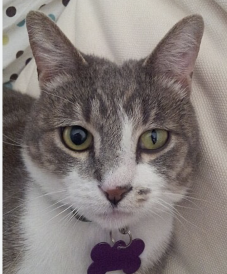
Horner’s Syndrome:
This condition can develop if the sympathetic nerves within the middle ear are affected, leading to ptosis (drooping eyelid), miosis (constricted pupil), enophthalmos (sunken eye), and protrusion of the third eyelid on the affected side.
Diagnostics
Accurate diagnosis of otitis media in cats requires a combination of clinical examination, imaging, and sometimes advanced diagnostics to confirm the presence of an infection and to evaluate the extent of the disease.
1. Otoscopic Examination:
Visual Inspection: An otoscopic examination can help visualize the tympanic membrane, assess its integrity, and detect signs of inflammation, discharge, or perforation.
Limitations: In some cases, the ear canal may be too inflamed or obstructed by debris or polyps to allow for a clear view of the tympanic membrane.
2. Imaging:
Radiography: Skull radiographs can provide some information about the bony structures of the middle ear, but they may not be sufficiently detailed for a definitive diagnosis.
Computed Tomography (CT): CT scans offer superior detail and are often used to assess the bulla and surrounding structures for evidence of fluid accumulation, bony changes, or masses.
Magnetic Resonance Imaging (MRI): MRI is particularly useful for evaluating soft tissue structures, including any nerve involvement or the extension of the disease to the inner ear or brain.
3. Myringotomy:
Procedure: Myringotomy involves making a small incision in the tympanic membrane to collect samples of the middle ear fluid for cytology and culture.
Indications: This procedure is indicated when imaging suggests fluid accumulation, and when a definitive diagnosis is needed to guide antimicrobial therapy.
4. Cytology and Culture:
Cytology: Examination of ear discharge or myringotomy samples under a microscope can reveal the presence of inflammatory cells, bacteria, or yeast.
Culture and Sensitivity: Bacterial culture of the middle ear fluid is crucial for identifying the causative organism and determining the most effective antibiotic treatment.
5. Neurological Examination:
Assessment: A thorough neurological examination can help identify signs of facial nerve involvement, Horner’s syndrome, or vestibular dysfunction, which are important indicators of the extent and severity of otitis media.
Treatment
The treatment of otitis media in cats involves addressing the underlying infection, managing pain and inflammation, and addressing any complications that arise.
1. Antimicrobial Therapy:
Systemic Antibiotics: Based on culture and sensitivity results, systemic antibiotics are prescribed. Commonly used antibiotics include amoxicillin-clavulanate, clindamycin, or enrofloxacin, depending on the identified pathogen.
Duration: Treatment is typically extended for 4-8 weeks to ensure the resolution of the infection, with follow-up evaluations to monitor progress.
2. Pain Management:
Analgesics: Pain management is crucial, and nonsteroidal anti-inflammatory drugs (NSAIDs) or opioids may be used to alleviate discomfort.
Corticosteroids: In cases of severe inflammation, corticosteroids may be used cautiously to reduce swelling, although their use should be carefully weighed against potential side effects.
3. Surgical Intervention:
Bulla Osteotomy: In cases where medical management fails, or in the presence of chronic otitis media with bony changes, a ventral bulla osteotomy (surgical opening of the bulla) may be necessary to drain and remove infected material.
Polyp Removal: If a polyp is identified, surgical removal may be required to prevent recurrence and improve drainage.
4. Supportive Care:
Nutritional Support: Cats with severe otitis media may require supportive care, including ensuring adequate nutrition and hydration.
Physical Therapy: If facial nerve paralysis or vestibular signs persist, physical therapy may be beneficial to aid recovery.
Seizure disorders
Seizures in cats, while less common than in dogs, present a significant diagnostic and therapeutic challenge in veterinary practice. The etiology of seizures in cats can range from primary epilepsy to secondary causes such as metabolic disorders, structural brain diseases, and infectious agents. This article provides a comprehensive overview of the pathophysiology, clinical signs, risk factors, diagnostic workup, and treatment options for feline seizures, with a focus on distinguishing hallmark signs and achieving an accurate diagnosis.
Pathophysiology
Seizures occur due to abnormal, excessive electrical activity in the brain, which can be focal (originating in one part of the brain) or generalized (involving both hemispheres). The underlying pathophysiology of seizures in cats is often multifactorial and can be categorized into the following:
Idiopathic Epilepsy: This refers to seizures with no identifiable cause. In cats, idiopathic epilepsy is less common than in dogs but still represents a significant portion of seizure cases, especially in younger animals. The exact mechanism is not well understood, but it is believed to involve an inherent imbalance between excitatory and inhibitory neurotransmitters in the brain.
Structural Epilepsy: Caused by identifiable brain abnormalities such as tumors, trauma, inflammation, or congenital malformations. Structural epilepsy in cats often involves focal seizures, with or without secondary generalization.
Reactive Seizures: Resulting from metabolic or toxic insults, such as hypoglycemia, hepatic encephalopathy, or exposure to toxins (e.g., permethrin, ethylene glycol). These seizures are not considered true epilepsy as they are provoked by transient disturbances in brain function.
Genetic Epilepsy: Although less studied in cats, certain breeds may be predisposed to genetic forms of epilepsy, similar to canine epilepsy syndromes.
Clinical Signs
Seizures in cats can vary widely in presentation, and recognizing the clinical signs is crucial for timely intervention. Common seizure types include:
Generalized Seizures: These involve the entire brain and are often characterized by loss of consciousness, tonic-clonic movements (rigid extension followed by rhythmic jerking), autonomic signs (salivation, urination, defecation), and postictal disorientation or blindness. Generalized seizures are more easily recognized due to their dramatic presentation.
Focal Seizures: These originate in a specific area of the brain and may present with subtle signs such as facial twitching, excessive salivation, chewing motions, or limb paddling. Focal seizures can progress to generalized seizures if the abnormal activity spreads across the brain.
Absence Seizures: Less common in cats, these involve brief episodes of staring or “blanking out” with no muscle activity. They may be easily missed or mistaken for behavioral abnormalities.
Status Epilepticus: A medical emergency characterized by continuous seizure activity lasting longer than five minutes or recurrent seizures without full recovery between episodes. It requires immediate intervention to prevent permanent brain damage or death.
Risk Factors
Several risk factors can predispose cats to seizures:
Age: Young cats (less than 1 year old) are more likely to suffer from idiopathic epilepsy, while older cats may develop seizures due to brain tumors or metabolic diseases.
Breed Predisposition: Although less defined than in dogs, certain breeds such as the Siamese and Burmese may have a higher incidence of epilepsy.
History of Trauma: Previous head injuries can lead to structural epilepsy due to scarring or other damage to brain tissue.
Infectious Diseases: Feline infectious peritonitis (FIP), toxoplasmosis, and other infectious agents can cause encephalitis, leading to seizures.
Toxic Exposures: Ingestion of neurotoxic substances such as permethrin (often found in flea treatments), lead, or organophosphates can induce seizures.
Metabolic Disorders: Conditions like hypoglycemia, hepatic encephalopathy, or severe electrolyte imbalances can trigger reactive seizures.
Metabolic disorders are systemic conditions that can significantly disrupt the normal physiological functions of the body, including the brain's ability to regulate and maintain stable electrical activity. When these disorders affect the brain, they can lead to what are termed "reactive seizures." Unlike primary epilepsy, which originates from intrinsic brain abnormalities, reactive seizures occur secondary to systemic metabolic disturbances. Here are some of the key metabolic conditions that can trigger reactive seizures in cats:
Hypoglycemia
Hypoglycemia refers to abnormally low blood glucose levels, which can starve the brain of its primary energy source—glucose. The brain is highly dependent on a constant supply of glucose to function correctly. When blood glucose levels drop too low, the brain cannot sustain normal electrical activity, leading to a variety of neurological signs, including seizures.
Hepatic Encephalopathy
Hepatic encephalopathy (HE) is a neurological syndrome resulting from liver dysfunction, where the liver's ability to detoxify blood is compromised. The accumulation of toxins, particularly ammonia, in the bloodstream can cross the blood-brain barrier and affect brain function, leading to seizures.
Electrolyte Imbalances
Electrolytes such as sodium, potassium, calcium, and magnesium play crucial roles in maintaining normal cellular function, including the generation and propagation of electrical impulses in neurons. Severe imbalances in these electrolytes can disrupt neuronal activity, leading to seizures.
Hyponatremia (Low Sodium Levels): Hyponatremia can cause brain cells to swell (cerebral edema), leading to increased intracranial pressure and seizures. It can occur due to excessive water intake, certain medications, or diseases like kidney failure or heart failure.
Hypernatremia (High Sodium Levels): Hypernatremia leads to cellular dehydration, including in brain cells, which can also trigger seizures. Causes include dehydration, salt poisoning, or inadequate water intake.
Hypocalcemia (Low Calcium Levels): Hypocalcemia can cause increased neuronal excitability, leading to tetany and seizures. It is often associated with conditions like eclampsia in pregnant or nursing queens, chronic renal disease, or hypoparathyroidism.
Hypercalcemia (High Calcium Levels): Hypercalcemia can cause neuromuscular excitability and seizures. It is often related to underlying conditions like hyperparathyroidism, certain neoplasms, or vitamin D toxicity.
Hypokalemia (Low Potassium Levels): While more commonly associated with muscle weakness, severe hypokalemia can also cause seizures due to altered membrane potentials.
Hyperkalemia (High Potassium Levels): Hyperkalemia is more likely to cause cardiac arrhythmias but can also affect neuronal function and potentially lead to seizures.
Diagnosis: Diagnosis involves blood tests to measure electrolyte levels. Depending on the specific imbalance, blood gas analysis, kidney function tests, and hormone assays (e.g., parathyroid hormone levels) may also be indicated.
Treatment: Treatment focuses on correcting the underlying electrolyte imbalance. For example, hypernatremia may require gradual rehydration, while hypocalcemia is treated with calcium supplementation. It’s crucial to correct these imbalances slowly and carefully to avoid complications such as cerebral edema or rebound electrolyte disturbances.
Diagnostics
A thorough diagnostic workup is essential to determine the underlying cause of seizures in cats. The following steps are typically involved:
History and Physical Examination: Collect a detailed history, including age at onset, frequency, duration, and type of seizures. Investigate possible exposures to toxins, recent trauma, and any concurrent illnesses. A comprehensive physical and neurological examination can reveal clues to the etiology, such as signs of systemic illness or focal neurological deficits.
Blood Work: Perform a complete blood count (CBC), serum biochemistry profile, and urinalysis to identify metabolic or systemic causes. Tests for liver function (e.g., bile acids test) and glucose levels are particularly important in ruling out reactive seizures.
Imaging: Advanced imaging techniques such as magnetic resonance imaging (MRI) or computed tomography (CT) are crucial for detecting structural brain abnormalities, including tumors, hydrocephalus, or encephalitis. MRI is the preferred modality for its superior soft tissue resolution.
Cerebrospinal Fluid (CSF) Analysis: CSF collection and analysis can help identify inflammatory or infectious diseases of the central nervous system (CNS), such as FIP or toxoplasmosis. Elevated protein levels, pleocytosis, or the presence of infectious agents can provide diagnostic clues.
Electroencephalography (EEG): While less commonly used in veterinary medicine, EEG can be helpful in characterizing the type of seizure and identifying the focus of epileptic activity.
Genetic Testing: In cases where a genetic basis is suspected, particularly in predisposed breeds, genetic testing may be considered.
Treatment
The treatment of seizures in cats depends on the underlying cause and the severity of the condition. The primary goals of treatment are to control seizure frequency and severity while minimizing side effects.
Antiepileptic Drugs (AEDs):
Phenobarbital: The most commonly used AED in cats, effective in controlling both generalized and focal seizures. Regular monitoring of blood levels is required to avoid toxicity.
Levetiracetam: An alternative to phenobarbital with fewer side effects, especially in cats with hepatic disease. It is often used as an adjunct therapy but it is given 3 times a day which is not very convenient for pet owners.
Zonisamide: Another option for cats that do not respond well to phenobarbital or levetiracetam. It has a longer half-life and is generally well-tolerated.
Diazepam: Used for emergency management of status epilepticus. Caution is advised as chronic use can lead to tolerance and hepatotoxicity in cats.
Treatment of Underlying Conditions: If the seizures are secondary to an underlying condition, such as a brain tumor or hepatic encephalopathy, treating the primary disease is essential. This may involve surgery, dietary management, or specific treatments such as antibiotics or antivirals.
Emergency Management of Status Epilepticus: Immediate administration of diazepam or midazolam intravenously or rectally is crucial. If seizures persist, general anesthesia may be required to control the activity.
Supportive Care: In cats with frequent or severe seizures, supportive care, including maintaining hydration, electrolyte balance, and nutritional support, is critical. In some cases, hospitalization may be necessary.
Vestibular Disease
Peripheral Vestibular Disease typically arises from issues in the inner ear, such as inflammation, infection, trauma, or neoplasia. Conditions like otitis media/interna, ear polyps, or idiopathic vestibular syndrome are common causes.
Central Vestibular Disease involves the brainstem or cerebellum and may result from more severe conditions such as neoplasia, infarcts, inflammatory diseases (e.g., Feline Infectious Peritonitis), or toxins.
When the vestibular system is compromised, the brain receives incorrect information about the body's position, leading to the characteristic signs of vestibular disease.
Central vestibular signs primarily arise from issues in the brainstem and cerebellum, which are the main components of the central vestibular system. Here's a more detailed breakdown:
1. Brainstem
The brainstem, particularly the medulla oblongata and pons, is a critical area for central vestibular function. It contains the vestibular nuclei, which process information from the inner ear and coordinate balance and eye movements. Lesions in the brainstem can disrupt these processes, leading to central vestibular signs such as:
Nystagmus (which may be horizontal, vertical, or rotary) Rotary nystagmus can be seen with both peripheral and central vestibular disease. In peripheral vestibular disease, rotary nystagmus often occurs along with horizontal nystagmus, and it usually maintains a consistent direction. In central vestibular disease, nystagmus can be more variable and may change direction depending on the position of the head. However, rotary nystagmus is not specific to either peripheral or central vestibular disease and can occur with lesions affecting either part of the vestibular system. Horizontal nystagmus is typically seen with peripheral and vertical nystagmus is typically seen with central.
Ataxia (lack of coordination)
Postural deficits (such as leaning or falling to one side)
Cranial nerve deficits (since many cranial nerves originate in the brainstem, other signs like facial paralysis, altered gag reflex, or changes in pupil size might also be seen)
2. Cerebellum
The cerebellum is responsible for fine-tuning motor movements and coordinating balance. It receives input from the vestibular system and plays a significant role in maintaining posture and coordinating voluntary movements. Lesions in the cerebellum can cause:
Intention tremors (shaking during purposeful movements)
Hypermetria (exaggerated limb movements)
Truncal ataxia (swaying of the body)
Head tilt and nystagmus (often more severe and potentially involving multiple directions)
Vestibular diseases
The clinical presentation of vestibular disease in cats can be acute and dramatic, causing significant distress to both the cat and its owner. Common clinical signs include:
Head Tilt: Often towards the side of the lesion, a hallmark sign of vestibular dysfunction.
Nystagmus: Involuntary rhythmic eye movements. The direction of nystagmus can provide clues to whether the issue is peripheral (horizontal or rotary nystagmus) or central (vertical nystagmus).
Ataxia: A staggering or uncoordinated gait, often accompanied by a wide-based stance.
Circling: Typically towards the side of the lesion, indicating a loss of balance.
Strabismus: Abnormal positioning of the eye, particularly ventral or ventrolateral, which may be present in more severe cases.
Vomiting and Nausea: Due to the involvement of the vestibular apparatus in the vomiting reflex.
Loss of Appetite: Secondary to nausea and vomiting.
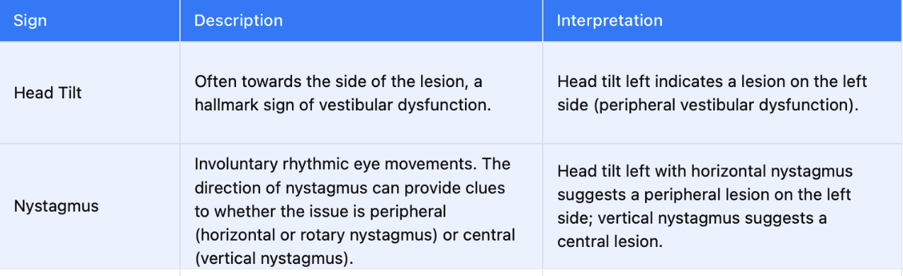
Head tilt left + horizontal nystagmus = Peripheral lesion (inner ear) on the left side.
Head tilt right + vertical nystagmus= Central lesion (brain) on the right side.
Diagnostics
Diagnosing vestibular disease in cats requires a thorough understanding of both the peripheral and central components of the vestibular system. A systematic approach is essential:
History and Clinical Examination:
Obtain a detailed history, focusing on the onset of signs, any previous ear infections, trauma, or exposure to ototoxic drugs.
Perform a thorough neurological examination to differentiate between peripheral and central vestibular disease.
Otoscopy:
Examine the ear canal for signs of infection, foreign bodies, or polyps.
Assess the tympanic membrane for integrity and signs of otitis media.
Advanced Imaging:
CT or MRI: Imaging is crucial for evaluating the middle ear and brain. CT is excellent for detecting bone involvement, such as bulla osteitis, while MRI is superior for assessing soft tissues like the brainstem and cerebellum.
Radiographs: Skull radiographs can be useful but are less sensitive than CT or MRI.
Cerebrospinal Fluid (CSF) Analysis:
Indicated if central vestibular disease is suspected. Analysis can reveal inflammation, infection, or neoplastic cells.Cerebrospinal fluid (CSF) analysis is one of the diagnostic tools used when central vestibular disease is suspected. This procedure involves collecting CSF from the spinal column to analyze it for abnormalities.
CSF Analysis:
Inflammation: The CSF may show an elevated white blood cell count (pleocytosis) and increased protein levels, suggesting an inflammatory condition.
Infection: The presence of infectious organisms (e.g., bacteria, fungi) or evidence of infection (e.g., elevated neutrophils, lymphocytes) can be detected in the CSF.
Neoplastic cells: In cases of brain tumors, abnormal cells may be present in the CSF, indicating a central nervous system neoplasm.
Bloodwork and Serology:
Complete blood count (CBC) and biochemistry to assess overall health.
Serological tests for infectious diseases, such as FIP, Toxoplasmosis, or Cryptococcus.
Electrodiagnostic Testing:
Brainstem auditory evoked response (BAER) testing can help assess the auditory pathways and differentiate between conductive and sensorineural deafness, which may accompany vestibular disease.
Treatment
The treatment of vestibular disease in cats depends on the underlying cause:
Idiopathic Vestibular Disease:
Often self-limiting, with signs improving within a few days to weeks. Supportive care includes anti-nausea medication (e.g., maropitant) and ensuring the cat remains hydrated and comfortable.
Otitis Media/Interna:
Systemic antibiotics based on culture and sensitivity results. Long courses (4-8 weeks) are often required.
Myringotomy or surgical intervention may be necessary in chronic or refractory cases.
Nasopharyngeal Polyps:
Surgical removal of the polyp is typically curative. Recurrence is possible, and postoperative care is essential.
Central Vestibular
Treatment focuses on the underlying cause, such as corticosteroids for inflammatory diseases, anticonvulsants for seizures, or surgical resection for tumors.
Prognosis depends on the severity and reversibility of the underlying condition.
Supportive Care:
Anti-emetics to control nausea.
Fluid therapy if the cat is dehydrated due to vomiting or inappetence.
Nutritional support if the cat is not eating.
Hallmark Clinical Signs and Diagnosis For Vestibular Disease
The hallmark clinical signs of vestibular disease in cats include a head tilt, nystagmus, and ataxia. Differentiating between peripheral and central vestibular disease is crucial:
Peripheral Vestibular Disease: Typically presents with a head tilt, horizontal nystagmus, and mild ataxia. These cats often have normal mentation.
Central Vestibular Disease: May present with vertical or rotary nystagmus, proprioceptive deficits, and altered mentation. These signs are often more severe and may be accompanied by other neurological deficits.
Diagnosis is made based on a combination of clinical signs, imaging studies, and, if necessary, CSF analysis. Early and accurate diagnosis is critical for initiating appropriate treatment and improving the cat’s prognosis.
Spinal Injuries
Cervical Spine (C1–C5):
Tetraparesis or tetraplegia.
Hyperreflexia in all limbs.
Proprioceptive deficits in all limbs.
Respiratory compromise in severe cases.
Cervicothoracic Spine (C6–T2):
LMN signs in thoracic limbs (flaccid paresis, hyporeflexia).
UMN signs in pelvic limbs (spasticity, hyperreflexia).
Horner’s syndrome if sympathetic innervation is affected.
Thoracolumbar Spine (T3–L3):
Normal thoracic limbs.
UMN signs in pelvic limbs (spastic paresis, hyperreflexia).
Proprioceptive deficits and ataxia in pelvic limbs.
Paraplegia in severe cases.
Lumbosacral Spine (L4–S3):
LMN signs in pelvic limbs (flaccid paresis, hyporeflexia).
Urinary and fecal incontinence due to disruption of the pudendal and pelvic nerves.
Tail paresis or paralysis.
Caudal Spine (Below S3):
Flaccid tail paralysis.
Fecal and urinary incontinence without significant limb involvement.
Hepatic Encephalopathy
Hepatic encephalopathy (HE) is a neurological syndrome resulting from liver dysfunction, where the liver's ability to detoxify blood is compromised. The accumulation of toxins, particularly ammonia, in the bloodstream can cross the blood-brain barrier and affect brain function, leading to seizures.
Causes: Hepatic encephalopathy is commonly associated with conditions like portosystemic shunts (abnormal blood vessels that bypass the liver), chronic liver disease, or acute liver failure. In these conditions, the liver cannot effectively remove toxins from the blood, leading to their accumulation in the brain.
Clinical Signs: Cats with hepatic encephalopathy may show a range of neurological signs, including behavioral changes, ataxia, hypersalivation, and seizures. These signs can be episodic, often triggered by high-protein meals, dehydration, or gastrointestinal bleeding.
Diagnosis: Diagnosis involves blood tests showing elevated liver enzymes, high blood ammonia levels, and imaging studies (ultrasound or CT) revealing liver abnormalities or portosystemic shunts.
Treatment: Acute management includes reducing ammonia levels with lactulose, dietary protein restriction, and antibiotics (like metronidazole or neomycin) to reduce ammonia-producing bacteria in the gut. Long-term management may require surgical correction of portosystemic shunts or medical management of chronic liver disease.
Hypoglycemia
Hypoglycemia refers to abnormally low blood glucose levels, which can starve the brain of its primary energy source—glucose. The brain is highly dependent on a constant supply of glucose to function correctly. When blood glucose levels drop too low, the brain cannot sustain normal electrical activity, leading to a variety of neurological signs, including seizures.
Causes: Hypoglycemia in cats can occur due to various reasons, such as insulin overdose in diabetic cats, prolonged fasting, severe liver disease (which impairs glucose production), or insulinoma (a pancreatic tumor that produces excess insulin).
Clinical Signs: Early signs of hypoglycemia include lethargy, weakness, disorientation, and ataxia. If untreated, these can progress to more severe symptoms like tremors, seizures, and even coma.
Diagnosis: Blood glucose measurement is crucial for diagnosing hypoglycemia. Glucose levels below 60 mg/dL are typically concerning and can be associated with seizure activity.
Treatment: Immediate treatment involves administering glucose either orally (if the cat is conscious) or intravenously (in emergency situations). Long-term management focuses on addressing the underlying cause and preventing future hypoglycemic episodes.