5- Prokaryotic cell structure and function

0.0(0)
studied byStudied by 0 people
0.0(0)
full-widthCall Kai
learnLearn
examPractice Test
spaced repetitionSpaced Repetition
heart puzzleMatch
flashcardsFlashcards
GameKnowt Play
Card Sorting

1/9

encourage image

There's no tags or description

Looks like no tags are added yet.

Study Analytics
Name
Mastery
Learn
Test
Matching
Spaced

No study sessions yet.

10 Terms

1
New cards

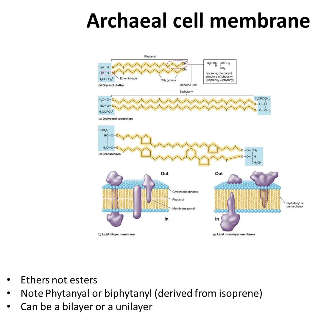
how are cell membranes in archaea unique?

  • phospholipids in archaea are joined by ether bonds, not ester bonds

  • diglycerols can be formed, so they can form monolayers as well as bilayers

<ul><li><p>phospholipids in archaea are joined by <strong>ether </strong>bonds, <strong>not ester</strong> bonds</p></li><li><p><strong>diglycerols</strong> can be formed, so they can form <strong>monolayers </strong>as well as bilayers</p></li></ul><p></p>
2
New cards

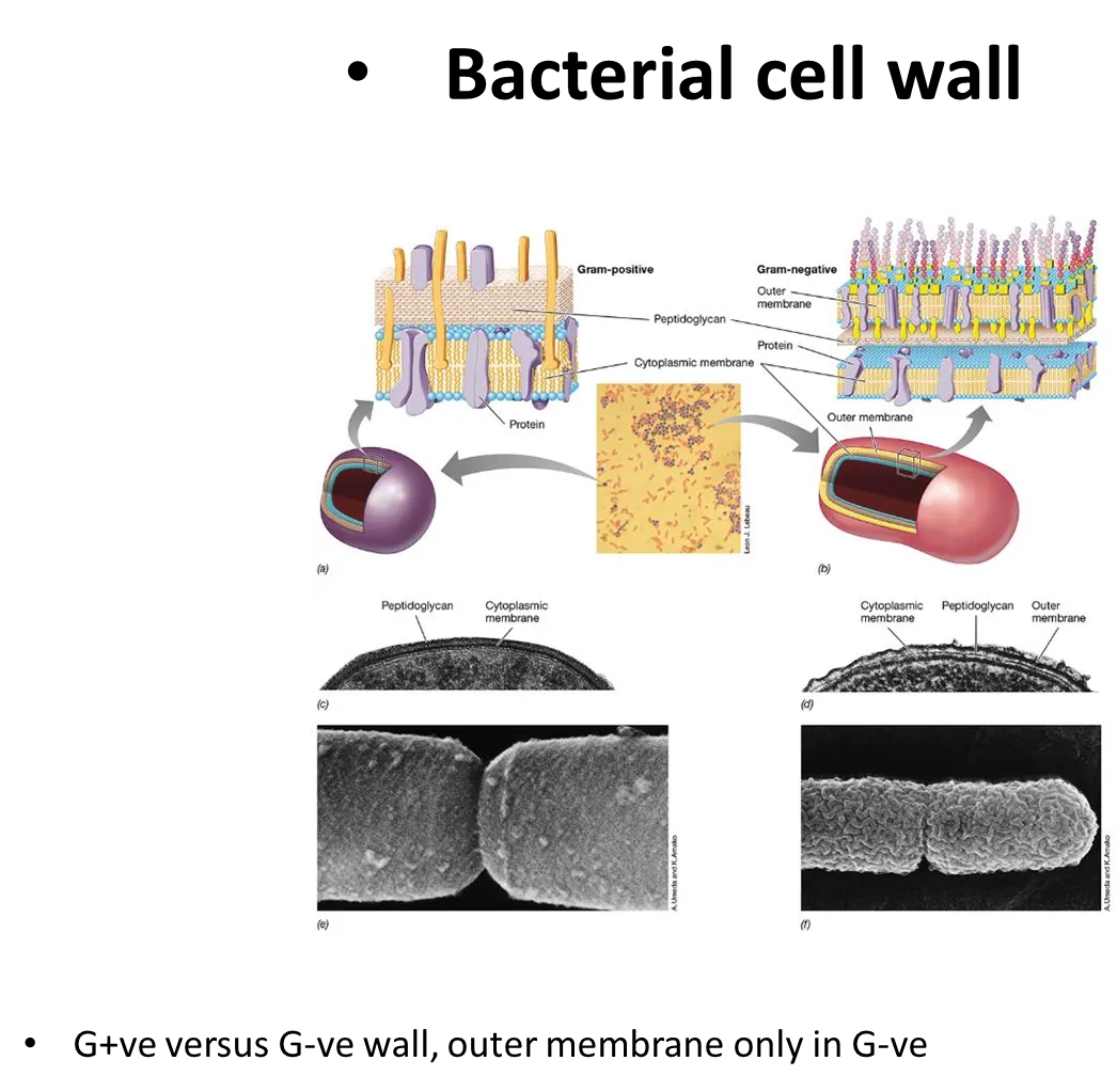
how are the structures of G+ve and G-ve bacterial cell walls different?

  • G+ve bacteria have a cytoplasmic membrane and a large layer of peptidoglycan over it

  • G-ve bacteria have a thin layer of peptidoglycan around the cytoplasmic membrane, followed by a second outer unit membrane (so they don’t take up gram stain), with large carbohydrate chains sticking off- inbetween the two membranes is the periplasm

<ul><li><p>G+ve bacteria have a cytoplasmic membrane and a large layer of peptidoglycan over it</p></li><li><p>G-ve bacteria have a thin layer of peptidoglycan around the cytoplasmic membrane, followed by a second outer unit membrane (so they don’t take up gram stain), with large carbohydrate chains sticking off- inbetween the two membranes is the periplasm</p></li></ul><p></p>
3
New cards

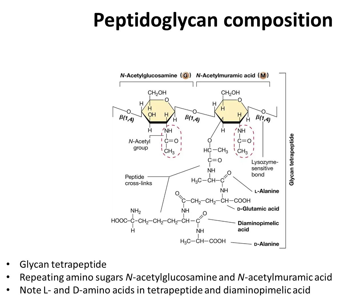
what is the structure of peptidoglycan?

  • the sugar backbone is composed of alternating monomers of N-acetylglucosamine and N-acetylmuramic acid (modified glucose residues) joined by a beta-1,4 linkage

  • a short peptide side chain is attached to N-acetylmuramic acid- the amino acids vary between species

  • strands of peptidoglycan cross-link by peptide bonds between the amino acids in these peptide side chains

<ul><li><p>the sugar backbone is composed of <strong>alternating </strong>monomers of <strong>N-acetylglucosamine and N-acetylmuramic acid </strong>(modified glucose residues) joined by a beta-1,4 linkage</p></li><li><p>a short <strong>peptide side chain </strong>is attached to<strong> N-acetylmuramic acid</strong>- the amino acids vary between species</p></li><li><p>strands of peptidoglycan <strong>cross-link</strong> by <strong>peptide </strong>bonds&nbsp;between the amino acids in these peptide side chains</p></li></ul><p></p>
4
New cards

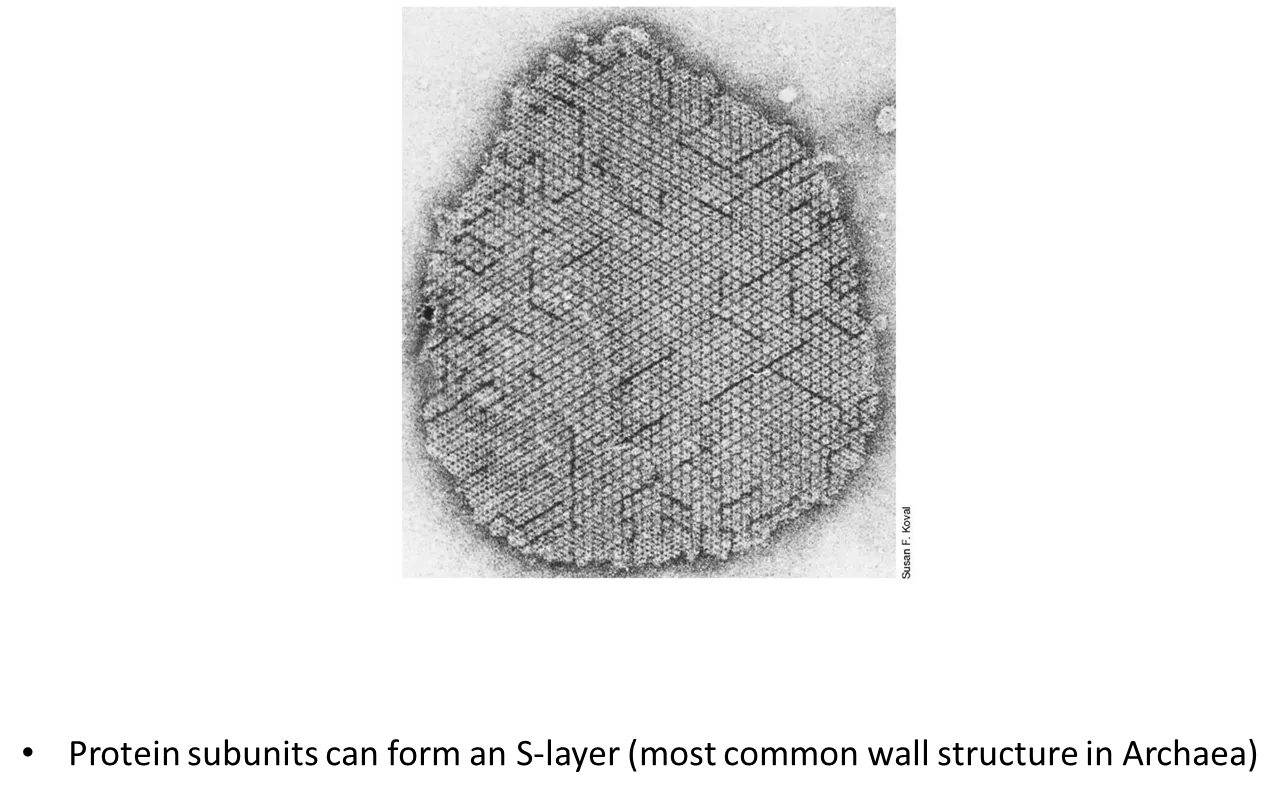
how are archaeal cell walls different to bacterial cell walls?

  • archaea don’t have peptidoglycan in their cell walls

  • they don’t have an outer membrane like G-ve bacteria

  • protein subunits can form an S-layer

  • the structure of the cell walls varies a lot between archaeal species

<ul><li><p>archaea <strong>don’t have peptidoglycan</strong> in their cell walls</p></li><li><p>they <strong>don’t have an outer membrane</strong> like G-ve bacteria</p></li><li><p>protein subunits can form an <strong>S-layer</strong></p></li><li><p>the structure of the cell walls varies a lot between archaeal species</p></li></ul><p></p>
5
New cards

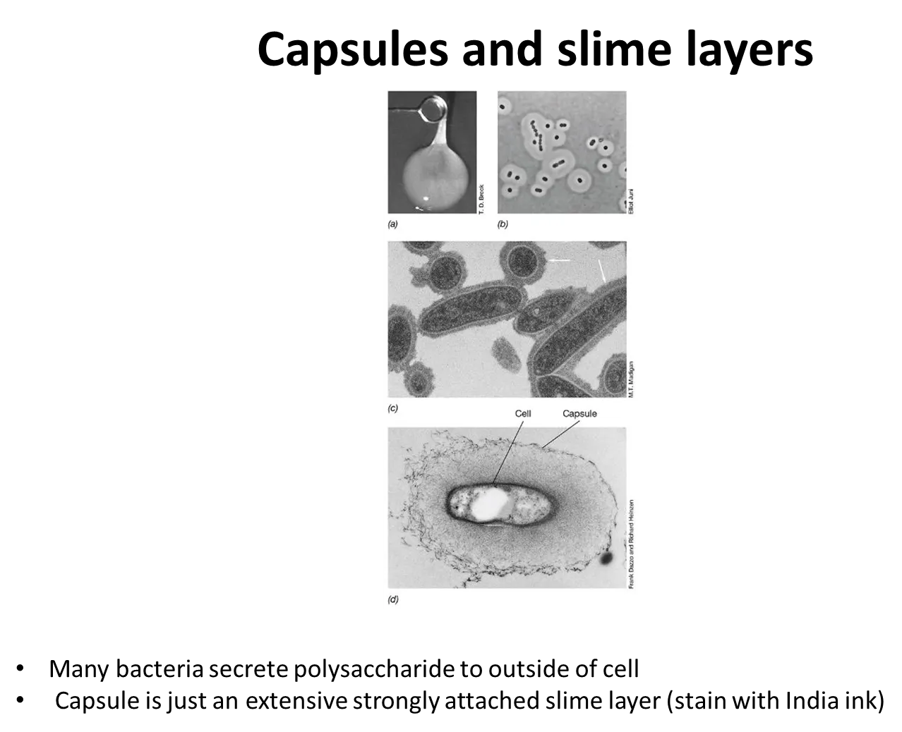
what are capsules and slime layers?

many prokaryotes secrete sticky/slimy polysaccharides on their cell surface, outside the cell envelope:

  • a capsule is a tight matrix that excludes small particles

  • a slime layer is easily deformed and loosely attached, and doesn’t exclude particles

these mediate attachment, protect the cell from attack and environmental stresses eg. dehydration

  • they are often used by nitrogen-fixing bacteria to exclude oxygen, because nitrogenase is destroyed by oxygen

<p>many prokaryotes secrete <strong>sticky/slimy polysaccharides</strong> on their cell surface, outside the cell envelope:</p><ul><li><p>a <strong>capsule </strong>is a <strong>tight matrix </strong>that excludes small particles</p></li><li><p>a <strong>slime layer</strong> is easily deformed and <strong>loosely attached</strong>, and doesn’t exclude particles</p></li></ul><p>these mediate attachment, protect the cell from attack and environmental stresses eg. dehydration</p><ul><li><p>they are often used by nitrogen-fixing bacteria to exclude oxygen, because nitrogenase is destroyed by oxygen</p></li></ul><p></p>
6
New cards

what are pili used for?

  • sticking to surfaces 

  • sticking to other cells (helpful for pathogens/conjugation)

  • forming thin sheets of cells: biofilms (on solid surfaces) and pellicles (on liquids)

  • movement via twitching motility (by attaching to a surface then retracting)

all G-ve have pili, some G+ve do too

7
New cards

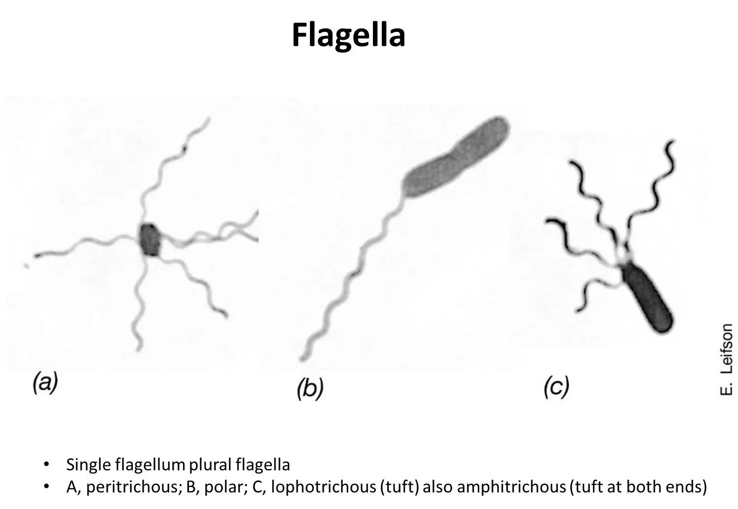
what are the four distributions of flagella?

  • peritrichous - scattered around the cell surface

  • polar- one flagella at one end

  • lophotrichous- bundle at one end

  • amphitricous- bundles at both ends

<ul><li><p><strong>peritrichous </strong>- scattered around the cell surface</p></li><li><p><strong>polar</strong>- one flagella at one end</p></li><li><p><strong>lophotrichous</strong>- bundle at one end</p></li><li><p><strong>amphitricous</strong>- bundles at both ends</p></li></ul><p></p>
8
New cards

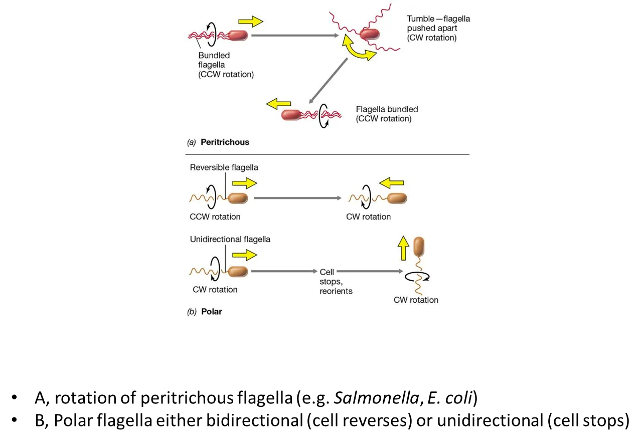
how do bacteria move using flagella?

in peritrichous bacteria:

  • all the flagella bundle together and rotate counterclockwise

  • to turn, one flagellum turns clockwise, causing a ‘tumble’ where it will change direction

  • then the flagella bundle back up and rotate counterclockwise in the new direction

in polar bacteria:

  • some cells can reverse by turning their flagella counterclockwise or clockwise (reversible flagella)

  • some cell have to stop and reorient to turn (unidirectional flagella)

<p>in <strong>peritrichous</strong> bacteria:</p><ul><li><p>all the flagella bundle together and rotate counterclockwise</p></li><li><p>to turn, one flagellum turns clockwise, causing a ‘tumble’ where it will change direction</p></li><li><p>then the flagella bundle back up and rotate counterclockwise in the new direction</p></li></ul><p>in <strong>polar</strong> bacteria:</p><ul><li><p>some cells can reverse by turning their flagella counterclockwise or clockwise (<strong>reversible</strong> flagella) </p></li><li><p>some cell have to stop and reorient to turn (<strong>unidirectional</strong> flagella)</p></li></ul><p></p>
9
New cards

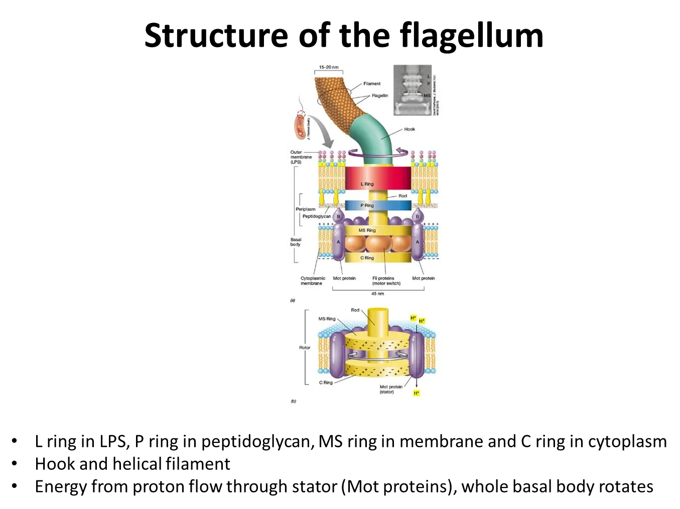
how do flagella rotate?

  • bacterial flagella are rigid and helical

  • the basal body is anchored in the cytoplasmic membrane

  • this contains a proton motor- proton movement through channels and electrostatic forces cause the rotation of the proteins

  • they change their speed in relation to the strength of the proton motive force

<ul><li><p>bacterial flagella are rigid and helical</p></li><li><p>the basal body is anchored in the cytoplasmic membrane</p></li><li><p>this contains a <strong>proton motor</strong>- proton movement through channels and electrostatic forces cause the rotation of the proteins</p></li><li><p>they change their speed in relation to the strength of the <strong>proton motive force</strong></p></li></ul><p></p>
10
New cards

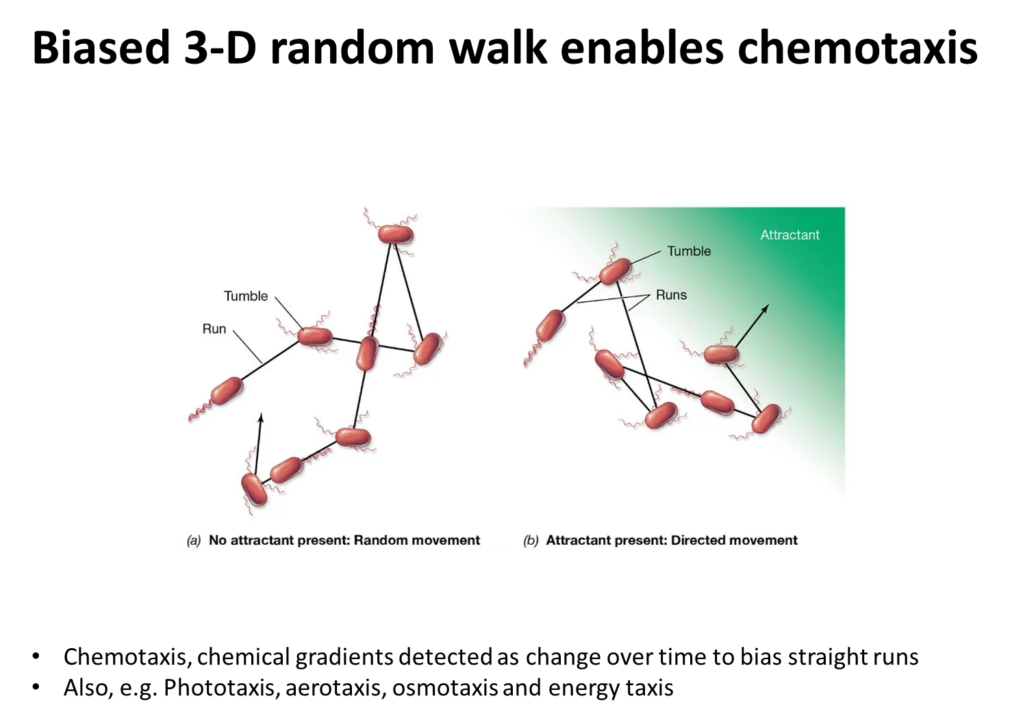
how do flagella enable chemotaxis?

positive chemotaxis towards an attractant (eg. sugar):

  • if the bacteria are swimming away from the attractant, the frequency of tumbling increases so that the bacteria turn towards it, and the frequency of tumbling then decreases

the opposite occurs for negative chemotaxis away from a repellent (eg. toxin)

this is called the biased 3D random walk

<p><strong>positive chemotaxis towards an attractant (eg. sugar):</strong></p><ul><li><p>if the bacteria are swimming away from the attractant, the frequency of tumbling increases so that the bacteria turn towards it, and the frequency of tumbling then decreases</p></li></ul><p>the <strong>opposite</strong> occurs for <strong>negative chemotaxis away from a repellent (eg. toxin)</strong></p><p></p><p>this is called the <strong>biased 3D random walk</strong></p><p></p>