A&P Lab 3
 Call Kai
 Learn
Learn Practice Test
 Spaced Repetition
 Match
Match Flashcards
 Knowt Play
1/166

There's no tags or description
Looks like no tags are added yet.
Name | Mastery | Learn | Test | Matching | Spaced | Call with Kai | Chat |
|---|
No analytics yet
Send a link to your students to track their progress
167 Terms
What is the difference between dendrites and axons?
Dendrites primarily receive signals; axons conduct signals away from the cell body.
What are the meninges?
The three protective coverings of the brain and spinal cord

In the meninges there are…..
3 layers with 2 spaces!


What is number 1?
Dura mater
outermost, tough meningeal layer
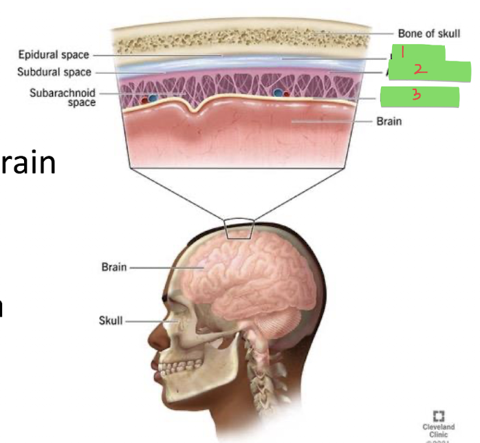
What is number 2?
Arachnoid mater
middle, web-like meningeal layer
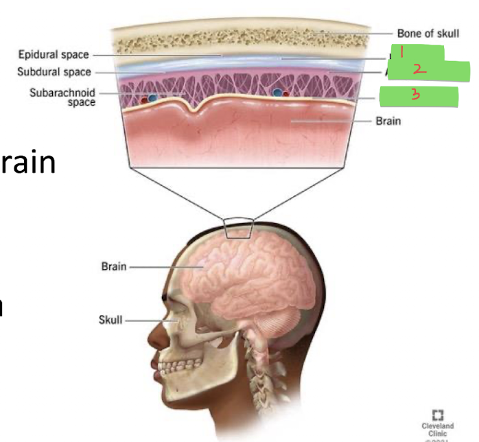
What is number 3?
Pia mater
innermost meningeal layer that closely follows the surface of the brain and spinal cord
What is the subarachnoid space?
space between the arachnoid mater and pia mater that contains CSF

What is the subdural space?
space between the dura mater and arachnoid mater

What is the epidural space?
space external to the dura mater; in the spinal cord region, it contains fat

The webby arachnoid matter…..
…holds cerebrospinal fluid
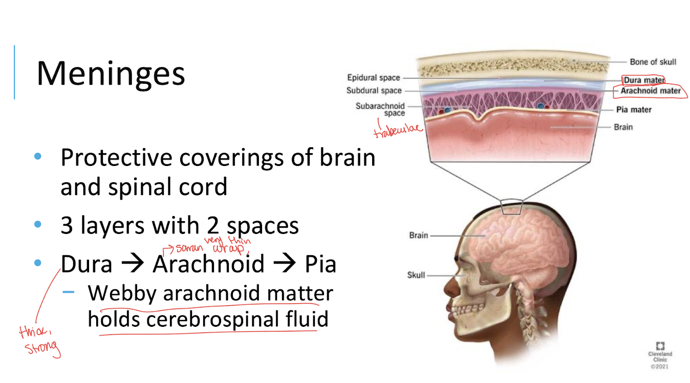

What are the blue arrows in this image pointing at?
the dura mater!
has some thinner & thicker portions
What kind of tissue is the dura mater made of?
Dense irregular connective tissue!

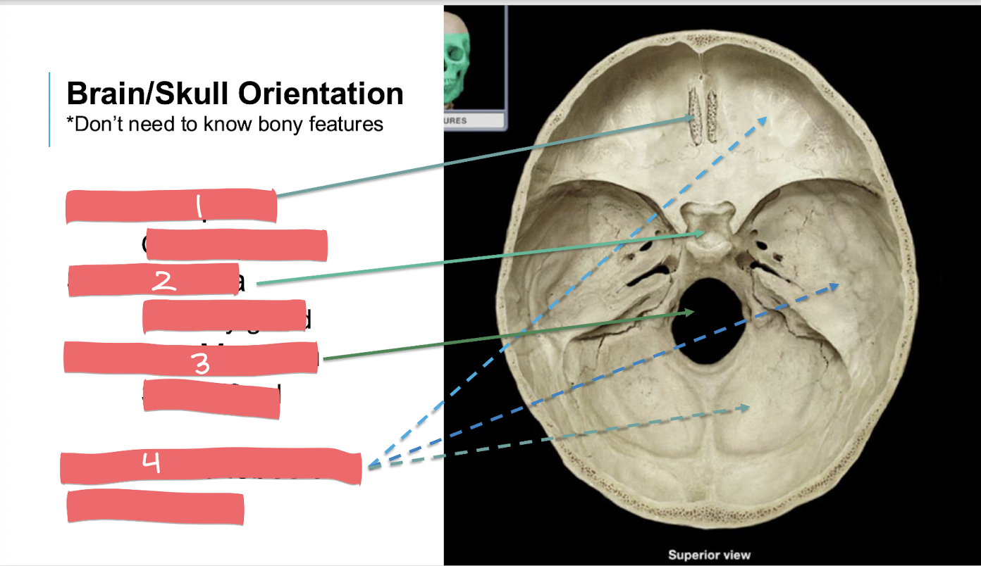
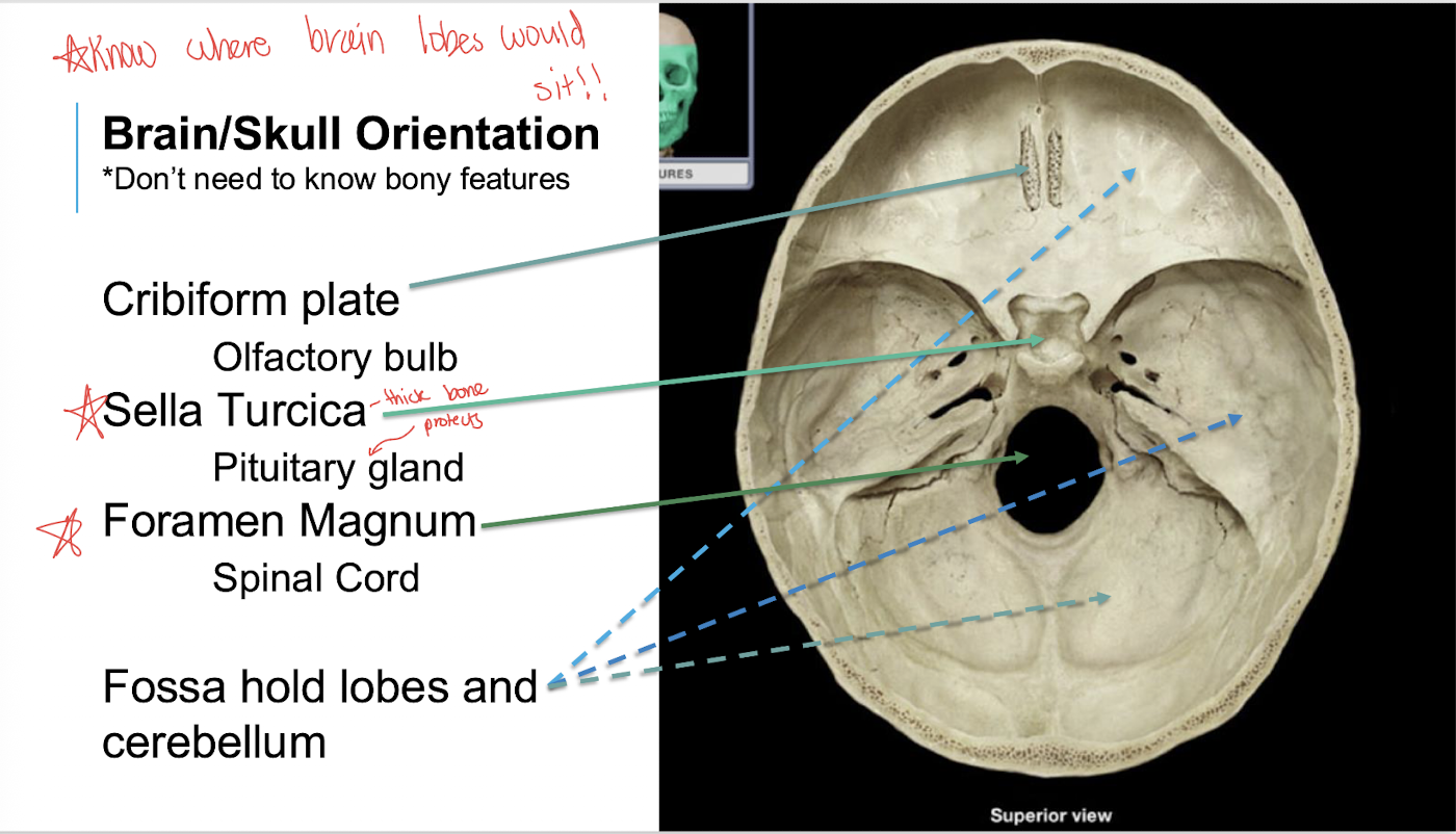
What is number 1?
cribiform plate

What is number 2?
sella turcica
saddle-shaped depression in the sphenoid bone

What is number 3?
foramen magnum

What is number 4?
Fossa
What does the cribiform plate hold?
the olfactory bulb!


What does the sella turcica hold?
Pituitary gland!!!

What does the foramen magnum hold?
spinal cord

What does the fossa hold?
holes lobes & cerebellum
Anterior cranial fossa: Holds the frontal lobes of the cerebral cortex, the olfactory bulbs (for smell), and the crista galli.
Middle cranial fossa: Holds the temporal lobes of the brain and the pituitary gland (which sits in a specific pocket called the sella turcica).
Posterior cranial fossa: Holds the cerebellum and the brainstem (pons and medulla oblongata).
What is gray matter?
Tissue primarily containing neuronal cell bodies, dendrites, and unmyelinated axons.
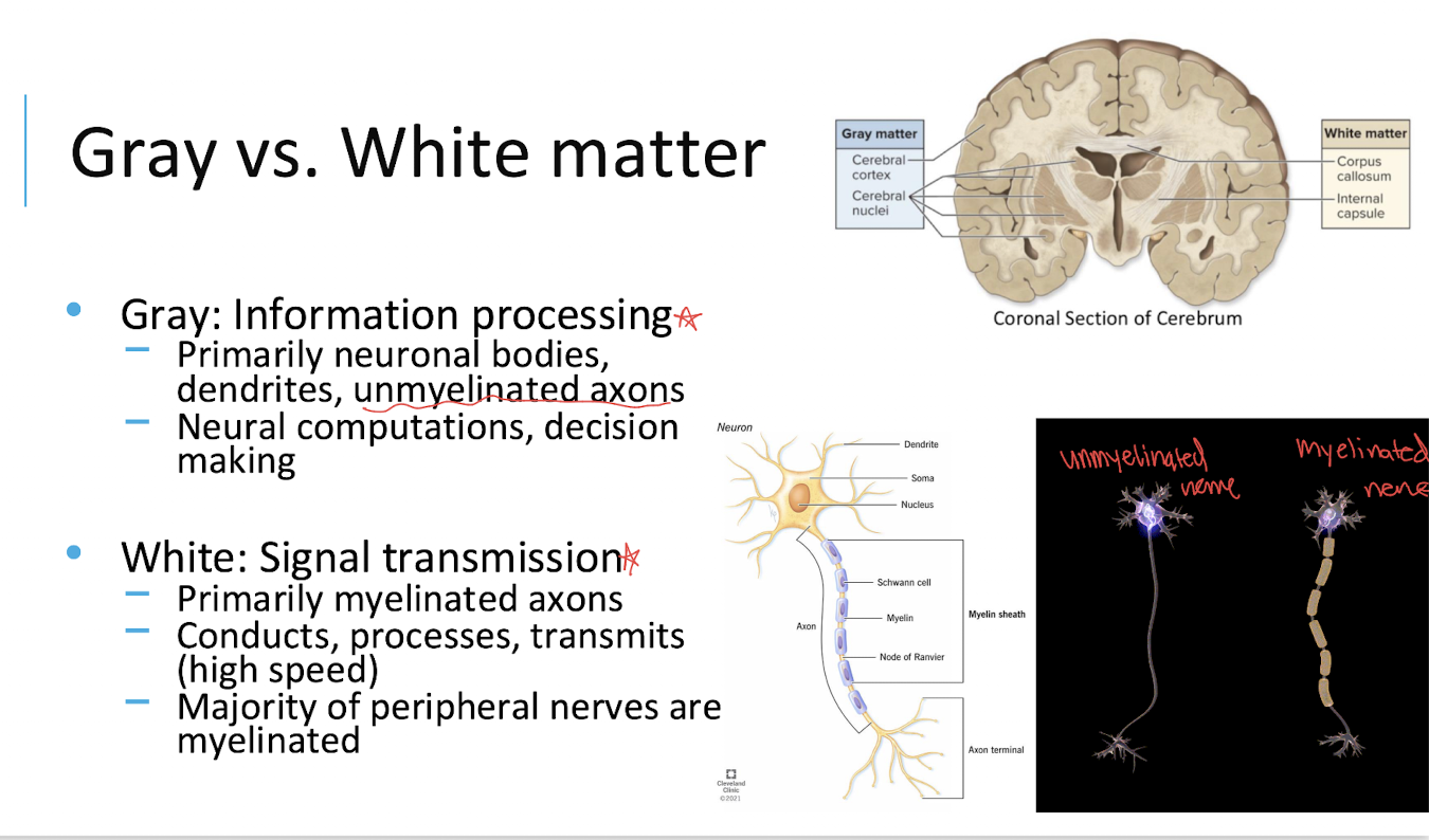
What is the main function of gray matter?
Information processing
including neural computations and decision-making.
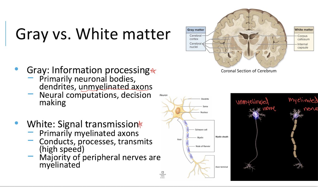
What is white matter?
Tissue primarily containing myelinated axons.

What is the main function of white matter?
Signal transmission through the nervous system.
conducts processes, transmits (high speed)
majority of peripheral nerves are myelinated

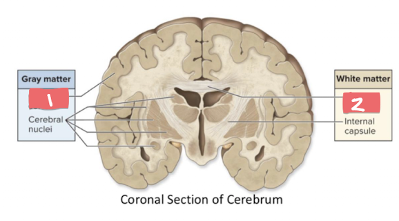
What is number 1?
Cerebral cortex
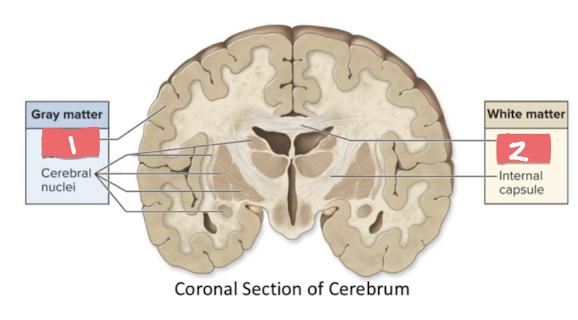
What is number 2?
Corpus callosum

What is number 1? *
White matter *
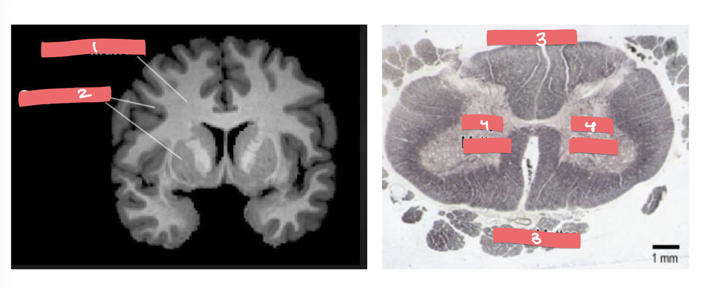
What is number 2? *
Grey matter *

What is number 3? **
White Matter **

What is number 4?
Grey Matter **
Where is gray matter located in the cerebral hemispheres?
In the cerebral cortex, surrounding the cerebral hemispheres.

Where is gray matter located in the spinal cord?
In the center of the spinal cord.

Where is white matter located in the spinal cord?
Around the outside of the gray matter
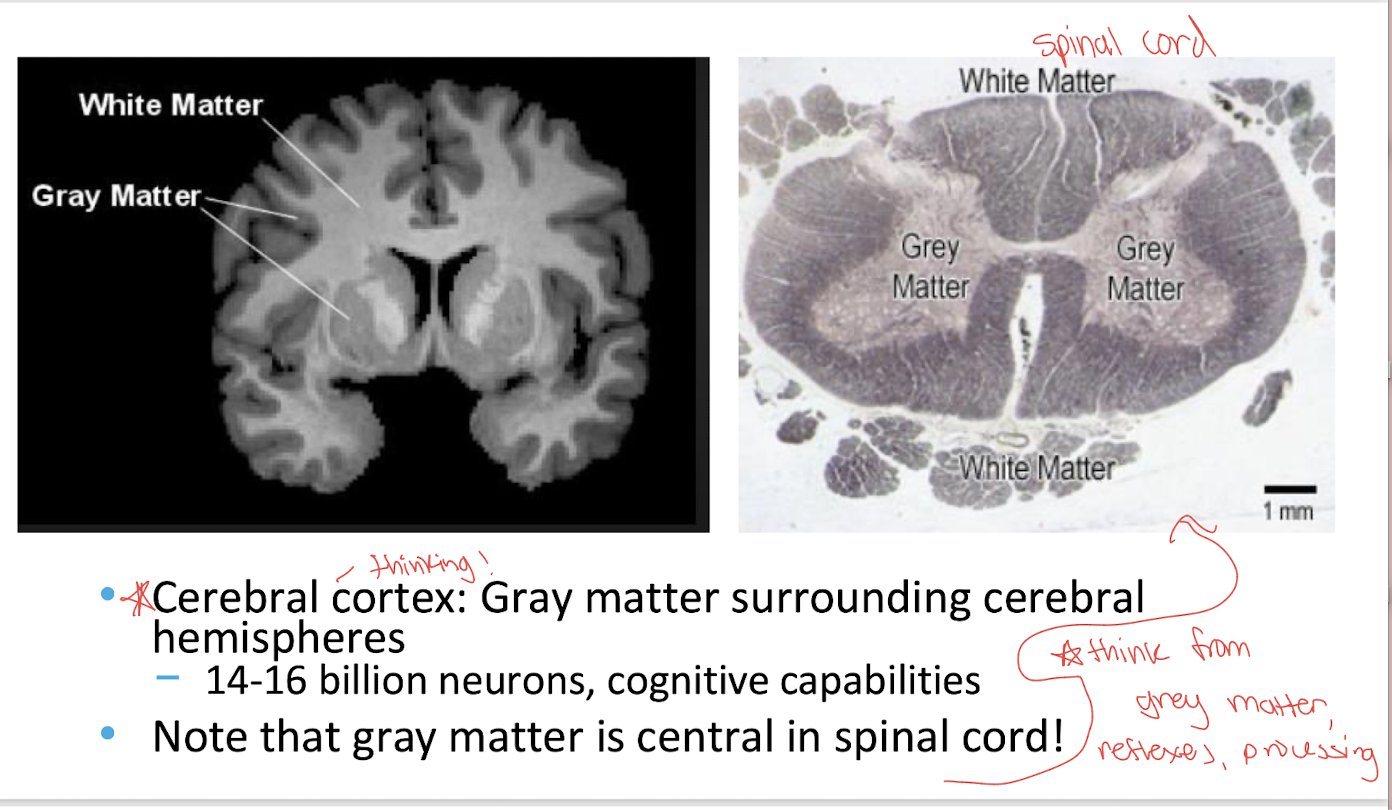
Why does white matter appear white?
Because it contains many myelinated axons.
Myelin allows axons to conduct signals more rapidly
Overall: What is the main difference between gray and white matter?
Gray matter processes information; white matter transmits information.
What is the corpus callosum?
A large white matter tract connecting the left and right cerebral hemispheres.
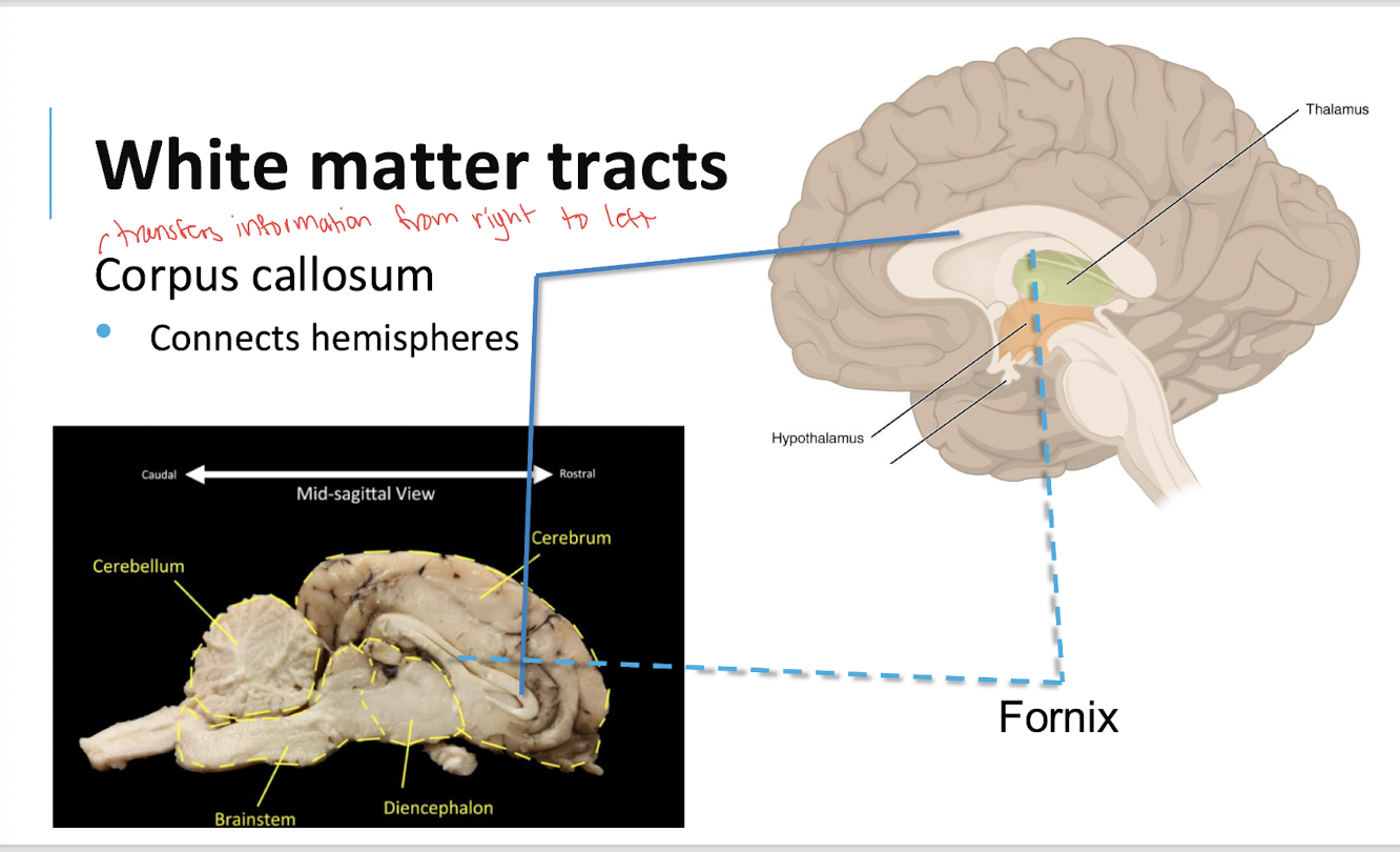
What is the function of the corpus callosum?
It allows information to be communicated between the two cerebral hemispheres.

What is the fornix?
A white matter tract associated with communication between structures involved in memory.
connects the hippocampus to other areas like the hypothalamus, playing a key role in memory and cognitive functions

What is the general function of white matter tracts?
They transmit information between regions of the nervous system.

What are the cerebral hemispheres?
The left and right halves of the cerebrum
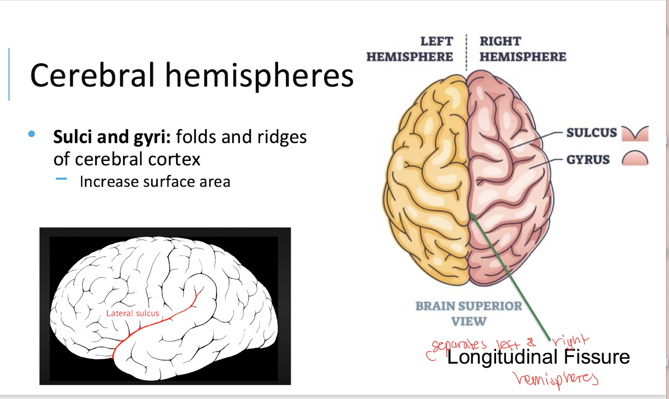
What is the cerebral cortex?
The outer layer of gray matter surrounding the cerebral hemispheres

What are gyri?
The ridges or folds of the cerebral cortex.
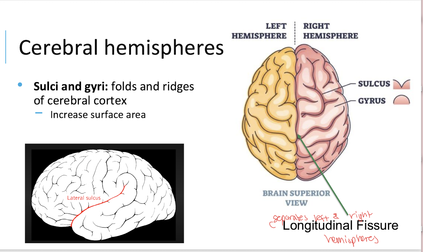
What are sulci?
The shallow grooves/depressions between gyri

What is the function of sulci and gyri?
They increase the surface area of the cerebral cortex.
What is the longitudinal fissure?
The deep groove separating the left and right cerebral hemispheres.

What is the central sulcus?
A prominent sulcus separating the frontal lobe from the parietal lobe.

What is the transverse fissure?
A deep groove separating the cerebrum from the cerebellum.

What is the lateral sulcus?
deep groove on the surface of the brain that separates the temporal lobe from the frontal and parietal lobes

What is the function of the frontal lobe?
thinking, memory, behaviour, & movement

What is the function of the parietal lobe?
language & touch

What is the function of the temporal lobe?
hearing, learning & feelings

What is the function of the occipital lobe?
sight

What is the function of the cerebellum?
balance & coordination
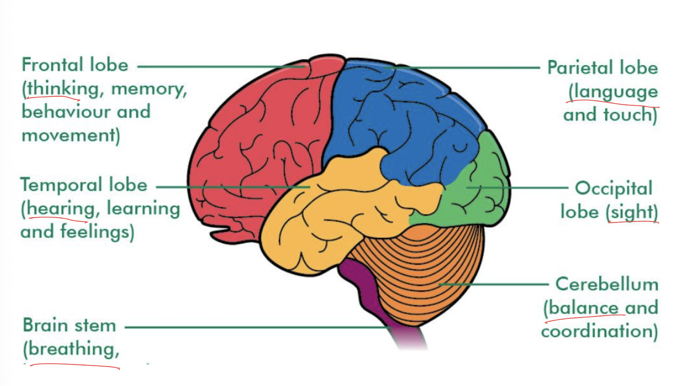
What is the function of the brain stem?
breathing

What is the insula?
A lobe of the cerebral cortex located deep within the lateral sulcus.

Why is the insula not visible on the external surface of an intact brain?
It is covered by portions of the frontal, parietal, and temporal lobes and requires dissection to view.

What are the three major parts of the diencephalon?
Thalamus, hypothalamus, and epithalamus (Pineal gland)

What is the thalamus?
A major region of the diencephalon that acts as a relay center for sensory information traveling to the cerebral cortex.
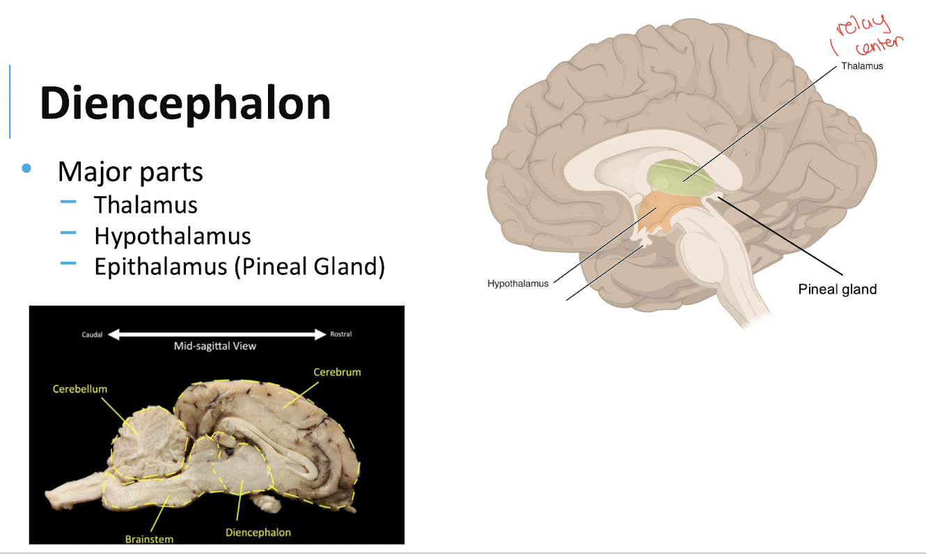
What is the hypothalamus?
A region of the diencephalon involved in homeostasis and regulation of autonomic and endocrine functions.

What is the epithalamus?
A region of the diencephalon that includes the pineal gland.
Pineal gland = endocrine gland associated with the epithalamus, melatonin
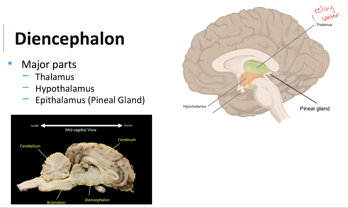
Is the pituitary gland part of the diencephalon?
No. It is connected to the diencephalon through the hypothalamus and infundibulum.

What are the three components of the brainstem?
Midbrain, pons, and medulla oblongata

What is the midbrain?
The superior portion of the brainstem

What is the pons?
The middle portion of the brainstem

What is the medulla oblongata?
The inferior portion of the brainstem, continuous with the spinal cord.

What is the cerebellum?
A brain region located posterior to the brainstem that is involved in movement coordination and motor control and cognitive function
attention, language
Emotional regulation
fear & pleasure

What is the arbor vitae?
The branching white matter inside the cerebellum.

Why is the arbor vitae important?
It contains myelinated axons that carry information through the cerebellum.


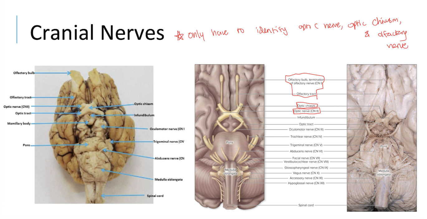
What are the two main structures associated with cranial nerve I?
The olfactory bulb and olfactory tract.


What is the optic chiasm associated with?
The optic nerve (II) and the visual pathway.

What is the general function of the oculomotor, trochlear, and abducens nerves?
They control eye movements
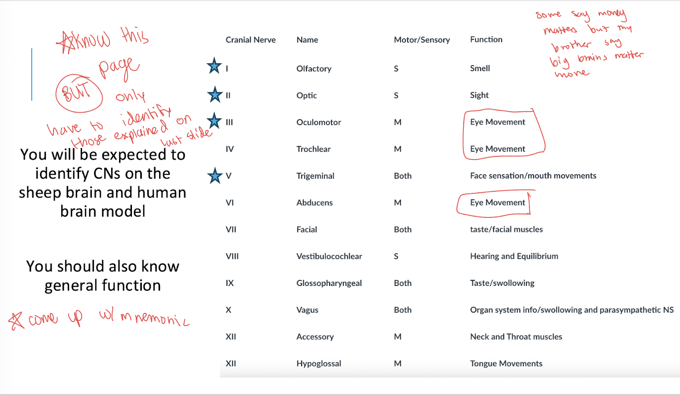
What is the general function of the trigeminal nerve?
Facial sensation and muscles of mastication

What is the general function of the facial nerve?
Facial expression and taste, along with some glandular functions

What is the general function of the vestibulocochlear nerve?
Hearing and balance

What is the general function of the vagus nerve?
It carries information and motor output associated with the head, neck, thoracic organs, and abdominal organs, including major parasympathetic functions.

What is number 1
olfactory bulb

What is number 2
olfactory tract

What is number 3
optic nerve (CN II)

What is number 4
optic tract

What is number 5
optic chiasm

What is number 1 *
olfactory bulb *
termination of olfactory nerve (CN I)

What is number 2 *
olfactory tract *

What is number 3 *
optic chiasm *
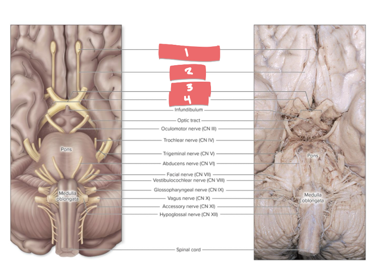
What is number 4 *
optic nerve (CN II)
What is cerebrospinal fluid (CSF)?
A clear fluid circulating through the ventricles and subarachnoid space.
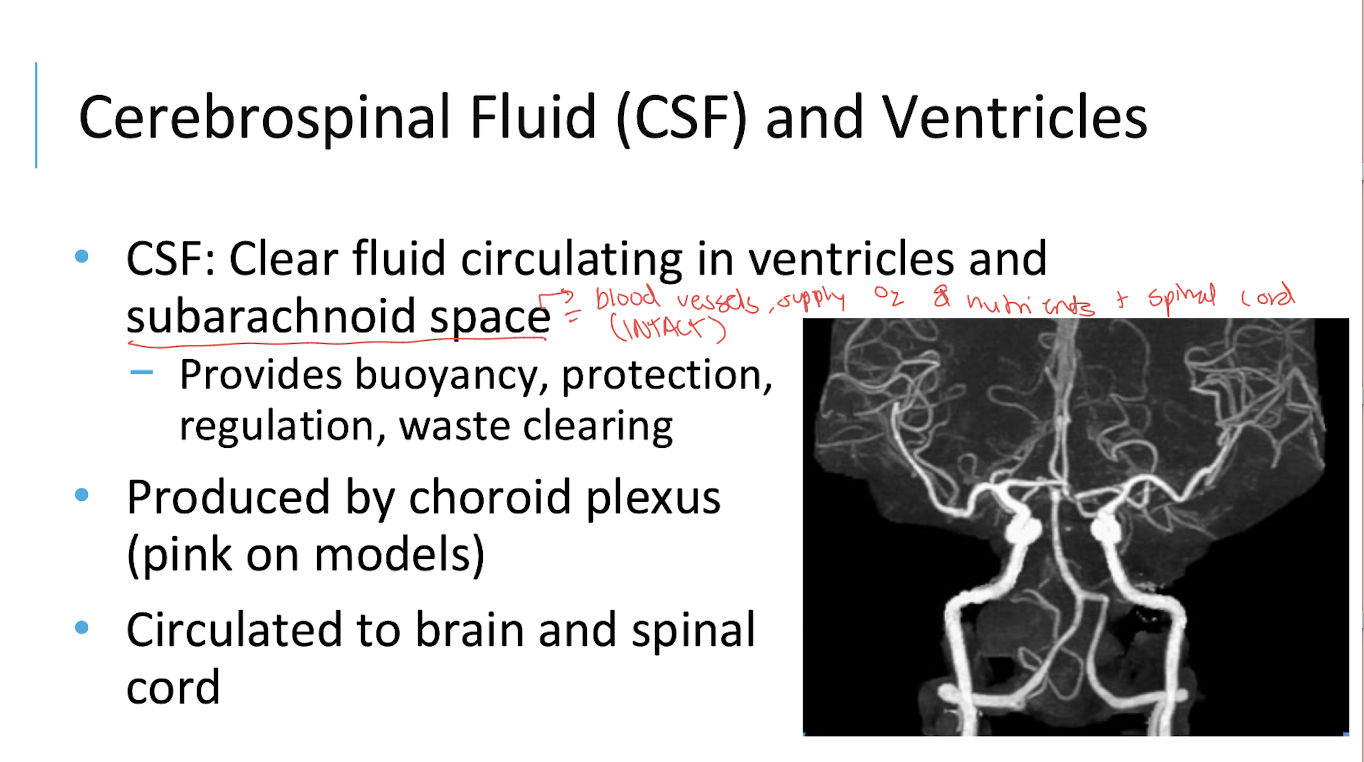
What are the functions of CSF emphasized in lecture?
Buoyancy, protection, regulation, and waste clearing.

What produces CSF?
The choroid plexus

Where does CSF circulate?
Through the brain’s ventricles, the central canal of the spinal cord, and the subarachnoid space.

What are the ventricles?
A series of connected spaces within the brain that contain and circulate CSF.

What are the lateral ventricles?
Two ventricles located within the cerebral hemispheres.

What is the third ventricle?
A ventricle located in the diencephalon, between the thalami.
sandwiched directly between the left and right thalami and hypothalami

What is the cerebral aqueduct?
A narrow channel connecting the third ventricle to the fourth ventricle.
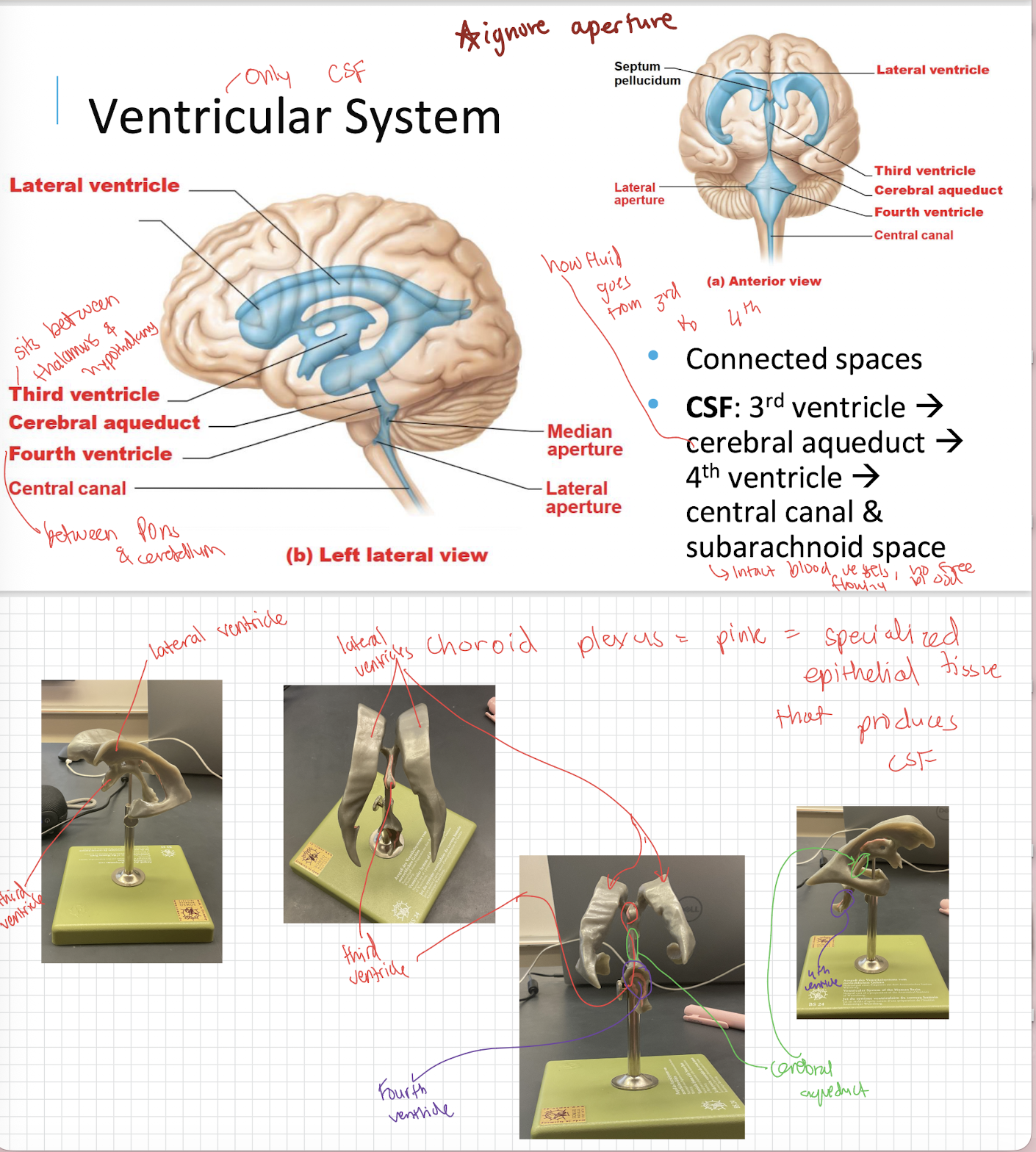
What is the fourth ventricle?
A ventricle located between the brainstem and cerebellum.

What is the central canal?
A narrow channel extending through the spinal cord that is continuous with the fourth ventricle.

What is the general flow of CSF through the ventricular system?
Third ventricle → cerebral aqueduct → fourth ventricle → central canal and subarachnoid space.

What is the spinal cord?
A CNS structure extending inferiorly from the brainstem that carries information between the brain and the peripheral nervous system.
single continuous cable of tightly packed nerve wires

What are the two enlargements of the spinal cord?
The cervical enlargement and lumbar enlargement.

Why are the cervical and lumbar enlargements present?
They contain increased nervous tissue associated with the nerves serving the upper and lower limbs, respectively.

What is the conus medullaris?
The tapered inferior end of the spinal cord
inferior tip of the solid cord
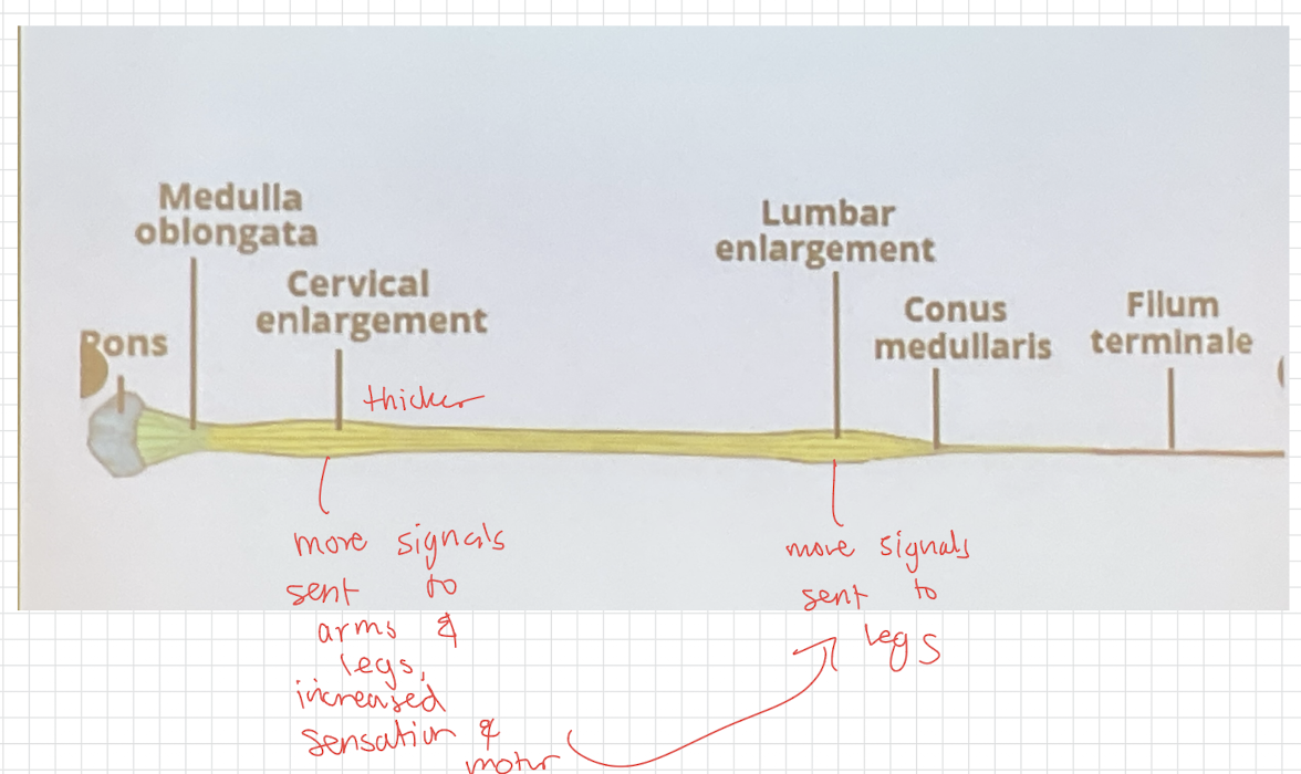
At approximately what vertebral level does the spinal cord end?
Around L1
