Anterior Abdominal Wall & Inguinal Canal
 Call Kai
 Learn
Learn Practice Test
 Spaced Repetition
 Match
Match Flashcards
 Knowt Play
1/19

There's no tags or description
Looks like no tags are added yet.
Name | Mastery | Learn | Test | Matching | Spaced | Call with Kai | Chat |
|---|
No analytics yet
Send a link to your students to track their progress
20 Terms
Identify the surface landmarks that define the abdominal quadrants , and recognize the position of the abdominal viscera in relation to those quadrants
Surface Landmarks
Xiphoid Process
Tendinous intersection of rectus abdominis
Linea semilunaris
Location of linea alba
Umbilicus
Pubic Symphysis
Inguinal Groove (location of inguinal ligament)
ASIS
Rectus Abdominis
External Oblique
Serratus Anterior
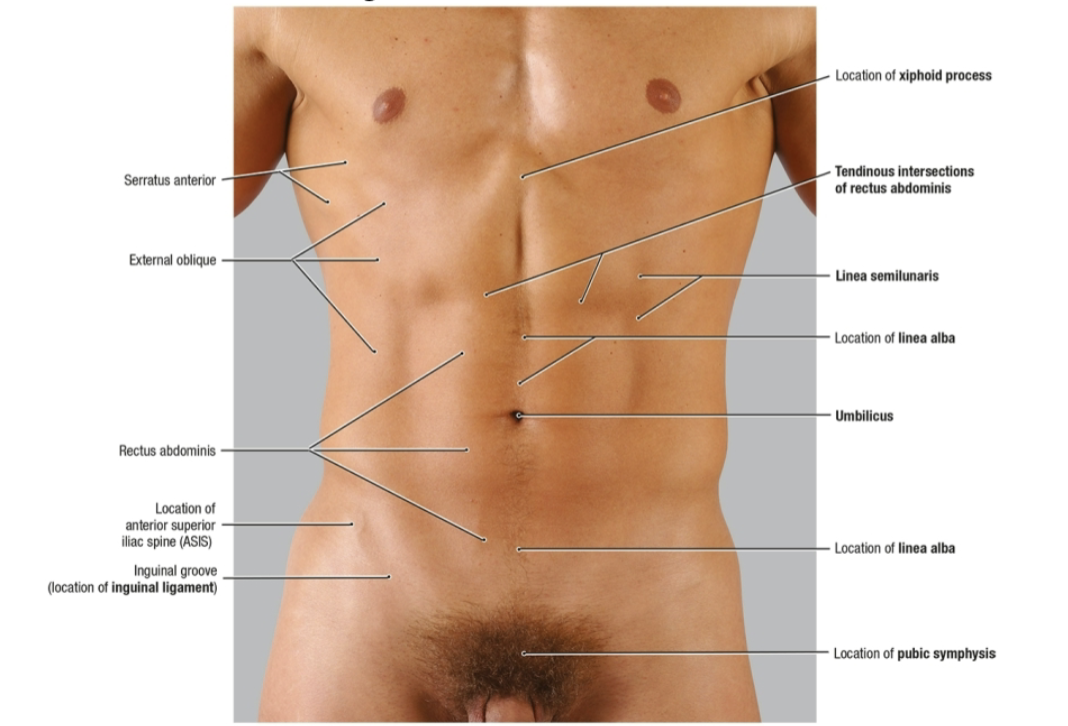
Describe the structural components that form the abdominal wall, its neurovascular supply , and lymphatic drainage
Describe the composition and organization of the anterior abdominal wall musculature and fascia
Describe the developmental origin and structure of the inguinal canal and its contents
Compare and contrast direct versus indirect inguinal hernias with respect to their development, populations, and causes
Overview of the Abdomen
Region of the trunk between the — and —
Bounded by the — superiorly
Continuous with the — inferiorly
Contains the — and parts of the —
thorax ; pelvis
diaphragm
pelvic cavity
alimentary canal ; urogenital system
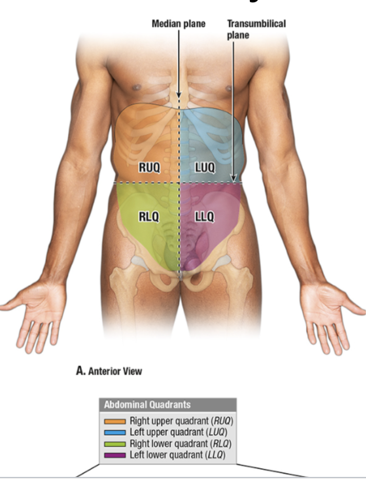
What are the abdominal quadrants and what planes are used to divide them?
Right Upper Quadrant (RUQ)
Right Lower Quadrant (RLQ)
Left Upper Quadrant (LUQ)
Left Lower Quadrant (LLQ)
Median Plane (going vertically down midsagittal plane)
Transumbilical Plane (going vertically across mid abdomin)
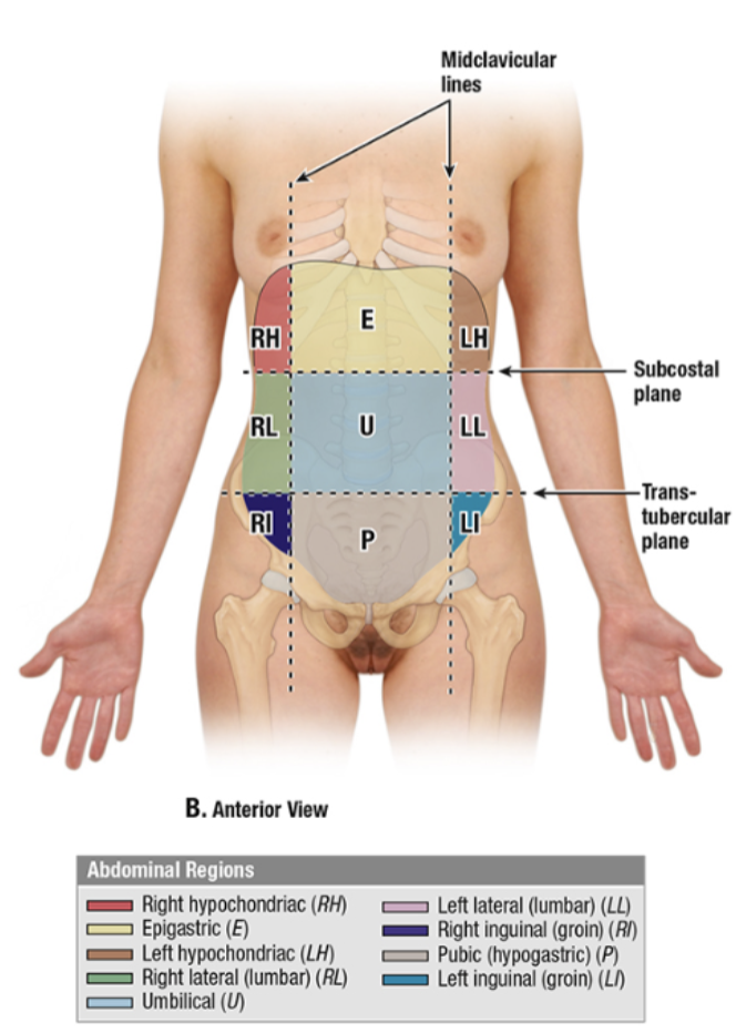
What are the abdominal regions and what planes are used to divide them?
The Right Upper Quadrant Contains
Liver: Right Lobe
Gallbladder
Stomach: pylorus
Duodenum: parts 1-3
Pancreas: head
Right suprarenal gland
Right kidney
Right Colic (hepatic) flexure
Ascending colon: superior part
Transverse color: right half
The Left Upper Quadrant Contains
Liver: Left lobe
Spleen
Stomach
Jejunum and proximal ileum
Pancreas: body and tail
Left kidney
Left suprarenal gland
Left colic (splenic) flexure
Transverse colon: left half
Descending Colon: superior part
The Right Lower Quadrant Contains
Cecum
Appendix
Most of Ileum
Ascending Colon: Inferior Part
Right Ovary
Right Uterine Tube
Right Ureter: abdominal part
Right Spermatic Cord: Abdominal part
Uterus (if enlarged)
Urinary Bladder (if very full)
The Left Lower Quadrant Contains
Sigmoid Colon
Descending colon: Inferior Part
Left Ovary
Left Uterine Tube
Left Ureter: Abdominal Part
Left Spermatic Cord: abdominal part
Uterus (if enlarged)
Urinary Bladder (if very full)
Superficial to Deep Layers of the Abdominal Wall
Skin
Superficial Fascia of subcutaneous tissue (fatty layer: camper fascia ; membranous later: scarpa fascia)
Muscles covered by deep (investing) fascia (external oblique, internal oblique, transversus abdominis)
Transversalis Fascia
Extraperitoneal Fat (variable)
Parietal Peritoneum
Abdominal Cavity
The — is the dividing line for superficial lymphatic drainage of the abdominal wall
Above Umbilicus: —
Below Umbilicus: —
umbilicus
axillary lymph nodes
superficial inguinal lymph nodes
Venous Drainage
Above Umbilicus: —
Below Umbilicus: —
Axillary Veins
External Iliac and Femoral Veins
Caput Medusae
Dilation of — radiating from the umbilicus
Caused by —, diversion of blood into hepatic portal vein —> — (within the falciform ligament)
superficial abdominal wall
hepatic portal hypertension ; paraumbilical veins
Superficial Fascia
Above umbilicus - superficial fascia is — layer
Below umbilicus - superficial fascia is divided into : — and —
only one
Superficial Fatty Layer (Camper’s Fascia) Continuous with superficial adipose tissue throughout the body
Deep Membranous Layer (Scarpa’s Fascia) Continuous with Colles Fascia around the scrotum , fuses deep fascia of the thigh
Superficial Fascia
There is a — between the deep membranous layer of superficial fascia and the deep fascia covering abdominal muscles
Fluid can accumulate here (in lower abdomen, scrotum, pelvis)
Fluid here will — spread into the thigh
potential space
not
Penile / Bulbar Urethral Injury
Tramatic — injury
Can — the penile / bulbar urethra
Results in — of blood / urine into the potential space (b/w scarpa’s fascia and deep investing fascia of abdominal muscles)
straddle
rupture
extravasation
Abdominal Wall Muscles
External Oblique
Internal Oblique
Transversus Abdominis
Rectus Abdominis
Tendinous Intersections subdivide rectus abdominis into multiple segements