Biology U4 AOS 1
 Call Kai
 Learn
Learn Practice Test
 Spaced Repetition
 Match
Match Flashcards
 Knowt Play
1/89

There's no tags or description
Looks like no tags are added yet.
Name | Mastery | Learn | Test | Matching | Spaced | Call with Kai | Chat |
|---|
No analytics yet
Send a link to your students to track their progress
90 Terms
What is the immune system responsible for?
Pathogens
Antigens + their forms
Antigens do not need to be attached to a pathogen or cell, they can simply be free-floating molecules. T or F?
The immune system protects our body by scanning for and destroying pathogens - disease causing agent that impairs the normal functioning of an organism
Antigen: any molecule that can trigger an immune response in the body (short for antibody generator)
Depending on their source, antigens can exist as many different types of molecules, including proteins (mostly), sugars, and DNA or RNA (nucleic acids) and even some metals
Antigens do not need to be attached to a pathogen or cell --> they can simply be free-floating molecules. T
Self-antigens
In vertebrates (an animal of a large group distinguished by the possession of a backbone or spinal column), what are the most important self-markers? —> what two classes can they be divided into + what’s the difference between these two classes
Self-antigens, which are located on the surface of cells, mark the cells of an organism as ‘self’ so that the immune system doesn’t attack them
In vertebrates (an animal of a large group distinguished by the possession of a backbone or spinal column), the most important self-markers are major histocompatibility complex (MHC) markers (also known as MHC proteins, MHC molecules or self-antigens) which bind antigen fragments (peptides) and present them on the surface of cells, and can be divided into two different classes
MHC Class I markers are expressed on all nucleated cells in the body --> so all cells in the human body except for those without a nucleus (e.g. red blood cells) express MHC Class I markers
MHC Class II markers are found on specialised cells of the immune system alongside MHC Class 1 markers --> these are known as antigen presenting cells
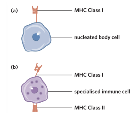
The MHC markers found on our cells differ between individuals
Explain how this creates problems during an organ transplant + how could this problem be fixed
T or F? Red blood cells have the same self-marking antigens which are glycoproteins on the surface
An organ transplant, the MHC Class I markers expressed on the donor organ will be different to the MHC Class I markers of the organ receiver, which can stimulate the receiver’s immune system to recognise the transplanted organ as non-self and launch an attack
Therefore, organ transplant recipients must routinely take immunosuppressants in order to prevent the immune system from attacking the donated organ
F. Red blood cells have different self-marking antigens which are glycoproteins on the surface of the cell and give the specific blood types
Non-self antigens + what happens if they are recognised
Outline what the plasma membrane of immune cells has
Non-self antigens are antigens that the immune system reads as ‘foreign’/ not belonging to that individual - If a non-self antigen is recognised within the body, the immune system is activated and attempts to eliminate it
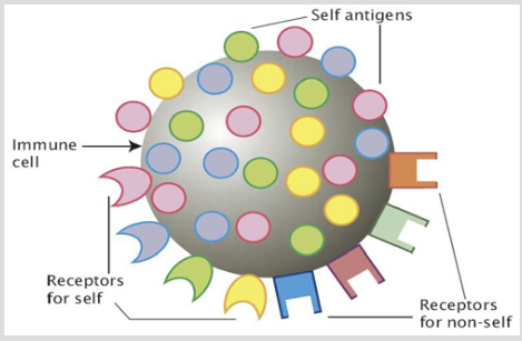
The plasma membrane of immune cells has:
1. Self-antigens that identify the immune cell as ‘self’
2. Receptors for self-antigens so that the immune cell can identify self-cells
3. Receptors for foreign antigens so that the immune cell can identify non-self-cells and signal other immune cells to destroy it
Autoimmune disease
How does it occur
Examples
Disease in which an individual’s immune system initiates an immune response against their own cells
A malfunction involving antigens can occur when an error in the immune system results in the recognition of self-antigens as non-self --> resulting in the immune system attacking self-cells
Autoimmune diseases include rheumatoid arthritis and lupus
Allergen + are they dangerous?
Allergic reactions
Mild symptoms
Severe symptoms
a subcategory of non-pathogenic/harmful antigens + no
an overreaction to the presence of an allergen which the immune system recognises as non-self and initiates a strong immune response towards
Mild symptoms of allergic reactions can include: an itchy rash, runny nose, sneezing, shortness of breath, and swelling
However, in more severe cases, constriction of airways, increased permeability of blood vessels, difficulty breathing, and decreased blood pressure may also be observed + death
Cellular pathogens + examples
Non-cellular pathogens + examples
Infection
Disease
Incubation period
Cellular pathogens have a cellular structure with organelles and are living organisms
e.g.
Bacteria
Fungi
Protozoa
Parasites
Worms
Non-cellular pathogens do not have a cellular structure and are non-living
e.g.
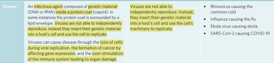
Viruses
Prions
An infection occurs when pathogens enter the human body and once in the body begin to multiply
A disease occurs when the body’s cells or organs are damaged by the pathogens/affects the normal functioning of them and signs and symptoms of the disease become obvious
The time after infection and before the first symptoms of a disease appear is called the incubation period
Extracellular threats + can they interfere with the cells’ functioning
Intracellular threats
Extracellular threats are threats found outside of a cell that can interfere with its functioning (e.g. bacteria)
Intracellular threats are threats found within a cell that can interfere with its functioning (e.g. viruses)
List examples of cellular pathogens
Bacteria
Fungi
Worms
Protozoa
Bacteria + are they a pathogen & if so, how can they damage cells/cause disease?
How they typically reproduce
Structure
What toxins do they release
Unicellular prokaryotes that can infect almost
any part of the body. Bacteria can cause disease
through the production of toxins and enzymes
which either affect the functioning of cells or cause
their death.
Asexually via binary fission
Structure
All bacteria have a cell wall made of peptidoglycan, a cell membrane and a single circular chromosome of DNA
Many bacteria have plasmids (small circular pieces of double stranded DNA)
Some bacteria produce toxins that damage host tissues and cause disease
- Exotoxins – are highly soluble proteins produced by bacteria as part of their metabolism
• released into surroundings as bacteria grow
• inhibit protein synthesis
• damage cell membranes or disrupt membrane transport
• interfere with nerve functions
--> Tetanus and cholera are 2 diseases caused by exotoxin producing bacteria
Endotoxins – are lipid based and are released when bacteria die
• fever, diarrhoea, chills, tissue death may result from endotoxin producing bacteria
--> Typhoid fever and meningitis are 2 diseases caused by endotoxin producing bacteria
Fungi
Worms
Protozoa
Plus how does each reproduce

List examples of non-cellular pathogens
Viruses
Prions
Viruses + how they reproduce
Prions
Which organisms do they only occur in + which organ/structures do they affect
How they reproduce + do they have genetic material
T or F? Prion diseases are both rare and always fatal
Do not contain genetic material
Prion protein can exist in 2 forms
1. a normal form of protein called PrPC produced by cells normally, usually in the central nervous system
2. a harmful infectious form of protein called PrPSc
--> difference between the normal and harmful forms is due to the secondary structure of the protein
Reproduction
Whenever a prion comes in contact with a normal prion protein, it somehow causes the normal protein to ‘flip’ into a twisted shape, thereby becoming a prion (abnormal protein) Any other normal protein that a twisted prion touches will also be converted, producing a chain reaction
Prion proteins are insoluble, infectious even at high temperatures, and are almost indestructible (thus, they cannot be fully broken down by proteases)
True. Prion diseases are rare but always fatal

Innate immune system/non-specific immune system
What is it composed of?
Non-specific response meaning
T or F? This system responds to injury and antigens extremely slowly
Innate immune system/non-specific immune system: a component of the immune system that is composed of generalised and non-specific defences and/or responses to pathogens
It is composed of two different defences known as the first and second lines of defences
--> Both of these mechanisms involve a non-specific response to foreign antigens, responding the same way regardless of the type of pathogen or antigen present
False. Another characteristic of these two mechanisms is that they respond to injury and antigens extremely quickly – within minutes to hours they already begin to limit the spread of infection and stimulate local changes at the site of injury
First line of defence
First line of defence: a component of the innate immune system characterised by the presence of physical, chemical, and microbiological barriers to keep pathogens out of the host organism
What are the two types of barriers present in the first line of defence of plants + describe them but don’t give examples
There are two types of barriers present in the first line of defence of plants – physical and chemical barriers
Physical barrier feature solid or fluid obstacles that block pathogen entry such as skin or mucus
Chemical barrier feature the use of chemicals such as enzymes, toxins, and acids to protect against pathogen invasion/protect against spread of disease (e.g. by destroying or slowing their growth)
Examples of barriers in plants (both physical and chemical)

What is a barrier that animals have that plants do not. Explain it and how it acts as a barrier.
While animals have physical and chemical barriers, they also have microbiological barriers - a component of the first line of defence in which the presence of normal flora (normal, harmless microorganisms) limits the growth of pathogenic bacteria
They can do this by:
They compete for space. If beneficial bacteria are already covering a surface (such as your skin or intestines), there is less room for harmful bacteria to settle.
They compete for nutrients. They use up the available food, making it harder for pathogens to survive.
They produce substances that inhibit pathogens. Some normal bacteria release acids or antimicrobial chemicals that slow or kill harmful microbes.
They help stimulate the immune system. Their presence helps your immune system stay prepared to respond to infections.
Why is the respiratory tract, gastrointestinal tract, and genitourinary tract, despite being inside the body, are in the first line of defence
They are still considered exposed to the outside environment because they have openings to the outside (such as the nose, mouth, and urethra)
Because the pathogens are stopped before they enter the body's tissues, these are all part of the first line of defence.
The second line of defence only begins if pathogens cross these barriers and invade the body's tissues or bloodstream
Examples of barriers in animals
Physical | Barriers that prevent or impede entry of pathogens | Intact skin and body surfaces This means that unbroken skin and the linings of certain body systems act as barriers to keep pathogens out. Examples include:
Memory tip: Think "Trap → Sweep → Swallow → Destroy."
|
Chemical | Barriers that act to inhibit the growth or development of pathogens and/or act to destroy pathogens |
|
Microbiological | The presence of non-pathogenic bacteria (known as normal flora) in the body can prevent the growth or colonisation of pathogenic microorganisms as they compete for space and resources |
|
Second line of defence
What are the two components of this line
All of the cells involved are called _____
Second line of defence a component of the innate immune system characterised by the non-specific and immediate response to injury that make it past the 1st line of barriers and pathogens by a variety of cells and molecules
There are two components of the second line of defence: cellular and non-cellular components
All of the cells involved are called leukocytes/white blood cells
Phagocytes
Examples of phagocytes (these are important later on) + which of these are antigen-presenting cells? And hence what does this mean? (Hint: MHC markers)
A group of leukocytes that engage in phagocytosis, a process in which they consume and destroy pathogens, foreign or dead material present in the body by engulfing it through the process of endocytosis
Phagocytes include neutrophils, macrophages (immature = monocytes), and dendritic cells
—> macrophages and dendritic cells – are also known as antigen-presenting cells/professional antigen-presenting cells where they do not only consume and destroy foreign material, but they also present antigens from consumed material on their surface using MHC Class II
Think of Neutrophils as - NA - Neutrophils & antigen-presenting cells - “NAH they’re not an APC”
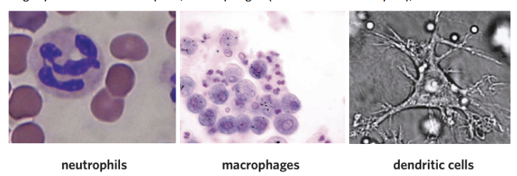
Diagram of phagocytosis

Cytokines
To communicate within the immune system, phagocytes release a number of substances such as cytokines – which are important cell signalling molecules which help protect against pathogens and can help guide immune cells to the site of infection or injury
Natural killer cells
How do they kill damaged/infected cells?
A type of leukocyte responsible for the recognition and destruction of damaged and/or infected host cells
This is achieved with the presence of two receptors – a killer inhibitory receptor and a killer activation receptor
Killer inhibitory receptor – examines the surface of cells for MHC Class I markers
Killer activation receptor – binds to certain molecules which appear on cells undergoing cellular stress (e.g. infected or cancerous cells)
The presence of MHC Class I markers can be altered due to a number of different disease processes
e.g.
MHC Class I markers may be absent due to the presence of a viral infection, which can either destroy or suppress the production of MHC Class I markers
Also the gene expression of MHC Class I markers may be affected in cancer cells, also leading to the absence of MHC Class I markers
If the killer inhibitory receptor detects a sufficient number of MHC Class I markers, then it overrides/disallow the killer activation signal, preventing cell death
Whereas, when the killer activation receptor is activated and the killer inhibitory receptor is unable to bind to a sufficient number of MHC Class I markers in infected or abnormal cells --> Cell death is initiated
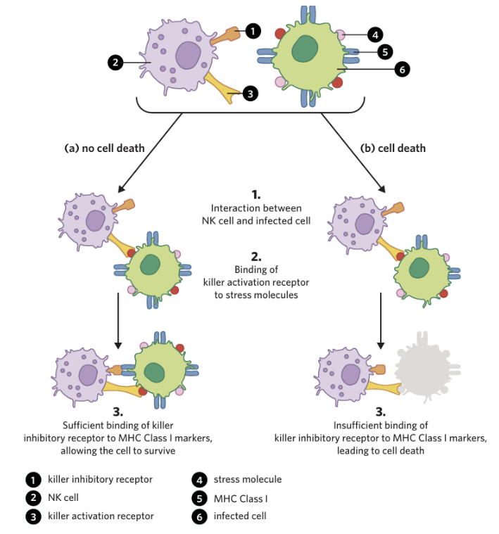
Mast cells + where do they reside
Eosinophils + which pathogens do they typically target
Mast cells
A type of leukocyte that detects injury to surrounding cells or are stimulated by antigens or allergens, where they become activated and degranulate, releasing histamine during allergic and inflammatory responses
They reside in connective tissues throughout the body
Eosinophils
A large granular leukocyte responsible for the release of toxic proteins such as DNases, RNases, and proteases which help destroy invading pathogens
They typically target pathogens which are too large to be phagocytosed
Are interferon and complement proteins cellular or non-cellular components of the second line of defence
Interferon
Non-cellular
Interferon - A cytokine released by virally infected cells which interact with receptors on neighbouring uninfected cells, causing them to undergo a number of changes that make them less susceptible/more resistant to viral infection --> this helps prevent the virus from spreading between cells
• produced by almost all cells of the immune system, but especially T cells
Complement proteins
Complement cascade
Complement proteins
A number of different types of proteins found in the blood that opsonise, cause lysis, and attract phagocytes to invading pathogens
In the presence of certain pathogens, these proteins begin reacting with each other in a series of reactions called the complement cascade
Describe the outcomes of the complement cascade
Memory trick (maybe)
Put a SIGN ON it - Opsonisation —> "Eat Me!" sticker
Chemical Taxi - Chemotaxis —> taxis following a GPS signal

Fever
Why does it occur + is it a specific response?
Do prolonged fevers have detrimental impacts?
Fever
A temporary increase in body temperature
A complex series of responses can raise the set temperature point of the body during a fever --> In response, the body initiates a number of countermeasures that increase core body temperature to reach this new setpoint, including shivering and heat-conserving behaviours (e.g. putting on a jumper)
= This an innate response to potential infection, as many pathogens cannot survive at the elevated temperatures created by a fever + fevers help the immune system by activating certain proteins in the body that make them work more efficiently
However, it is important to note that prolonged fevers can be detrimental to the body due to your own cells also have difficulty functioning, proteins and enzymes work best around normal body temperature, prolonged high temperatures put stress on the body's tissues
What is the inflammatory response + symptoms
Is it specific?
List the three main aspects of the inflammatory response
The process of inflammation increases blood flow to an injured area, bringing a greater number of immune cells and components to help clear debris and fight pathogens that may have entered the body
--> this increase in blood and fluid to the affected tissue causes swelling, pain, heat, and redness
It is a complex, non-specific process that always occurs in the same way regardless of the pathogen present or the injury that has occurred
There are three main aspects of the inflammatory response – initiation, vasodilation, and migration
🧠 “Inflammation Visits Me”
Go over the three stages of the inflammatory response
Initiation – damage/infection occurs
Infection occurs which causes the damaging of cells and introduces pathogens such as bacteria into the body
Macrophages (a phagocyte) situated in the tissue become activated and, along with damaged cells, release cytokines + mast cells degranulate, releasing histamine
Vasodilation
The histamine released from mast cells travels to nearby blood vessels and binds to specific receptors, causing vasodilation which causes increased diameter and permeability of blood vessels, increasing blood flow to the injury site --> this is the reason behind the swelling, redness, pain and warmth
Migration
The vasodilation allow for a number of innate immune system components to leave the bloodstream and enter the site of injury
This includes:
Phagocytes, including macrophages and neutrophils, leave the blood and enter the tissue due to increased permeability of blood vessels, where they are guided by the cytokines secreted by activated macrophages and damaged cells to the site of injury --> where they phagocytose pathogens and digest them using enzymes such as lysozymes
Complement proteins are attracted to pathogens and make it easier for phagocytes to destroy them
What is pus + when does the inflammatory response finish
Pus contains a large amount of dead immune cells and pathogens
The response continues until the site has been cleared of pathogens and debris, and the site of injury has been healed and will eventually return to normal
Third line of defence/adaptive immune system/specific immune response
Its features which separate it from the second line of defence
Third line of defence/adaptive immune system/specific immune response a subset of the immune system within vertebrates that is composed of the humoral and cell-mediated responses which create a specific immune response and form immunological memory
The two features of the adaptive immune system which separate it from the second line of defence:
Specificity – the adaptive immune system responds to each distinct pathogen in a unique manner
Immunological memory – the adaptive immune system results in the production of cells that allow the body to respond to future re-infections by a previously encountered pathogen quickly and effectively
Describe the process of antigen presentation
After antigen-presenting cells (APCs) engulf and digest pathogens via phagocytosis, displaying pathogenic antigens on their MHC Class II markers --> they then travel via the lymphatic system to lymph nodes
T-Helper cell with complementary receptor on its surface binds to foreign antibody and is ‘selected’
The T helper cell then becomes activated/selected
--> The activated T helper cell can then help initiate the adaptive immune response through either the humoral or cell-mediated immune responses

Humoral immunity (do not describe its response)
B lymphocytes
An adaptive immune response in which extracellular pathogens are neutralized or destroyed by the production and secretion of antibodies produced by plasma cells
You can think of it as ‘eh’ —> extracellular - humoral
B lymphocytes are a type of white blood cell whose surfaces are covered with B cell receptors (known as membrane-bound antibodies) --> these cells circulate throughout the body in the bloodstream and are found in high numbers within lymph nodes
Describe the humoral immune response + Clonal selection theory
A pathogen with an antigen that is complementary in shape to the antigen-binding site on a B cell receptor binds to the receptor on the B cell --> when this occurs, the B cell is said to have been ‘selected’
Selected B cell and T-helper cell with a matching receptor to the antigen on the pathogen interact and secrete cytokines --> these cytokines cause the B cell to undergo clonal expansion where many copies of the selected B cell are produced
The T helper cell also stimulates the selected B cell via cytokines to undergo the process of differentiation, in which the clones of the selected B cell differentiate into two different types of B cells: B memory cells and plasma cells
The plasma cells secrete antibodies into the bloodstream in order to destroy selected pathogen
Clonal selection theory: the process in which B and T cells encounter an antigen that matches their antigen binding site, and then generate many copies of themselves
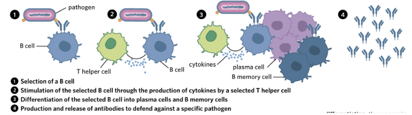
What are B memory cells responsible for?
A differentiated B lymphocyte that reside in the body for a prolonged period of time and are responsible for providing long-lasting immunological memory of an antigen
Antibodies/immunoglobulins:
The type of molecule they are + what structure of that molecule
Describe its structure
T or F? There are different classes of antibodies - the type of light chain determines the class to which the antibody belongs
How many antigen binding sites present?
Sketch a labelled diagram
Antibodies released by plasma cells are proteins with a quaternary structure
Structure
--> They are composed of four polypeptide chains, including two heavy chains and two light chains, arranged into a ‘Y’ shape
The two heavy chains are joined by a disulphide bond - a strong covalent bond occurring between two sulphur atoms
Each antibody has two regions --> the ‘stem’ of the antibody is known as the constant region which does not vary, and the tops of the ‘arms’ are known as the variable region that differ between antibodies
False. There are different classes of antibodies - the type of heavy chain determines the class to which the antibody belongs
There are two antigen binding sites present --> hence an antibody can bind with two pathogens at once

What are the standard five types of antibodies
Are antibodies specific?
There are five types of antibodies – IgA, IgD, IgE, IgG, IgM --> which are all secreted at different times in the immune response
"A Doctor Eats Green Marshmallows."
A = IgA
Doctor = IgD
Eats = IgE
Green = IgG
Marshmallows = IgM
Due to the process of clonal selection, these antibodies are specific and have an antigen-binding site that is complementary to the antigens located on the pathogen

Describe the different ways antibodies interact with pathogenic antigens in a number of key ways in the humoral immune response

Cell-mediated immunity (don’t explain its response)
Cytotoxic T cells
Cell-mediated immunity
Infected or abnormal cells are destroyed by cytotoxic T-cells (T cell immunity). This relates to intracellular threats where the virus has entered the cell
Cytotoxic T cells, a type of T lymphocyte, primarily carry out their role by assessing the MHC Class I marker of infected cells (nucleated cells)
Where in addition to their role of self-recognition, MHC Class I can also display foreign antigens that have been broken down in a cell on its surface
Describe cell-mediated immune response
T-helper cell releases cytokines + APC MHC class I markers that present complementary antigens to receptors on naive Cytotoxic T cell —> stimulates/activates naïve Cytotoxic-T cell = selected
The selected cytotoxic T cell to undergo clonal expansion and differentiation
The clones of the selected T cell differentiate into two types of T cells – effector cells called cytotoxic T cells, and T memory cells
T memory cells reside in the body for extended periods of time and help form immunological memory
The majority of selected T cells differentiate into cytotoxic T cells, which leave the lymph node and travel throughout the body, reaching the site of infection
Once the cytotoxic T cell has found an abnormal cell that is presenting complementary foreign antigens on its MHC Class I complex, it binds to it
Chemicals, such as perforin, are then secreted by the cytotoxic T cell to induce apoptosis in the infected or abnormal cell

Compare humoral and cell-mediated response
| Antibody-mediated (Humoral) response | Cell-mediated response |
Similarities | Both involved T helper cells -both are specific -both involve recognition of presented antigens Involve WBC that reside in lymph nodes |
|
Differences | -occurs in blood, lymph and tissue fluids
-involves B cells and antibodies | -infected cells -doesn’t involve B cells/antibodies |
Immunological memory
Explain how B and T memory cells respond to the same pathogen entering
The ability of the immune system to quickly and aggressively combat a previously encountered pathogen due to the presence of T and B memory cells
If the same pathogen enters again:
The B memory cells recognise the antigen straight away because they have receptors that match it
They rapidly divide (clonal expansion) into new antibody-producing plasma cells
The plasma cells produce large amounts of antibodies much faster than during the first infection
(B memory cells also create immunological memory by constantly secreting low amounts of their antibody)
When the same pathogen enters again:
An antigen-presenting cell (APC) displays the familiar antigen.
The T memory cells recognise it immediately.
They rapidly divide into:
T helper cells (which release cytokines)
Cytotoxic T cells (which kill infected cells)
Sketch a labelled diagram comparing the processes of humoral and cell-mediated immunity:

Functions of the lymphatic system
What is the lymphatic system comprised of
Act as a transport network for the antigen presenting cells – location of clonal selection and expansion
Production of leukocytes/WBC, including lymphocytes in primary lymphoid tissues
Removal of fluid from tissues around the body
Absorption of fatty acids from the digestive system
The lymphatic system is comprised of a series of lymphatic vessels throughout the body that function to transport lymph to a number of primary and secondary lymphoid tissues
Primary lymphoid tissues
What are the main primary lymphoid tissues + their function
Primary lymphoid tissues
Components of the lymphatic system that are responsible for the production and maturation of lymphocytes
Where the main primary lymphoid tissues include the bone marrow and the thymus
The production of B and T lymphocytes occurs in the bone marrow, which is primarily found inside long bones such as the femur and humerus --> while B lymphocytes remain in the bone marrow to mature further (think of it as B cells = bone marrow), T lymphocytes travel to the thymus to mature (think of it as T cells = thymus)
secondary lymphoid tissues
What are the main secondary lymphoid tissues + their function
Why do lymph nodes swell when one is sick
Secondary lymphoid tissues
Components of the lymphatic system that are responsible for the maintenance of mature lymphocytes and the activation of the adaptive immune response
The main secondary lymphoid tissues include the lymph nodes (that act as filters), tonsils and the spleen
In these tissues, mature lymphocytes are clustered together and ‘scan’ passing lymph for the presence of any pathogens or foreign antibodies on antigen-presenting cells
--> If a foreign antigen matches the receptors of specific lymphocytes, these lymphocytes then undergo clonal selection and differentiation which results in a large number of B and T cells being created within these tissues --> resulting in the characteristic swelling of lymph nodes when you’re sick
Lymphatic drainage
Lymphatic capillaries + what do they collect
Lymph
Lymphatic capillaries are extremely small vessels that exist throughout the tissues of the body
They collect:
Excess tissue fluid
Proteins
Dead cells
Pathogens
Immune cells, including antigen-presenting cells (APCs)
Once the clear fluid enters these capillaries – it is called lymph where it is carried away into the lymphatic system, where it eventually arrives at a lymph node
Lymphatic flow
The small lymphatic capillaries throughout the body gradually join together to form larger vessels that contain an increasing amount of lymph
Describe what type of walls these vessels have and how squeeze lymph fluid through the system
T or F? These vessels are one-way valves.
These vessels have thin walls and rely on surrounding muscle movements to squeeze lymph fluid through the system (note: the heart is not responsible for pumping lymph)
True. These vessels feature a number of one-way valves --> so fluid moves in one direction only – away from the tissues and towards the lymph nodes
Lymphatic surveillance
The fluid drained from tissues arrives at lymph nodes via afferent lymphatic vessels – where the lymph travels through clusters of B and T cells
--> As it drains through these clusters, antigen-presenting cells and pathogens are most likely to meet a lymphocyte with a matching antigen receptor which stimulates the process of clonal selection + expansion
What happens once an adaptive immune response is initiated? (hint: antibodies & activated cytotoxic T cells)
T or F? This lymph is then returned into circulation near the heart, where the lymphatic vessels join with the large veins returning blood back to the heart
Is the adaptive immune system slower to activate than the innate immune system?
If an adaptive immune response is initiated, antibodies and activated cytotoxic T cells will be transported in the lymph away from the lymph nodes via efferent lymphatic vessels
T
Unlike the innate immune system, which provides an immediate source of protection, the adaptive immune system is much slower to activate
Immunity
Natural immunity
Artificial immunity/induced immunity
Active immunity
Passive immunity + is it long-term?
Immunity is resistance to particular infectious disease or pathogen
Natural immunity protection against a disease formed without medical intervention
Artificial immunity/induced immunity protection against a disease formed as a result of medical intervention
A person's immunity to a disease is further classified based on the strategy used to develop it:
Active immunity protection against a disease created by antibodies and memory cells formed by a person’s own adaptive immune system
Passive immunity protection against a disease created by antibodies from an external source
--> Passive immunity is temporary and there is no memory involved
Natural active immunity
Natural active immunity protection against a disease created by antibodies and memory cells produced by an individual’s own immune system without medical intervention
Natural passive immunity
Methods of acquiring natural passive immunity include:
Natural passive immunity protection against a disease created by antibodies from a natural external non-medical source
Two methods of acquiring natural passive immunity include:
Breastfeeding – human breast milk contains many nutrients and proteins essential for healthy growth and development, including antibodies generated from the mother’s own immune system --> once ingested, these antibodies are absorbed into the baby’s bloodstream and protect them against pathogens
Placenta – during pregnancy, some antibodies produced by the mother are able to cross the placenta and enter the foetus’ bloodstream via the umbilical cord
These are important, as babies have poorly developed adaptive immune systems and aren’t able to fully protect themselves against pathogens for the first few months of life – however the duration of protection is shorter as antibodies are quickly degraded
Artificial active immunity
Outline a way to achieve artificial active immunity
Artificial active immunity protection against a disease created by antibodies and memory cells produced by an individual’s own immune system after medical intervention
A way of achieving artificial active immunity is vaccines
Explain how vaccines help achieve artificial active immunity (don’t talk about the two main phases yet)
Vaccines are medical treatments that contain components that resemble a certain pathogen’s antigens, but these components are not able to cause disease
These components can be:
Attenuated (weakened) or inactivated (dead) pathogens
Toxoids (toxins that have been altered so they can’t cause disease)
Specific proteins from the surface of pathogens
RNA that enters immune cells and causes them to make pathogen-like proteins
A person’s adaptive immune system recognises these components as foreign and develops a response to them --> this means that when this individual encounters the actual pathogen in the future, their immune system has antibodies and memory B cells that can target its antigens and rapidly attack it
There are two main phases to forming artificial active immunity in vaccines – the primary immune response and the secondary immune response.
Explain both and draw a diagram:
After a person receives their first vaccination, there is a delay in the adaptive immune system’s response because the adaptive immune response is relatively slow --> it takes time for antigen-presenting cells to find T and B cells complementary to the vaccine’s antigen and for the process of clonal selection to occur
Once the primary immune response had taken place - a moderate number of antibodies and memory cells are formed
Upon receiving a second vaccination, the memory cells created by the first vaccine quickly recognise the antigen in the vaccine and mount a rapid, large secondary immune response --> resulting in the generation of a large number of antibodies and memory cells that go on to create long-lasting immunity

Note that more than one vaccination typically needs to take place in order for immunity to be formed
T or F? With subsequent immune responses each generating smaller, less rapid responses until long-lasting immunity has been achieved
What ways can vaccines be administered?
False. With subsequent immune responses each generating larger, more rapid responses until long-lasting immunity has been achieved
Vaccines can be administered via an injection or orally in the form of a liquid
Explain what booster vaccines are and why are they used?
It is normal for memory T and B cells to die after a long period of time --> this means that in individuals who were immunised against a disease many years ago, their immunity may start to decline and they may become susceptible to a disease they were previously immune to
In these individuals, a booster vaccine/shot is administered, which is simply another injection of the vaccine they received earlier --> By doing so, any remaining memory cells are stimulated to activate an immune response, generating more antibodies and memory cells to restore their immunity
Artificial passive immunity
How is it typically achieved
Artificial passive immunity protection against a disease created by antibodies from an external medical source
This occurs through an injection or an infusion (e.g. antiserum for a snake bite)
Explain why passive immunity is not long-term:
Antibody treatments immediately increase the number of antibodies in the blood, but over time these antibodies degrade until they’ve all disappeared and the immunity they created has gone
As if someone is only given antibodies, they will not develop active immunity because the antibodies they receive will not trigger production of the memory cells responsible for immunological memory

Herd immunity
How is it achieved
It is particularly crucial for protecting people who?
And how does it protect those people?
Herd immunity
Protection against a disease given to non-immune individuals when a high percentage of a population/herd is immune to the same disease
It is often achieved through high rates of vaccination
It is particularly crucial for protecting people who cannot be vaccinated --> including children who are too young to be vaccinated, people with immune system problems, and those who are too ill to receive vaccines (such as some cancer patients)
This occurs as:
When a sufficiently large proportion of people in a population are immune to a disease via vaccination --> then the pathogen causing that disease cannot easily reproduce and spread throughout
This then protects the people who aren’t immune as they are now highly unlikely to come into contact with a person harbouring the pathogen and therefore won’t become sick
Non-infectious diseases
Infectious diseases
Contagious diseases
Non-infectious diseases aren’t caused by pathogens
There are many causes of non-infectious diseases
Some non-infectious diseases, such as cystic fibrosis, are caused by abnormal genes
While others, like cardiovascular disease, are largely caused by lifestyle factors such as diet and exercise
Infectious diseases are diseases caused by pathogens
These pathogens harm their host, and cause the symptoms that patients present with
A subset of infectious diseases are contagious diseases, which are infectious diseases that can be transmitted and spread between different people
Contagious vs Virulence
Contagious a property of a pathogen or disease meaning that it can be transmitted from one organism to another
Virulence the potential of a pathogen or disease to cause serious illness or harm
Emerging diseases
Re-emerging diseases
Emerging diseases are diseases that have not occurred in humans before, have occurred previously but only affected particular populations in isolated places, or have occurred throughout history but have only recently been recognised as being caused by pathogens
Re-emerging diseases are diseases that were once major public health problems and then declined dramatically in incidence (frequency in population), but are again becoming health problems for a large number of people
Factors contributing to the emergence and re-emergence of diseases
Factor | What it means |
Evolution of the causative organism | Pathogens causing disease can evolve to either infect humans or, if previously capable of infecting humans, evolve to evade treatments by acquiring resistance |
Globalisation and travel | Due to people being able to quickly travel around the world - an infectious disease that starts in one place can quickly spread around the world |
Increased exposure of humans to animals | As people expand into forests, farm more land, or climate change changes animal habitats, humans have more contact with animals
|
Increasing human population | More people means more crowded cities and communities. Diseases spread more easily when many people live close together. |
Changing technology | New technologies or human-made systems can sometimes create environments where pathogens grow and spread
e.g. Legionnaires’ disease is caused by a pathogen that inhabits air conditioning systems |
Insufficient vaccination of the population | If fewer people are vaccinated, herd immunity decreases --> This means there are more people who can catch and spread the disease, allowing diseases that were once under control to return/spread more easily |
Outbreak
Epidemics
Pandemics
Endemic
Outbreak a sudden and unexpected increase in the occurrence of a disease
Disease outbreaks can be classified into one of two categories based on the geographic spread of the disease:
Epidemics involve a sudden, widespread increase in the occurrence of an infectious disease among a specific population in a specific location at a particular time
Pandemics involve epidemics that have spread to different countries and/or continents in different regions of the world --> so they typically affect a greater number of people when compared to epidemics and are much more difficult to control
Endemic when a disease occurs at a relatively constant baseline level in a population
So it is always present in a particular population or area at a predictable, relatively constant level --> It doesn't disappear completely, but it also isn't causing unusually large outbreaks all the time
e.g. the flu (influenza)
The arrival of the first convicts and settlers from England to Australia in 1788 brought about the introduction of disease and dispossession among the Indigenous population
Major diseases in Europe during the late 18th century were smallpox, syphilis, tuberculosis, influenza, and measles --> when colonists arrived in Australia, they brought these diseases with them, unleashing them on the local population and causing widespread illness (morbidity) and death (mortality)
Explain some reasons why these diseases particularly affected the Indigenous population:
Lack of immunity in the Indigenous population
Many Europeans had caught diseases like measles as children.
They survived the infection.
Their bodies developed natural active immunity, which protected them later in life.
If they caught the disease again, it was usually less severe.
However for many Indigenous Australians:
It was their first exposure to these diseases.
Many became infected as adults, when diseases such as measles can cause more serious illness.
They had no existing immunity, so diseases spread quickly and caused many deaths.
A lack of knowledge and experience with European diseases
Before colonisation, Indigenous Australians had extensive knowledge of:
local diseases
traditional medicines
However, European diseases were completely new.
This meant they:
did not know how these diseases spread,
did not know effective ways to prevent them,
had no previous experience treating them.
At the same time, many Indigenous communities were prevented from practising their traditional medicine, leaving people with little or no medical care
The disruption caused by colonisation
Before colonization
Most Aboriginal and Torres Strait Islander communities:
lived in small, uncrowded, spread-out groups,
had access to clean food and water
had healthy diets (rich in carbs and protein) —> These conditions generally supported good health.
After colonization
European settlement caused major disruptions:
People were removed from their traditional lands.
Access to food and clean water became limited.
Traditional medicine and cultural practices were disrupted.
Many people were forced to live in crowded camps or settlements - made it much easier for infectious diseases to spread.
Poor nutrition and stress also weakened people's immune systems, making infections more severe.
Outline the methods of identifying pathogens
Method | Explanation | Example |
Physical | Uses microscopes to directly observe pathogens - examine their size, shape, arrangement, and structures to help identify them. |
|
Phenotypic | Identifies pathogens based on their observable characteristics, such as how they grow, their appearance, and their biochemical properties. | Selective media: An agar plate designed to allow certain pathogens to grow and multiply to test for their presence in a sample can be used
e.g. buffered charcoal yeast extract agar allows Legionnaires' disease-causing Legionella pneumophila to grow --> and so if this bacteria is present in a sample and combined with buffered charcoal yeast it will grow and multiply, allowing scientists to identify it
Biochemical tests panels: a series of tests designed to specify a sample’s genus and species
e.g. scientists perform tests such as the Gram stain to determine whether bacteria are Gram-positive or Gram-negative, followed by tests for oxygen requirements (aerobic or anaerobic) and other tests until the species is identified. |
Immunological | They identify a pathogen by detecting its antigens (proteins on the pathogen) or the antibodies your immune system makes against it --> These methods rely on the specific binding between antibodies and antigens | ELISA (enzyme-linked immunosorbent assay):
Serology the study of blood serum, typically to determine the presence of antibodies and/or antigens
There are four main types of ELISA tests – direct, indirect, sandwich, and competitive
This is sandwich method Step 1: Put antibodies on a plate Antibodies specific to a certain pathogen are attached to a plate Step 2: Add the patient's sample The serum sample to be tested is then applied to the plate If the pathogen is present, its antigens stick to the antibodies
Step 3: Add a second antibody A second detection antibody, linked to a colour-changing enzyme, is added to the plate, binding to any antibody-antigen complexes present
Now the pathogen is trapped between two antibodies. This is why it's called a sandwich ELISA. Step 4: Add the substrate A substrate is then added, reacting with the enzyme on the second antibody and changing colour/emitting a signal to reveal whether any pathogenic antigens were present in the sample |
Molecular | Detects or analyses a pathogen's genetic material (DNA or RNA). | Hybridisation-based detection Labelled segments of genetic material that are complementary to a pathogen’s genetic material are added to a sample
If the pathogen is present: The segment sticks to the pathogen DNA. Signal = pathogen present No signal = pathogen absent
Whole-genome sequencing: Provides detailed information about the pathogen |
Direct transmission
Indirect transmission
Direct transmission involves contact between an infected person and a susceptible person, either via actual physical contact between them or via close proximity between them
Indirect transmission is disease transmission that occurs without any form of contact or proximity between an infected person and a susceptible person
What are the different modes of disease transmission
Transmission route | Description | Examples
|
Airborne transmission | The spread of pathogens through air via small particles (traditionally <5 µm) that stay in the air for prolonged periods of time after a person sneezes, coughs, exhales, or talks
| Influenza virus – the causative agent of the flu SARS-CoV-2 – the causative agent of COVID-19 Rhinovirus – the causative agent of the common cold
(These are both for airborne transmission and droplet transmission) |
Droplet transmission | The spread of pathogens through air and contaminated surfaces via respiratory droplets (droplets (traditionally >5 µm) produced by breathing, talking, vomiting, and coughing that could contain pathogens)
|
|
Direct physical contact transmission | The spread of pathogens through physical contact between a host and another individual
| Tinea pedis – the causative organism of athlete’s foot Human immunodeficiency virus (HIV) – the causative agent of acquired immunodeficiency syndrome (AIDS) Epstein-Barr virus (EBV) – the causative agent of infectious mononucleosis (glandular fever) |
Indirect physical contact transmission | The spread of pathogens via fomites (an inanimate object that, when contaminated with a pathogen, can transmit that pathogen to a new host) (e.g. food, water, tissues, needles) or vectors (an organism that is not affected by a disease but spreads it between hosts) (e.g. Mosquitoes) | Plasmodium – the causative agent of malaria, spread by mosquitoes |
Faecal-oral transmission | The spread of pathogens via oral consumption of contaminated faeces
If proper hygiene or sanitation is lacking, the faeces can contaminate:
These droplets may:
| Vibrio cholerae – the causative organism of cholera Rotavirus – the causative agent of diarrhoea, typically in young children |
Key strategies in controlling disease transmission
Prevention |
|
Screening |
|
Quarantine and isolation |
|
Antibiotics
Antivirals
Fungicides
Antibiotics are medicines that can be used to treat diseases caused by bacteria by killing or slowing their growth - they selectively affect bacterial cells by targeting specific biochemical pathways or components unique to bacteria, without damaging the patient’s cells
Antivirals are prescription medicines (pills, liquid, an inhaled powder, or an intravenous solution) that can be used to treat diseases caused by viruses/viral infections --> where they are designed to specifically target viruses, interfering with their ability to attach to, replicate in, and exit from a host cell
Fungicides are medicines that can be used to treat diseases caused by fungi/fungal infections --> where they specifically target fungal cells
Sometimes antibiotics are used inappropriately, for example by:
Taking antibiotics for viral infections (such as the common cold or flu), even though antibiotics do not work against viruses.
Not finishing the full course of antibiotics.
Taking antibiotics when they are not needed.
Using antibiotics too often
How can this lead to antimicrobial resistance in bacteria
When antibiotics are used incorrectly, not all bacteria are killed.
Most bacteria die.
A few bacteria may have mutations that make them naturally more resistant to the antibiotic
The resistant bacteria survive and reproduce.
They pass their resistance genes to their offspring (and sometimes to other bacteria).
Over time, this can leads to antimicrobial resistance in more bacteria where they are no longer affected by antibiotics
Eventually, the antibiotic no longer works well against that type of bacteria.
Immunotherapy
List and explain the two broad categories of immunotherapy
mmunotherapy is a category of medical treatments that treat disease by modulating/adjusting the immune system
Examples include:
dendritic cell therapy
-CAR-T cell therapy
Antibody therapy
Cytokine therapy
There are two broad categories of immunotherapy:
Activation immunotherapies, which aim to induce or amplify an immune response
Suppression immunotherapies, which aim to prevent or reduce an immune response
Monoclonal antibodies (mAbs)
T or F? Monoclonal antibodies (mAbs) can only treat diseases in a immunotherapeutic way
Monoclonal antibodies (mAbs) laboratory-made antibodies produced by plasma cell clones that bind to a specific antigen
Because of their specificity to one antigen, monoclonal antibodies can be used to target specific types or parts of cells --> e.g. monoclonal antibodies can be used to treat cancer and autoimmune diseases due to their ability to trigger the killing of cancerous or self-recognising cells respectively
False. Monoclonal antibodies can also be used to treat diseases in non-immunotherapeutic ways by treating diseases without modulating the immune system
How are monoclonal antibodies produced? (the traditional way - the process that scientists used when they were first producing monoclonal antibodies)
Scientists identify and isolate an antigen that is present on a desired target cell --> this cell will typically be one that is responsible for causing the disease that scientists want to treat
Scientists vaccinate an animal, usually mice, with an antigen --> this vaccination stimulates an immune response against the antigen and results in the selection and proliferation (expansion) of a B lymphocyte that matches the antigen
Scientists extract these B lymphocytes from the spleen of the mice
The extracted B lymphocytes are fused with rapidly-dividing cancerous human plasma cells known as myeloma cells. The products of this fusion are called hybridomas
The reason why these myeloma cells are chosen to fuse with the B lymphocytes is because B lymphocytes do not grow well in vitro (outside living organisms), whereas myeloma cells have the ability to grow indefinitely and produce large quantities of antibodies
Hybridomas are screened so that only the cells with the appropriate antibody are selected --> The hybridomas that produce the specific antibody are cloned, which results in the mass production of these antibodies
Antibodies are then collected and purified before being administered to a patient
What is cancer
How does it occur/spread
Cancer is a complex group of diseases caused by the uncontrolled and unregulated replication of cells that then invade other sites of the body
The most important process in the development of cancer is the accumulation of mutations in a cancer cell’s DNA that allow it to bypass normal regulatory checkpoints of the cell cycle and provide it with survival advantages
Whilst the immune system is normally capable of recognising cells that have developed mutations and destroys them before they have a chance to replicate --> cancerous cells are sometimes able to evade the immune system or develop mutations that allow them to suppress the immune response against them
There are two types of monoclonal antibodies used in immunotherapy - identify + describe them
naked monoclonal antibodies - monoclonal antibodies that do not have any other molecules attached to them
conjugated monoclonal antibodies - monoclonal antibodies with other molecules (e.g. chemotherapy drugs or radioisotopes) attached to them
Naked monoclonal antibodies have three main mechanisms of action against cancer cells (just identify it)
Antibody-dependent cell-mediated cytotoxicity (ADCC)
Complement activation
Checkpoint inhibition
Antibody-dependent cell-mediated cytotoxicity (ADCC)

Complement activation

Checkpoint inhibition
Immune checkpoints are regulators in the immune system that, when activated, suppress the immune system
Whilst suppressing the immune system at times is a normal part of bodily function, some cancer cells secrete molecules that stimulate immune checkpoints, reducing the immune system’s ability to recognise and destroy them
Monoclonal antibodies can be used to block immune checkpoints, meaning the immune system is able to function at a greater capacity and destroy cancer cells more easily
What are the other uses of monoclonal antibodies

Note:
Immunotherapy Vs traditional cancer treatment
Traditional forms of cancer therapy such as chemotherapy and radiotherapy work by directly targeting and killing cells that are rapidly dividing, rather than stimulating the immune system
A large problem, however, is that many other cells of the body – such as hair follicles, and cells lining the mouth and gut – also divide quickly and are killed by these treatments --> this is one of the reasons why people on chemotherapy can suffer from side effects such as hair loss, nausea, and vomiting
Whereas antibody-based immunotherapies tend to be more specific and targeted in their attack as monoclonal antibodies have variable regions that bind with cancer antigens specifically, there is a lower chance that other cells in the body will be affected by the treatment and experience side effects
However immunotherapy still can cause a wide array of side effects, and is currently only available as a treatment for very specific types of cancer + it is still typically used in conjunction with traditional cancer treatments such as chemotherapy and radiotherapy
Autoimmune disease
How does it occur
Examples
What do B cells release and T cells become?
Autoimmune disease a disease in which an individual’s immune system initiates an immune response against their own cells
Body cells express major histocompatibility complex (MHC) proteins that mark them as ‘self’
--> If a person’s immune system is functioning normally, their lymphocytes should recognise these markers and not launch an attack against a cell expressing them
--> Sometimes, however, lymphocytes fail to recognise these self-markers and end up inducing an immune response against self-cells - resulting in an autoimmune disease
Examples of autoimmune diseases include: rheumatoid arthritis, multiple sclerosis, type 1 diabetes, and coeliac disease, lupus
In autoimmune diseases, B cells release autoantibodies (antibodies directed against an organism’s own tissues) and T cells become autoreactive (where they recognise a self-tissue or self-antigen as non-self)
Suppression immunotherapies can be used to reduce the immune system's ability to attack self-cells, leading to immunosuppression which is a reduction in the ability of the immune system to generate an immune response
How can this be done:
This can be done in a few ways, including:
Cytokine inhibition – Cytokines are messenger molecules used by the immune system to coordinate its response --> monoclonal antibodies that bind to and inhibit cytokines can be used to reduce the immune response
B cell and T cell depletion and inhibition – Monoclonal antibodies that bind to autoreactive B and T cells can be used to either inhibit these cells or stimulate other immune cells to destroy them, which reduces the immune system's attack on the body's own tissues

Note:
Immunotherapy Vs traditional autoimmune treatment
The majority of autoimmune diseases have no cure at this point in time --> instead, doctors try to reduce the symptoms experienced by patients
Treatments for autoimmune diseases have normally involved suppressing a patient’s whole immune system via immunosuppressant medications such as non-steroidal anti-inflammatory drugs (NSAIDs) or corticosteroids
This type of broad immunosuppression has meant that these patients become immunodeficient (state which the immune system is no longer able to protect the body against infection or disease) and hence can be more susceptible to developing infections and cancer.
Unlike traditional treatments, which suppress the entire immune system, immunosuppression via immunotherapy would be far more specific suppressing only autoreactive cells (immune cells causing the disease), allowing the rest of the immune system to function normally
Although some immunotherapy treatments are available, they are currently most often used alongside traditional treatments