Ch 17: Neck, post-surgical neck, and cervical lymph nodes
 Call Kai
 Learn
Learn Practice Test
 Spaced Repetition
 Match
Match Flashcards
 Knowt Play
1/20

There's no tags or description
Looks like no tags are added yet.
Name | Mastery | Learn | Test | Matching | Spaced | Call with Kai | Chat |
|---|
No analytics yet
Send a link to your students to track their progress
21 Terms
What is a Thyroglossal Duct Cyst
Congenital anomally that develops in remnant of thyroglossal duct, which courses from base of the tongue to suprasternal region
Thyroglossal Duct Cyst Sonographic Appearance
Anechoic, homogeneously hypoechoic with internal debris (within mid region of neck), pseudosolid (fluid-filled structure (like a cyst) that visually mimics a solid lump/mass because fluid isn’t perfectly clear), could be heterogeneous, and posterior enhancement
Branchial Cleft Cyst
Also termed lateral cervical cyst, is formed during development and its a cyst within clefts or pouches on medial neck
Branchial Cleft Cyst Sonographic Appearance
Anechoic, homogeneously hypoechoic with internal debris, heterogeneous, posterior enhancement, and may be difficult to distinguish from other neck cysts/masses (so its important to take multiple pictures, with and without color doppler, and cine loops)

Varying Locations of Branchial Cleft Cyst
Posterior to submandibular gland, superficially to CCA and IJV, or closely related to sternocleidomastoid muscle
Cystic Hygroma
Benign congenital modification in cervical lymphatic system. over 60% associated with chromosomal abnormality.
What chromosomal abnormalities are associated with cystic hygroma
turner syndrome and trisomy 18 and trisomy 21
Where does cystic hygroma most often appear
from posterior occipital region
Cystic Hygroma Sonographic Appearance
Large cystic mass on lateral aspect of neck, thin-walled, multilocular, and multiseptated
Hematoma appearance/cause
Appear cystic, solid, or complex depending on age of it and result from surgery or trauma
Deep Neck Space Infections definition and origins
Are abscesses with purulent (pus) fluid collection. originate from odontogenic (adults), tonsilitis (children), drug use, and post Bx
Deep Neck Space Infections symptoms
They present with pain, dysphagia (difficulty swallowing), immunosuppression, recent trauma, or surgery
Deep Neck Space Infections locations
Submandibular (lower part of jaw), and retropharyngeal or parapharyngeal (area around the tonsils)
Deep Neck Space Infection Sonographic Appearance
Partially or full fluid-filled, thick walled, enlarged lymph nodes (from trying to fight infection)
Cervical Lymph Nodes location
Located along lymphatic channels (run along blood vessels) and are normally found in submandibular, parotid, upper cervical, and posterior triangular regions
Cervical Lymph Node Normal Sonographic Appearance
Hypoechoic with echogenic hilus (should see some blood flow in hilus region) and predominantly oval shaped
Common sites/causes of pathologic involvement with cervical lymph node
Metastasis, lymphoma, lymphadenitis, or TB
Cervical Lymph Node Map
Know this

Cervical Lymphadenopathy definition and causes
Abnormal enlargement of lymph nodes, usually >1cm. caused by malignancies, infections, autoimmune conditions, or anything that activates immune system will activate lymph nodes and then they’ll enlarge
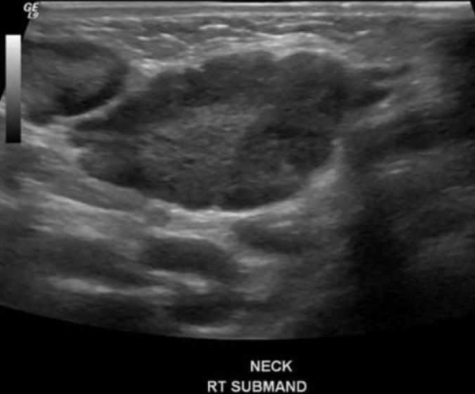
Cervical Lymphadenopathy Sonographic Appearance
Hypoechoic, rounded shape, absent echogenic hilus, intranodal necrosis (dead cells/breakdown), increasing size of serial exams, and peripheral vascularity. could have lobulated edges
Post Surgical Neck scan protocol
evaluate for residual thyroid tissue after thyroidectomy. thyroid bed should have images in sagittal and transverse, and should evaluate for abnormal lymph node and document by location using 7-region breakdown of neck