PSIO 2.1 Pre Lab
 Call Kai
 Learn
Learn Practice Test
 Spaced Repetition
 Match
Match Flashcards
 Knowt Play
1/35

There's no tags or description
Looks like no tags are added yet.
Name | Mastery | Learn | Test | Matching | Spaced | Call with Kai | Chat |
|---|
No analytics yet
Send a link to your students to track their progress
36 Terms
Skeletal System
organized into two divisions - axial skeleton and appendicular skeleton
Axial skeleton
composed of bones along the central axis of the body
three regions - the skull, the vertebral column, and the thoracic cage
main function of axial skeleton is to form a framework that supports and protects organs
80 bones - skull, hyoid bone, auditory ossicles, vertebra, sternum, and ribs
Appendicular skeleton
includes the bones of the upper and lower extremities and the girdles of bones that attach the upper and lower limbs of the axial skeleton
Pectoral girdles - consist of bones that hold upper limbs in place (clavicle and scapula)
Pelvic girdle consists of bones that hold the lower limbs in place (hip bone)
Divisions of the skeletal system
(a) Anterior view. (b) Posterior view. The appendicular skeleton is colored green, and the rest is axial skeleton.

Skull
two major regions - cranium and the face
most complex region of the skeletal system - houses the brain and a significant number of sense organs
cranium consists of mostly flat bones
Face is composed of irregular bones
Vertebral Column
five regions - cervical, thoracic, and lumbar vertebrae (sacrum + coccyx)
33 individual vertebrae that form the vertebral column
some fuse into larger structures such as the - sacrum
Thoracic Cage
consists of 12 pairs of ribs and the sternum - protects the lungs and heart yet provide flexibility during breathing
Hyoid bone
small floating bone between the floor of the moth and the upper anterior neck (only bone that doesn’t touch another bone)
clinical - if broken signals possible strangulation
Auditory ossicles
are three tiny bones located in the middle ear that transmits and amplify sound waves from the outer ear to the inner ear
Anatomical Features Marking of Bones Examples
Articular Surfaces
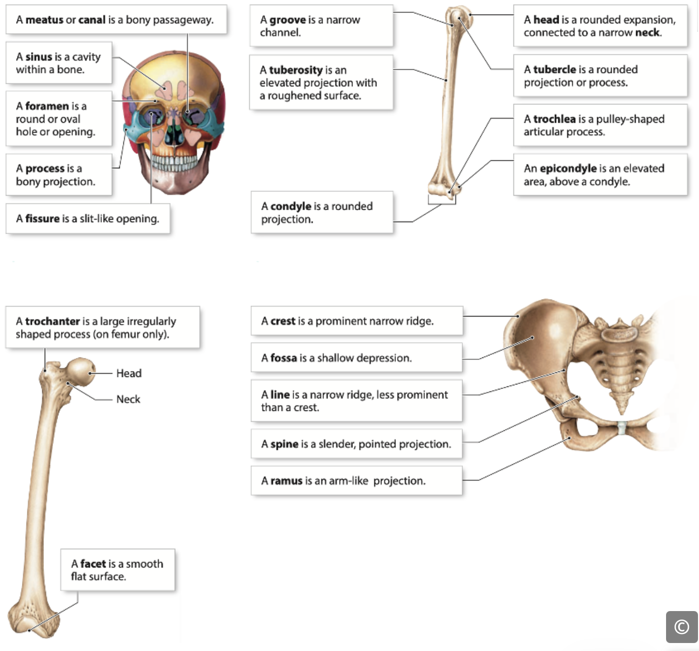
Condyle - a rounded articular projection; often articulates with a corresponding fossa ex; Occipital condyles of occipital bone
Facet - A smooth nearly flat articular surface ex; articular facts of vertebrae
Head - A prominent, rounded expansion carried on a narrow neck ex; head of the radius
Trochlea - A smooth grooved, pulley like process ex; trochlea of the humerus
CFTH - CANT F***N TAKE HER
Depression
Alveolus - a deep pit or socket ex: alveoli of the maxillae
Fossa - a shallow depression, often serving as an articular surface
Sulcus - a narrow grove ex; sigmoid sulcus of the temporal bone
Fovea - a small pit ex; fovea capitis of the femur
AFFS - A FAKE FRIEND SUCKS
Projections (for Tendons and Ligament attachment)
Crest - a narrow prominent ridge like projection ex; iliac res of the ilium
Epicondyle - a raised area on or above a condyle ex: lateral epicondyle of the humerus
Line - a narrow ridge of bone less prominent then a crest
Process - any bony prominence ex; lines aspera of the tibia
Protuberance - a bony outgrowth or protruding part ex; transverse processes of vertebrae
Ramus - an angular extensions of a bone relative to the rest of the structure ex; external occipital protuberance of the occipital bone
Spine - a sharp slender often pointed projection ex; spine of the scapula
Trochanter - a large irregularly shaped projection found only on the femur ex; greater trochanter of the femur
Tubercle - a small round process ex; tubercle of the rib
Tuberosity - an elevated projection with roughed surface ex; deltpid tuberosity of the humerus
Openings and Spaces
Canal/Meatus - A passageway (or canal) through a bone ex; external auditory meatus of the temporal bone
Fissure - a narrow slit like opening ex; orbital fissure behind the eye
Notch - an indentation at the edge of a structure ex; trochlear notch of the ulna
Foramen - a round or oval opening through a bone ex; olfactory foramen of the ethmoid bone
Sinus -an air filled cavity within a bone ex; nasal sinus
Vertebral Column
lies at the core of the human skeleton, the “backbone” that anchors nearly every major component of the skeletal support system.
five major regions: cervical, thoracic, lumbar, sacral, and coccygeal
vertebrae themselves change size and shape rather drastically from the cervical region to the sacral region. These changes reflect the different demands placed on the vertebrae in each region.
Cervical vertebrae
small and light because they are not supporting a lot of weight (relatively speaking), and they are specialized to allow a lot of movement of the neck, particularly rotation.
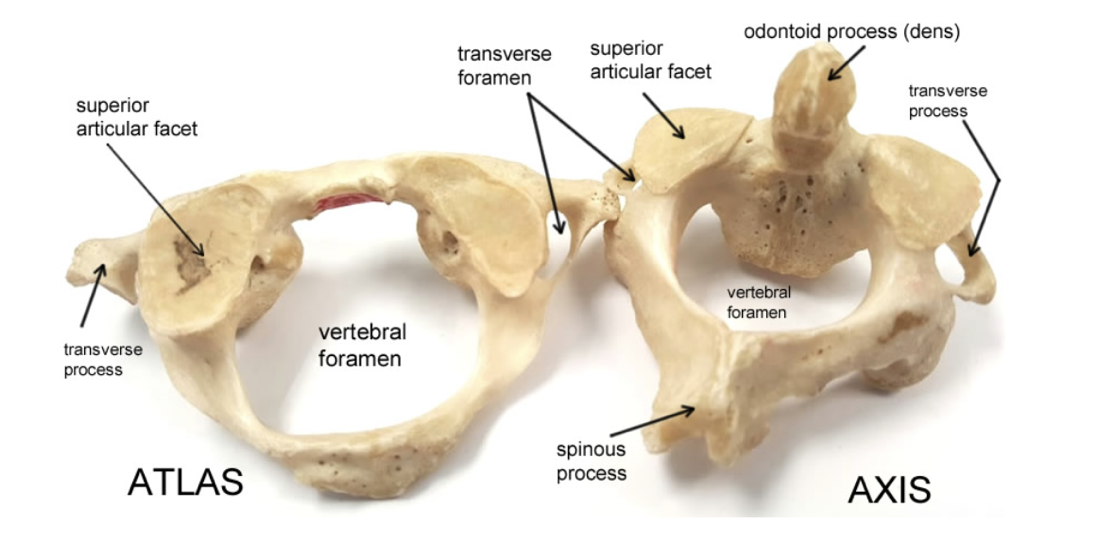
A unique feature - presence of transverse foramina which allow the passage of the arteries, veins, and nerves that supply the brain.
Due to their limited load-bearing function, cervical vertebrae are lightweight and have relatively small bodies.
Thoracic vertebrae
are specialized to provide articulation points for the ribs
The 12 TV are unique because they articulate with the 12 pairs of ribs.
As you move inferiorly down the thoracic spine, the vertebrae increase in size to bear more weight.
MISSING INFO pg 7
Lumbar vertebrae
very large, bulky vertebrae that are specialized for supporting the weight of the entire vertebral column and body structures above them.
They do not allow much movement, but instead are designed to keep the vertebral column STABLE.
MISSING INFO
Sacrum
consists of fused vertebrae, is specialized to provide a stable anchoring point for the bones of the pelvic girdle.
MISSING INFO
Coccyx
consists of three to five small vertebrae, which fuse together during development.
It serves as an attachment point for several ligaments and for muscles of the pelvic floor.
MISSING INFO pg 7
Spinal Curvatures
The vertebral column is not straight or rigid. When viewed from the side, it shows four natural curves: (1) the cervical curve, (2) the thoracic curve, (3) the lumbar curve, and (4) the sacral curve.
Primary curves
In newborn, the spine forms a C-shape, with the back curving posteriorly due to the thoracic and sacral curves - they develop during fetal growth
also called accommodation curves - provide space for the internal organs of the thoracic and abdominopelvic regions.
These curves are present at birth and help the infant maintain balance while lying down.
Secondary curves
cervical and lumbar curves - develop after birth.
These compensation curves help the body shift its center of gravity to allow for an upright posture.
The cervical curve begins to form when a baby learns to hold up their head
lumbar curve develops as the child starts to sit, crawl, and eventually walk.
These curves support balance and help distribute body weight and absorb shock as we move.
By age 10, all four curves are fully developed and contribute to a healthy, flexible spine.
Vertebra Structure
General Structure
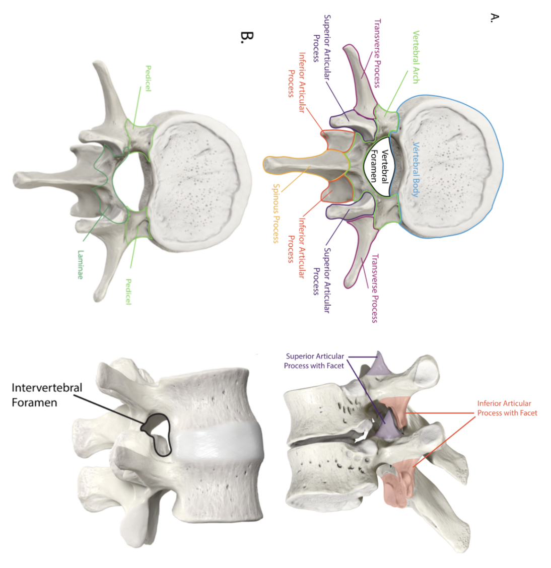
(Vertebral) Body
- Located anteriorly; is the largest part of the vertebra.
Pedicle
(pediculus = little foot)
- Short, thick projections that extend posteriorly from either side of the vertebral body.
Transverse Processes
Two lateral projections extending from each pedicle; serve as attachment points for muscles and ligaments.
Lamina (plate)
- A flat plate of bone that connects the transverse processes to the spinous process.
Spinous process
- A posterior projection from the fused laminae; provide attachment for muscles and ligaments.
Vertebral arch
- Formed by the fusion of the pedicles and laminae, creating the posterior part of the vertebra.
Vertebral Foramen
- Large opening formed by the vertebral arch. The central opening through which the spinal cord passes. The series of vertebral foramina forms the vertebral canal.
Superior and Inferior Articular Processes (with Facets):
Extend from the junction of the pedicle and lamina.
The superior articular processes face upward to articulate with the vertebra above
inferior articular processes face downward to articulate with the vertebra below.
The facets are the smooth surfaces on these processes where the articulation occurs.
Intervertebral Foramina
- Openings between adjacent vertebrae through which spinal nerves exit the spinal column.
Atlas C1 - Cervical Vertebrae
The first cervical vertebra (C1)
Unlike other vertebrae, the atlas has no vertebral body or spinous process.
Its large superior articular facets face superiorly and articulate with the occipital condyles of the occipital bone, enabling the head to move up and down in a nodding motion. (as in "yes"). This
articulation is called the atlanto-occipital joint.
Axis C2 - Cervical Vertebrae
The second cervical vertebra. (C2),
features a tooth-like projection called the dens or odontoid process
The dens fits into the vertebral foramen of the atlas, allowing it to pivot and enabling side-to-side "no" motions.
C3 - C6 and C7
C3 to C6 have bifurcated or bifid (forked) spinous processes
C7, known as the vertebra prominens, has a prominent single spinous process that can be easily seen and felt at the base of the neck.