Anna ch 2
 Call Kai
 Learn
Learn Practice Test
 Spaced Repetition
 Match
Match Flashcards
 Knowt Play
1/23

Earn XP
Description and Tags
cyto
Name | Mastery | Learn | Test | Matching | Spaced | Call with Kai | Chat |
|---|
No analytics yet
Send a link to your students to track their progress
24 Terms
Terminology / concept |
Cytology |
Cell |
Eukaryotic cell |
Three major cell regions |
Intracellular |
Extracellular |
Organelle |
Structure–function relationship |
anatomical meaning , function , where it fits?
Terminology / concept | Anatomical meaning / structure | Function + important information | Where it fits / flow |
Cytology | Study of cells. cyto- = cell; -ology = study of | This entire chapter is microscopic anatomy at the cellular level. | Anatomy → microscopic anatomy → cytology → cell |
Cell | Smallest living structural and functional unit of the body. | Obtains nutrients, synthesizes molecules, eliminates waste, maintains itself, and can reproduce when appropriate. | Tissues are built from cells. |
Eukaryotic cell | Cell containing a membrane-enclosed nucleus. Human cells are eukaryotic. | Separates DNA into a nuclear compartment while many cellular reactions occur in the cytoplasmic compartment. The professor uses the presence of a nucleus as the defining distinction for this course. | Cell → nuclear compartment + cytoplasmic compartment |
Three major cell regions | Plasma membrane + cytoplasm + nucleus | The basic map she should use whenever looking at a cell diagram or micrograph. | Plasma membrane → Cytoplasm → Nucleus |
Intracellular | Inside the cell. | Used constantly to describe locations/processes. | extracellular environment → membrane → intracellular environment |
Extracellular | Outside the cell. | Extracellular fluid surrounds cells. | Extracellular fluid → plasma membrane → cytoplasm |
Organelle | Specialized subcellular structure; literally a “little organ.” | Different organelles perform different jobs. Different cell types contain different amounts of them depending on function. | Cytoplasm → organelles → specialized functions |
Structure–function relationship | Cell shape and organelle abundance vary with what the cell does. | Very important anatomy principle: function predicts anatomy. Example: high-energy cells → many mitochondria; protein-secreting cells → abundant RER/Golgi. The textbook explicitly tests these relationships. | Cell function → required machinery → characteristic cellular anatomy |
Terminology / concept |
Plasma membrane / cell membrane / plasmalemma |
Unit membrane |
Phospholipid |
Hydrophilic |
Hydrophobic |
Polar |
Nonpolar |
Phospholipid bilayer |
Fluid mosaic model |
Cholesterol |
Integral protein |
Transmembrane protein |
Peripheral protein |
Glyco- |
Glycolipid |
Glycoprotein |
Glycocalyx / cell coat |
Receptor |
Selective permeability |
pm flow
Terminology / concept | Anatomical meaning / structure | Function + important information | Where it fits / flow |
Plasma membrane / cell membrane / plasmalemma | Thin flexible outer boundary of the cell. -lemma = layer/sheath in the terminology emphasized by the professor. | Separates intracellular from extracellular fluid; controls passage, receives signals, participates in recognition and anchors cells/cytoskeleton. | Extracellular fluid → PLASMA MEMBRANE → cytoplasm |
Unit membrane | Professor’s term for the common basic lipid-bilayer organization of biological membranes. | Plasma membrane and many organelle membranes share the phospholipid-bilayer principle, although their exact proteins/lipids differ. | Phospholipids → bilayer → plasma membrane / organelle membrane |
Phospholipid | Membrane lipid with a polar hydrophilic head + two nonpolar hydrophobic fatty-acid tails. | Amphipathic nature causes phospholipids to arrange themselves into bilayers in water. | hydrophilic head — hydrophobic tails |
Hydrophilic | “Water loving”; attracted to/interacts with water. | Phospholipid heads face the watery cytoplasm and extracellular fluid. | Water ↔ head |
Hydrophobic | “Water fearing”; avoids water. | Phospholipid tails point inward, away from water, creating the membrane’s hydrophobic core. | head → **tails |
Polar | Molecule/region with separated electrical charge that interacts with water. | Phospholipid head is polar. | polar → hydrophilic |
Nonpolar | Lacks strong separated electrical charges; generally poorly interacts with water. | Fatty-acid tails are nonpolar. | nonpolar → hydrophobic |
Phospholipid bilayer | Two layers of phospholipids arranged tail-to-tail. | Fundamental membrane barrier. Hydrophobic interior helps exclude many charged/water-soluble molecules. | water → heads → tails : tails → heads → water |
Fluid mosaic model | Membrane viewed as a flexible lipid bilayer in which proteins and other molecules are embedded and can move laterally. | Fluid = components can move within membrane. Mosaic = mixture of lipids, proteins, carbohydrates. Professor emphasizes that the membrane can remain intact while components move. | phospholipid bilayer + proteins + carbohydrates → fluid mosaic membrane |
Cholesterol | Lipid interspersed among phospholipids. | In Marieb, increases membrane rigidity and decreases permeability to water/water-soluble substances. | phospholipids + cholesterol → membrane properties |
Integral protein | Protein embedded firmly in the bilayer. | May act as receptor, carrier, channel, enzyme, adhesion molecule. | bilayer → embedded protein |
Transmembrane protein | Integral protein extending completely through the membrane. | Can create a connection/pathway between extracellular and intracellular environments. | outside → transmembrane protein → inside |
Peripheral protein | Protein attached to a membrane surface rather than buried across the bilayer. | Can provide structural support and connect membrane with cytoskeleton. | membrane → peripheral protein → cytoskeleton |
Glyco- | Word root meaning sugar/carbohydrate. | Helps decode glycolipid, glycoprotein, glycocalyx. | glyco = sugar |
Glycolipid | Membrane lipid + carbohydrate chain. | Contributes to external glycocalyx and cell recognition. | membrane lipid → sugar extends outside |
Glycoprotein | Membrane protein + carbohydrate chain. | Important in recognition, adhesion, receptors/signaling. | membrane protein → sugar extends outside |
Glycocalyx / cell coat | Carbohydrate-rich coating on the external membrane surface formed by glycoproteins + glycolipids. | Different cell types have distinctive carbohydrate patterns, helping cells bind to and recognize one another. | glycolipids + glycoproteins → glycocalyx → recognition |
Receptor | Usually membrane protein with a specific binding site for a signaling molecule. | Converts information outside the cell into a cellular response; membrane receptors are part of intercellular communication. | signal → receptor binds → cellular response |
Selective permeability | Ability of membrane to allow certain substances to cross more readily than others. | Central function of plasma membrane; makes different intracellular and extracellular compositions possible. | membrane composition → determines what enters/leaves |
Phospholipids form the barrier→ proteins provide specialized functions→ carbohydrates provide recognition→ cholesterol modifies membrane properties
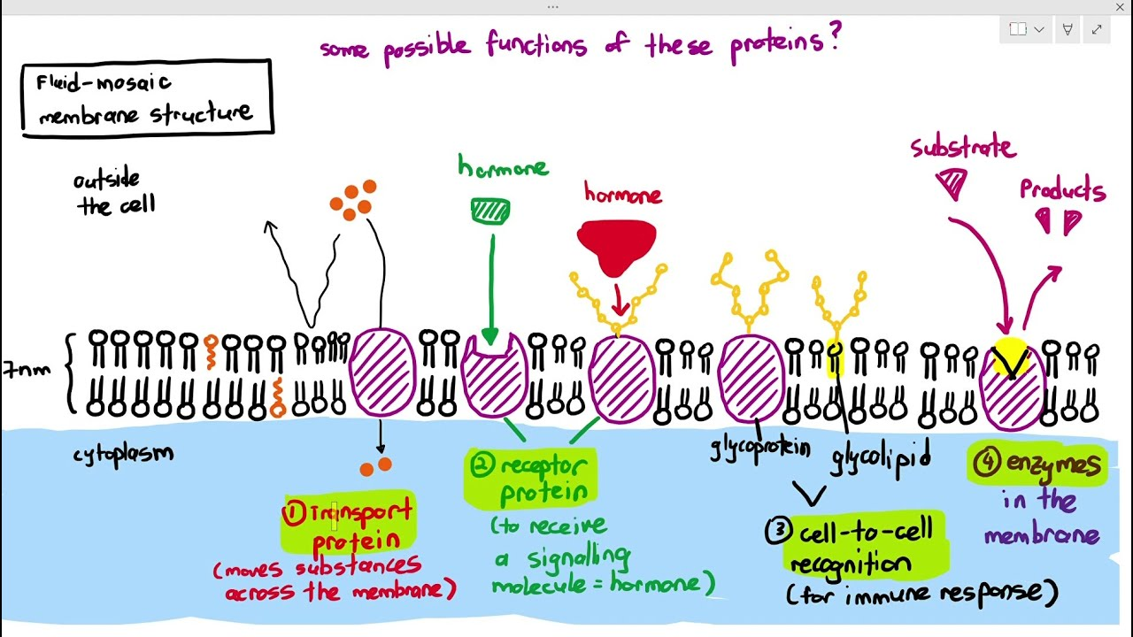
Why can the membrane do so many different jobs?
same basic phospholipid bilayer + different inserted/attached molecules = different membrane functions.
The professor emphasizes that membranes share a basic structure but can differ greatly according to which proteins and lipids are added or anchored.
membrane transport
Concentration gradient |
Diffusion |
Simple diffusion |
Osmosis |
Aquaporin |
Facilitated diffusion |
Channel protein |
Ion channel |
Carrier / transport protein |
Active transport |
ATP |
Passive transport concept |
Bulk / vesicular transport |
Vesicle |
Invagination |
Endocytosis |
Phagocytosis |
Pseudopod / pseudopodium |
Phagosome |
Pinocytosis |
Receptor-mediated endocytosis |
Clathrin |
Coated pit / coated vesicle |
Receptor recycling |
Exocytosis |
v-SNARE |
t-SNARE |
Secretion |
Excretion |
Terminology / concept | Anatomical meaning / structure | Function + important information | Where it fits / flow |
Concentration gradient | Difference in concentration of a substance between two regions. | Provides the driving force for diffusion. | HIGH concentration → LOW concentration |
Diffusion | Net movement down a concentration gradient. | Does not require the cell to directly spend ATP. | high → low |
Simple diffusion | Substance passes directly through lipid bilayer. | Used particularly by small uncharged/lipid-soluble molecules. | high → bilayer directly → low |
Osmosis | Diffusion of water across a membrane. | Water movement specifically. The textbook explicitly asks this in Chapter 2 review. | water high → membrane → water low |
Aquaporin | Membrane channel specialized for passage of water. | Allows much more rapid water movement through membranes. Professor specifically introduces aquaporins. | water → aquaporin → water |
Facilitated diffusion | Passive transport through a specific integral membrane protein. | Still moves down concentration gradient; no ATP required for the movement itself. | high → channel/carrier → low |
Channel protein | Transmembrane protein containing a selective passage. | Allows selected water-soluble or charged substances to cross. | solute → protein pore → other side |
Ion channel | Channel selective for ions. | Channel size/shape allows selectivity; not every molecule passes through every channel. Professor emphasizes this specificity. | ion → appropriate channel → across membrane |
Carrier / transport protein | Protein that binds a substance and changes configuration to move it through membrane. | Can participate in facilitated diffusion or active transport depending on direction/energy use. | molecule binds → protein changes shape → molecule crosses |
Active transport | Protein-mediated movement against a concentration gradient. | Requires energy; professor emphasizes ATP-driven protein conformational changes. | low → HIGH + ATP |
ATP | Adenosine triphosphate; major immediately usable cellular energy molecule. | Powers active transport and many other cellular processes. | ATP → energy released → cellular work |
Passive transport concept | Transport without direct cellular ATP expenditure. | Includes simple diffusion, osmosis and facilitated diffusion. | high → low |
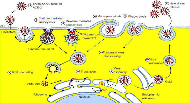
Bulk / vesicular transport | Movement of large materials by membrane-bound vesicles. | Used when material is too large to simply pass through membrane transport proteins. | membrane bends/fuses → vesicle moves cargo |
Vesicle | Small membrane-bound sac. | Carries substances between compartments or into/out of cell. | membrane → buds → vesicle → destination |
Invagination | Inward folding of a membrane. | First physical step in many forms of endocytosis. | membrane → bends inward → pit |
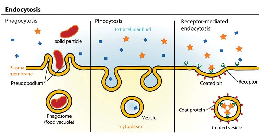
Endocytosis | Vesicular movement into the cell. endo = in/within | Plasma membrane folds inward and pinches off around extracellular material. | outside → invagination → vesicle → inside |
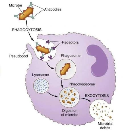
Phagocytosis | “Cell eating”; endocytosis of large particles. | Cells form pseudopods around material; commonly associated with protective phagocytic cells. | particle → pseudopods → phagosome → lysosome |
Pseudopod / pseudopodium | Temporary cell projection; “false foot.” | Surrounds material during phagocytosis and can participate in amoeboid movement. | membrane/cytoskeleton extends → surrounds object |
Phagosome | Vesicle containing material engulfed by phagocytosis. | Typically combines with lysosome so contents can be digested. | particle → phagosome + lysosome → digestion |
Pinocytosis | “Cell drinking”; uptake of extracellular fluid and dissolved solutes. | Nonspecific sampling using small vesicles in the textbook description. | extracellular fluid → tiny vesicle → cytoplasm |
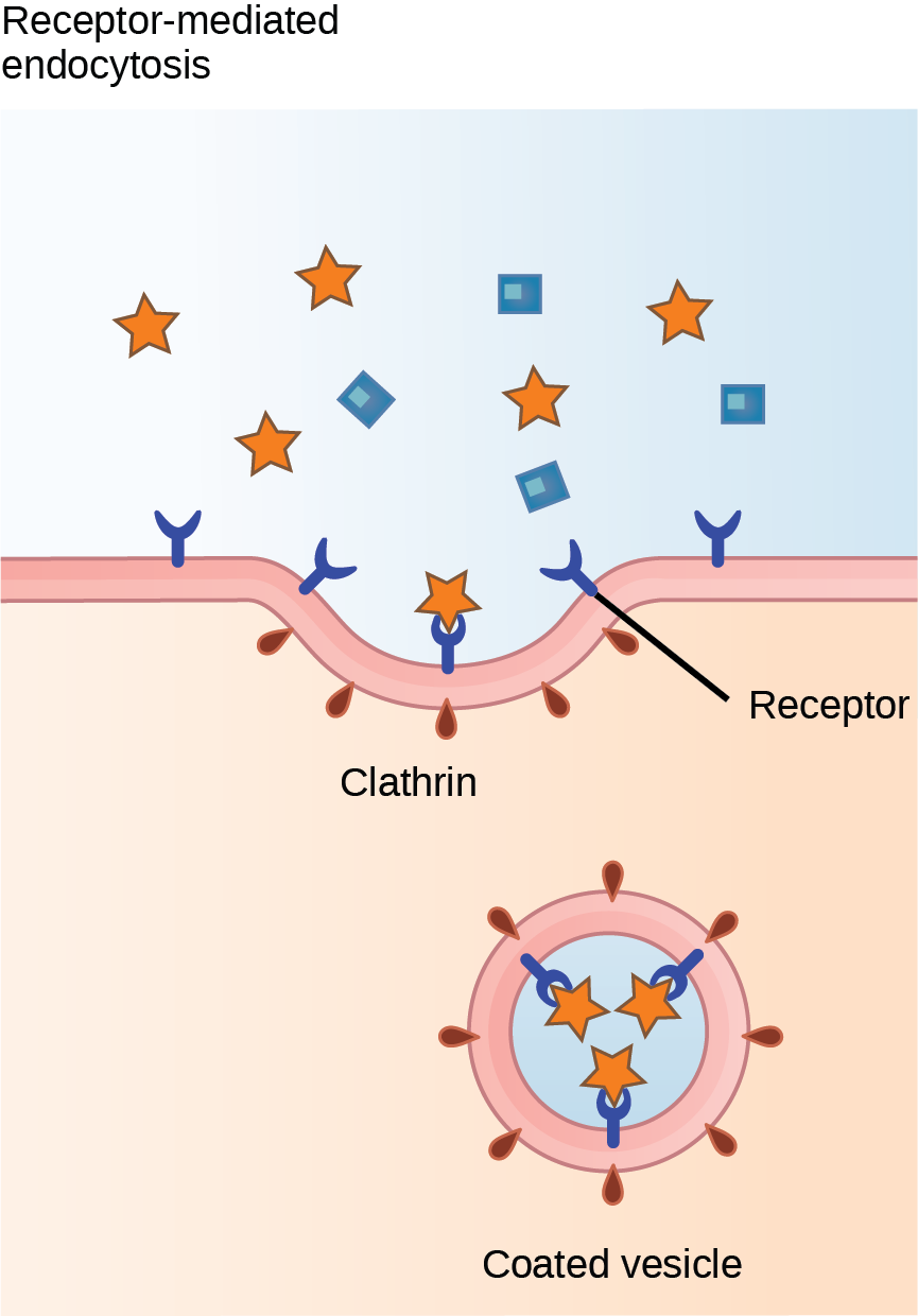
Receptor-mediated endocytosis | Selective uptake after extracellular material binds specific membrane receptors. | Allows cells to concentrate particular substances rather than indiscriminately engulfing fluid. | ligand → receptor → coated pit → coated vesicle |
Clathrin | Intracellular coat protein emphasized by professor during receptor-mediated endocytosis. | Clathrin molecules aggregate beneath membrane and help curve membrane into a coated pit/vesicle. | receptor binding → clathrin coat → pit → vesicle |
Coated pit / coated vesicle | Membrane region/vesicle with associated coat proteins. | Promotes selective receptor-mediated uptake. | receptor-ligand → coated pit → coated vesicle |
Receptor recycling | Return of endocytosis receptors to plasma membrane. | Receptors can be reused rather than permanently lost. | endocytic vesicle → receptor separates → recycled to membrane |
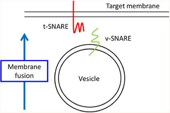
Exocytosis | Vesicular movement out of the cell. exo = out | Vesicle fuses with plasma membrane and releases contents outside. Major secretion pathway. | vesicle → membrane fusion → contents outside |
v-SNARE | Vesicle-associated SNARE protein shown in the book’s exocytosis figure. | Recognizes/binds appropriate target membrane machinery. | vesicle v-SNARE ↔ t-SNARE membrane |
t-SNARE | Target/plasma-membrane SNARE in the book’s exocytosis figure. | Helps dock/fuse vesicle with target membrane. | v-SNARE + t-SNARE → fusion → pore |
Secretion | Release/movement of a substance from a cell into another body compartment. | May be useful product rather than “waste,” e.g. mucus or hormone. | Golgi → secretory vesicle → exocytosis → secretion |
Excretion | Elimination from the body/region. | Professor distinguishes it from secretion: secretion can remain within the body; excretion leaves it. | cellular/body waste → outside body |


Transport different materials
what needs to cross the membrane
small+ lipid soluble
water
water soluble / charged materials
moving against gradient
very large material
endocytosis
very large solid
fluid
specific receptor bound cargo
Small + lipid soluble
→ simple diffusion
Water
→ osmosis, often through aquaporins
Water-soluble/charged + going high → low
→ facilitated diffusion
Moving against gradient
→ active transport + ATP
Very large material
→ vesicular transport
→ INTO = endocytosis
→ OUT = exocytosis
And endocytosis:
large solid → phagocytosis
fluid → pinocytosis
specific receptor-bound cargo → receptor-mediated endocytosis
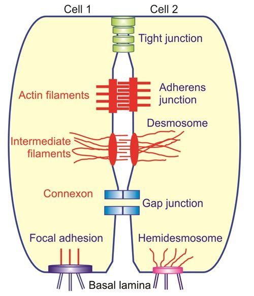
cell junctions
Cell junction |
Tight junction |
Desmosome |
Cadherin |
Gap junction / nexus |
Connexon |
Terminology / concept | Anatomical meaning / structure | Function + important information | Where it fits / flow |
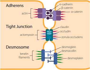
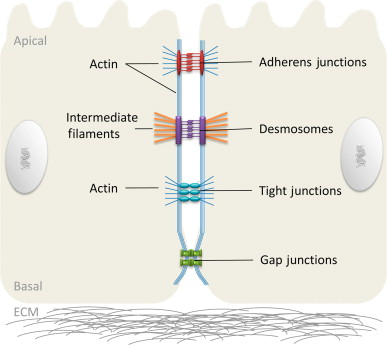
Cell junction | Specialized protein connection between neighboring cells. | Allows cells in tissues to seal, anchor or communicate with one another. | cell membrane ↔ junction ↔ neighboring cell membrane |
Tight junction | Junction where neighboring membranes are tightly linked along a region. | Creates a seal that restricts passage between cells. Professor compares repeated junctions to stitching fabric together. | cell A ║ SEAL ║ cell B |
Desmosome | Strong point-like anchoring junction connected to cytoskeletal intermediate filaments. | Resists pulling/mechanical stress; professor compares it to a rivet. | intermediate filament → desmosome ↔ desmosome ← intermediate filament |
Cadherin | Adhesion protein discussed by professor as part of strong cell-cell attachment at desmosomes. | Helps link neighboring cells. | cell A → cadherin ↔ cadherin ← cell B |
Gap junction / nexus | Channel junction between neighboring cells. | Allows ions and other small molecules to pass directly cell-to-cell, enabling communication/electrical coupling. | cytoplasm A → channel → cytoplasm B |
Connexon | Hollow protein channel unit composing a gap junction. | Connexons of neighboring cells align to form continuous passage. | connexon A ↔ connexon B → gap junction |
Tight junction = SEAL
Desmosome = STRENGTH
Gap junction = SIGNAL/COMMUNICATION

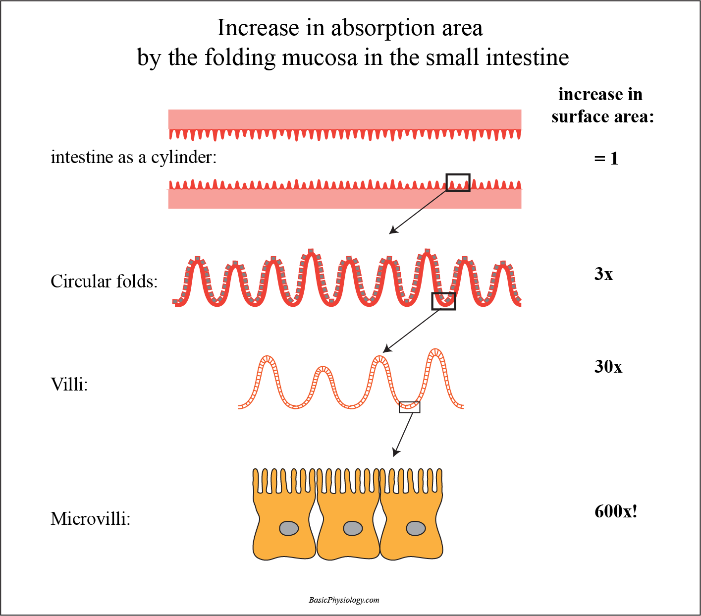
cell membrane projections
Terminology / concept |
Microvillus |
Microvilli |
Actin core of microvillus |
Cilium |
Cilia |
9 + 2 arrangement |
Dynein |
Flagellum |
Terminology / concept | Anatomical meaning / structure | Function + important information | Where it fits / flow |
Microvillus | One short fingerlike extension of plasma membrane. | Increases membrane surface area. | plasma membrane → folds outward → microvillus |
Microvilli | Plural of microvillus; many closely packed projections. | More surface area means room for more channels/transporters → enhanced absorption/transport. | more membrane → more transport machinery → more absorption |
Actin core of microvillus | Microfilaments inside microvillus. | Provides internal structural support to the projection. | membrane projection → actin microfilaments inside |
Cilium | Singular, longer hairlike motile membrane projection. | Moves extracellular material along cell surface. | cell → one cilium |
Cilia | Plural; numerous coordinated motile projections. | Coordinated beating moves mucus/fluid like a conveyor belt. | ATP-powered beat → fluid/mucus moves |
9 + 2 arrangement | Internal ciliary organization: 9 peripheral microtubule doublets surrounding 2 central microtubules. | Characteristic architecture of motile cilia taught by professor. | cilium → membrane surrounding 9+2 microtubules |
Dynein | Microtubule motor protein in cilia. | ATP-dependent dynein activity produces movement/bending. | ATP → dynein → microtubule movement → cilium beats |
Flagellum | Long cilia-like motile projection; sperm is the major human example discussed. | Propels the cell itself rather than moving material over a surface. | microtubule machinery → flagellum → cell movement |
Microvilli → increase SURFACE AREA
Cilia → MOVE material
And internally:
Microvilli → actin
Cilia → microtubules + dynein


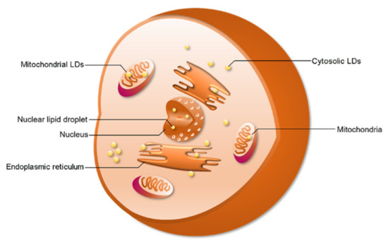
Cytoplasm
Terminology / concept |
Cytoplasm |
Cytosol |
Cytoplasmic organelle |
Cytoplasmic inclusion |
Lipid droplet |
Glycosome |
Glycogen |
Terminology / concept | Anatomical meaning / structure | Function + important information | Where it fits / flow |
Cytoplasm | Everything between plasma membrane and nucleus. | Site of most cellular activities. | plasma membrane → CYTOPLASM → nucleus |
Cytosol | Jellylike fluid portion of cytoplasm. | Contains water, ions and enzymes; organelles/inclusions are suspended in it. | cytoplasm = cytosol + organelles + inclusions |
Cytoplasmic organelle | Specialized functional structure in cytoplasm. | Metabolic machinery of the cell. | cytosol → organelles |
Cytoplasmic inclusion | nonpermanent/ Temporary stored material. | Not equivalent to a metabolically active organelle. | cytoplasm → storage material |
Lipid droplet | Non-membrane-bound drop of stored fat. | Energy/nutrient storage; can resemble lysosome in size but lacks surrounding membrane. | nutrients → fat → lipid droplet |
Glycosome | Cytoplasmic inclusion containing glycogen and enzymes involved in glycogen metabolism. | Stores carbohydrate energy as glycogen. | glucose → glycogen → glycosome |
Glycogen | Branched storage polymer of glucose. | Stored carbohydrate fuel. | glucose units → glycogen storage |



Ribosomes and the endomembrane/protein-processing system
distinguishes free ribosomes from RER-bound ribosomes based on the destination of the proteins they make.
Terminology / concept |
Ribosome |
rRNA |
Free ribosome |
Bound ribosome |
Endoplasmic reticulum / ER |
Rough ER / RER |
Cistern / cisternae |
Smooth ER / SER |
Calcium storage in SER |
Transport vesicle |
Golgi apparatus |
Cis face |
Trans face |
Secretory vesicle / secretory granule |
Endomembrane relationship |
Terminology / concept | Anatomical meaning / structure | Function + important information | Where it fits / flow |
Ribosome | Dense nonmembranous particle with large + small subunit, composed of rRNA and proteins. | Site of translation/protein synthesis. | mRNA → ribosome → protein |
rRNA | Ribosomal RNA. | Structural/functional RNA making up much of ribosome. | nucleolus → rRNA → ribosomal subunits |
Free ribosome | Ribosome floating in cytosol. | Produces proteins primarily used within cytosol. | mRNA → free ribosome → cytosolic protein |
Bound ribosome | Ribosome attached to rough ER during protein production. | Produces proteins destined for secretion, membranes, and endomembrane pathways. | mRNA → RER ribosome → secreted/membrane protein |
Endoplasmic reticulum / ER | Extensive membranous network of sacs and tubes; literally “network within cytoplasm.” | Major intracellularproduction/processing system. | nuclear envelope ↔ ER → Golgi |
Rough ER / RER | ER with ribosomes attached to cytosolic surface; much of it consists of flattened cisterns. | Synthesizes proteins for secretion/membranes; textbook also calls it a major | nucleus → mRNA → ribosome/RER → protein |
Cistern / cisternae | Fluid-filled flattened membranous cavity/cavities. | Structural units of rough ER and Golgi. | RER/Golgi → flattened sacs |
Smooth ER / SER | Tubular ER lacking ribosomes. | Lipid metabolism/synthesis, steroid hormone production, detoxification of lipid-soluble drugs, Ca²⁺ storage. | lipids/steroids/detox/Ca²⁺ |
Calcium storage in SER | SER stores Ca²⁺ and keeps resting cytosolic Ca²⁺ relatively low. | Released Ca²⁺ can participate in events such as muscle contraction and glandular secretion. | SER Ca²⁺ store → release → cellular response |
Transport vesicle | Small membrane sac carrying products from one organelle to another. | Transfers products from RER to Golgi, among other destinations. | RER → transport vesicle → Golgi |
Golgi apparatus | Stack of approximately 3–10 disc-shaped membrane cisterns. | Modifies, sorts and packages products received from RER. | RER → Golgi → destinations |
Cis face | Convex receiving side of Golgi. | Receives transport vesicles from RER. | RER → vesicle → CIS Golgi |
Trans face | Concave shipping side of Golgi. | New vesicles bud away toward final destinations. | cis → Golgi processing → TRANS → vesicle |
Secretory vesicle / secretory granule | Golgi-derived vesicle containing a product for secretion. | Travels toward plasma membrane and releases product by exocytosis. |
|
Endomembrane relationship | Nuclear envelope, ER, Golgi, vesicles, lysosomes and plasma membrane are structurally/functionally connected through membrane production and vesicle trafficking. | Helps explain why membrane-bound protein processing is a pathway, not isolated organelles. | nuclear envelope ↔ RER → vesicle → Golgi → vesicle → membrane/lysosome |

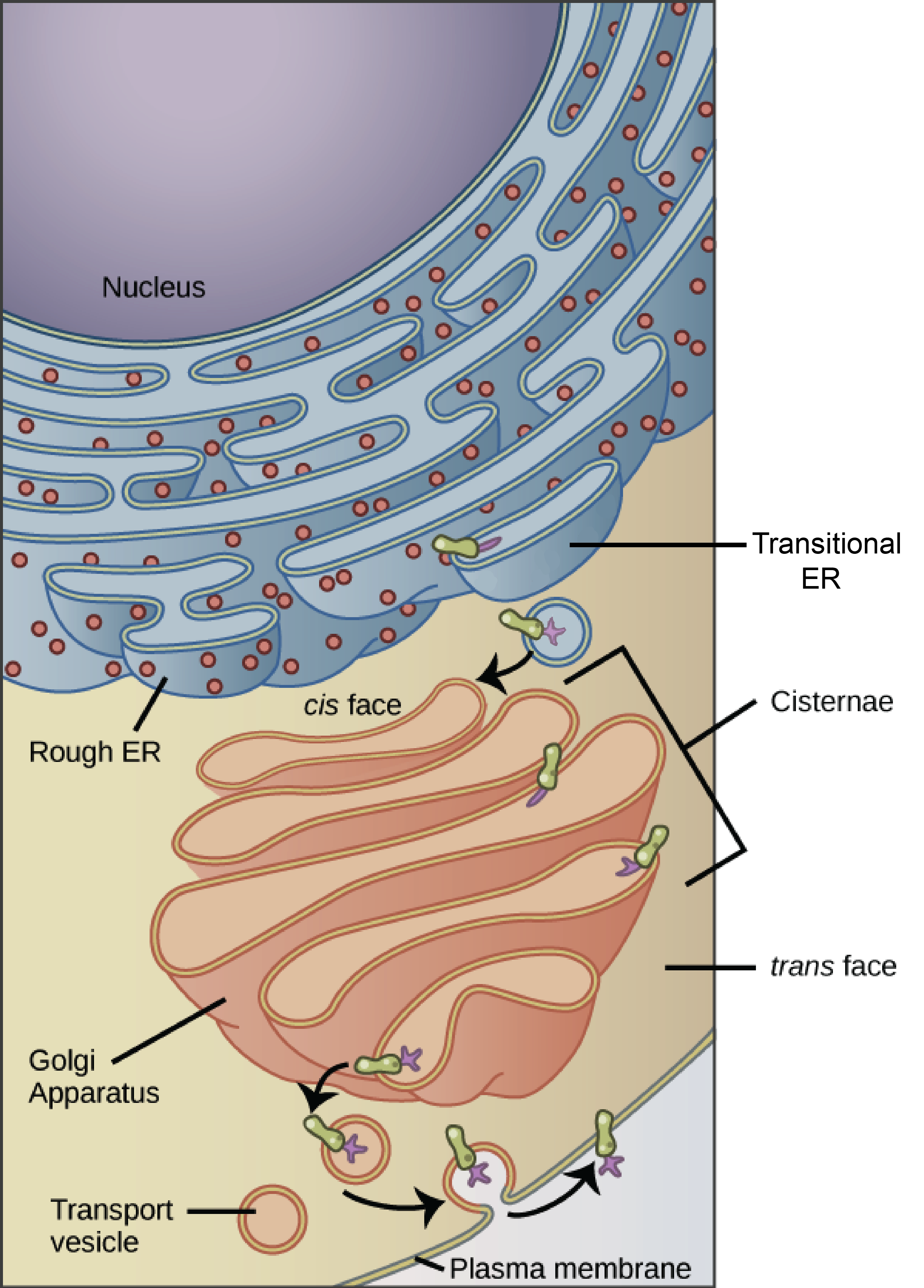
Secreted-protein pathway
DNA in nucleus→ transcription→ mRNA→ nuclear pore→ RER-bound ribosome→ protein enters RER→ transport vesicle→ cis Golgi→ modification/sorting→ trans Golgi→ secretory vesicle→ plasma membrane→ exocytosis
Lysosomes and peroxisomes
Lysosome |
Acid hydrolase |
Autodigestion/recycling concept |
Phagosome–lysosome relationship |
Peroxisome |
Oxidase |
Free radical |
Hydrogen peroxide / H₂O₂ |
Catalase |
Terminology / concept | Anatomical meaning / structure | Function + important information | Where it fits / flow |
Lysosome | Membranous sac containing digestive enzymes/acid hydrolases. | Intracellular digestion; destroys ingested substances and deteriorated cellular components. | material → lysosome → digestion/recycling |
Acid hydrolase | Digestive enzyme functioning in lysosome. | Breaks down macromolecules. | lysosome → hydrolysis → smaller components |
Autodigestion/recycling concept | Lysosomes can digest worn cellular structures as well as incoming material. | Explains lysosome as cellular recycling center. | damaged organelle → lysosome → recycled components |
Phagosome–lysosome relationship | Phagosome carrying engulfed material joins lysosomal digestive machinery. | Connects membrane transport with organelle function. | phagocytosis → phagosome → lysosome → digestion |
Peroxisome | Small membrane-bound organelle containing oxidative enzymes. | Detoxification and other oxidative metabolic processes. | toxic/reactive molecule → peroxisome |
Oxidase | Enzyme in peroxisomes that oxidizes substrates and can form hydrogen peroxide. | Helps neutralize reactive substances. | substrate → oxidase → H₂O₂ |
Free radical | Highly reactive molecule produced during metabolism or exposure to damaging agents. | Can damage proteins, membranes and DNA. | metabolism → free radicals → cellular damage unless controlled |
Hydrogen peroxide / H₂O₂ | Reactive peroxide produced during some peroxisomal reactions. | Must itself be detoxified. | oxidase → H₂O₂ → catalase |
Catalase | Important peroxisomal enzyme. | Breaks hydrogen peroxide down to safer products; textbook emphasizes this function. | H₂O₂ → catalase → H₂O + O₂ |
Lysosome = DIGEST
Peroxisome = DETOXIFY/OXIDIZE
Mitochondria
Terminology / concept | Anatomical meaning / structure | Function + important information | Where it fits / flow |
Mitochondrion | Singular organelle; generally rod/thread/bean-shaped with two membranes. | Main site of ATP production. | nutrients → mitochondrion → ATP |
Mitochondria | Plural. | Number varies with cellular energy requirement. Muscle/high-metabolic-demand cells contain many. | higher energy demand → more mitochondria |
Outer mitochondrial membrane | Outer boundary membrane. | Encloses mitochondrion. | cytoplasm → outer membrane |
Inner mitochondrial membrane | Membrane internal to outer membrane, heavily folded. | Contains important energy-producing machinery. | outer membrane → inner membrane |
Crista / cristae | Fold/folds of inner mitochondrial membrane. | Increase inner-membrane surface area for energy-producing machinery. | more folds → more membrane surface area → greater ATP machinery capacity |
Mitochondrial matrix | Innermost compartment enclosed by inner membrane. | Contains mitochondrial components involved in energy metabolism. | inner membrane/cristae → matrix |
ATP production | Principal functional association of mitochondria. | Immediate link she should make if shown a mitochondrion. | food-energy pathways → mitochondrion → ATP |
Mitochondrial DNA | Mitochondria contain some of their own genetic material. | One observation supporting their unusual/semi-autonomous evolutionary history. | mitochondrion → own DNA |
Endosymbiotic concept | Professor explains mitochondria as thought to derive evolutionarily from bacteria engulfed by ancestral cells. | Basic evidence emphasized: bacterial resemblance, own DNA, replication characteristics. | ancestral cell + engulfed bacterium → mitochondrion |
Structure–function link | Inner membrane folds extensively because important energy-production proteins are membrane associated. | Very anatomy-style question: why cristae? → increased surface area. | cristae ↑ → membrane surface ↑ → energy machinery ↑ |
cytoskeleton
cytoskeleton is not one structure. It contains three major protein-fiber systems with different anatomy and jobs
Terminology / concept | Anatomical meaning / structure | Function + important information | Where it fits / flow |
Cytoskeleton | Internal network of nonmembranous protein rods/fibers through cytosol. | Gives shape, mechanical support, organizes organelles and enables movement. | cytoplasm → cytoskeletal framework |
Microfilament / actin filament | Thinnest major cytoskeletal fiber; made of actin. | Contraction, cell movement, cytokinesis, membrane changes, support of microvilli. | actin → movement/contraction |
Actin | Protein making microfilaments. | Interacts with myosin to generate force. | actin + myosin → force |
Myosin | Motor protein interacting with actin. | Produces contractile movement. | actin ↔ myosin → contraction |
Pseudopod movement | Cell extension/retraction involving cytoskeletal rearrangement. | Supports amoeboid movement and phagocytosis. | cytoskeleton rearranges → pseudopod |
Intermediate filament | Tough fiber intermediate in diameter between microfilament and microtubule. | Most stable cytoskeletal element; resists tensile/pulling forces. | mechanical stress → intermediate filaments resist |
Tensile strength | Resistance to being pulled apart. | Major defining property of intermediate filaments. | pulling force → intermediate filament → structural integrity |
Microtubule | Largest major cytoskeletal element; hollow tube made of tubulin. | Cell shape, organelle positioning, intracellular tracks, mitotic spindle, cilia/flagella. | centrosome → microtubules radiate through cell |
Tubulin | Protein subunit composing microtubules. | Tubulin assembly/disassembly changes microtubule length. | tubulin units ↔ microtubule |
Dynamic microtubule behavior | Microtubules can assemble and disassemble. | Lets cell rapidly reorganize its internal framework. | tubulin added → grows; removed → shrinks |
Intracellular transport track | Microtubules act as routes along which cargo can move. | Positions/moves mitochondria, vesicles and other organelles. | cargo + motor protein → microtubule track → destination |
Kinesin | Microtubule-associated motor protein. | Professor describes kinesin “walking” vesicular cargo along microtubules. | vesicle → kinesin → microtubule → movement |
Dynein | Another microtubule-associated motor protein. | Involved in intracellular transport and particularly emphasized in cilia. | microtubule + dynein → movement |
Centrosome | Nonmembranous microtubule-organizing region near nucleus. | Site from which many microtubules originate; organizes mitotic spindle. | nucleus nearby → centrosome → microtubules |
Centrosome matrix | Cloudlike protein material forming outer part of centrosome. | Anchors/seeds microtubule growth. | centrosome matrix → microtubules radiate |
Centriole | Barrel-shaped structure inside centrosome. | Involved in organization of mitotic apparatus and formation of cilia/flagella bases. | centrosome → pair of centrioles |
9 triplets | Each centriole contains nine groups of three microtubules. | Important structural recognition point. | centriole cross-section → 9 × 3 |
cytoskeleon comparison
microfilaments
intermediate filaments
microtubules
Microfilaments
→ smallest
→ actin
→ movement/contraction
Intermediate filaments
→ middle
→ strongest/stable
→ resist tension
Microtubules
→ largest
→ tubulin tubes
→ shape + intracellular transport + spindle + cilia
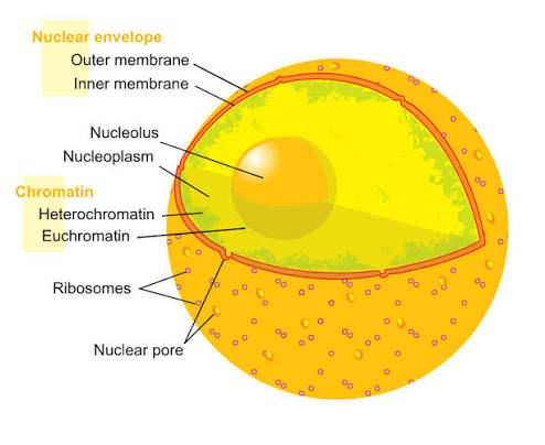
Nucleus
nuclear anatomy chart
Nucleus |
Anucleate |
Nuclear envelope |
Outer nuclear membrane |
Nuclear lamina |
Nuclear pore / nuclear pore complex |
Nucleoplasm |
Nucleolus |
Nucleus vs nucleolus |
nuclear anatomy is nuclear envelope → pores → nucleoplasm → chromatin → nucleolus.
Terminology / concept | Anatomical meaning / structure | Function + important information | Where it fits / flow |
Nucleus | Large membrane-enclosed cellular compartment containing most cellular DNA. | Controls cellular activity largely through gene expression. | cytoplasm → NUCLEUS → genetic information |
Anucleate | Having no nucleus. | Mature red blood cells are a textbook human example. | specialized exception |
Nuclear envelope | Double membrane surrounding nucleus. | Separates nucleoplasm from cytoplasm and regulates exchange. | cytoplasm → nuclear envelope → nucleoplasm |
Outer nuclear membrane | Outer of the two nuclear-envelope membranes. | Continuous with rough ER and can have ribosomes on its external surface. | nuclear envelope ↔ RER |
Nuclear lamina | Network of intermediate-filament proteins (lamins) lining inner nuclear membrane. | Supports and maintains nuclear shape. | inner envelope → lamina → nuclear support |
Nuclear pore / nuclear pore complex | Protein-lined opening where inner/outer nuclear membranes join. | Allows regulated transport of large molecules such as RNA and proteins. | nucleus ↔ pore ↔ cytoplasm |
Nucleoplasm | Jellylike fluid within nucleus. | Suspends chromatin and nucleolus; nuclear equivalent conceptually to cytosol. | nuclear envelope → nucleoplasm → chromatin/nucleolus |
Nucleolus | Dense, dark-staining nonmembranous region inside nucleus. | Produces rRNA and assembles large/small ribosomal subunits. | DNA genes → nucleolus → rRNA → ribosome subunits → nuclear pore |
Nucleus vs nucleolus | Nucleus = entire DNA-containing compartment; nucleolus = structure inside nucleus. | Textbook explicitly warns students not to confuse them. | cell → nucleus → nucleolus |
DNA, chromatin, and chromosome anatomy
DNA |
Nucleotide |
Sugar-phosphate backbone |
Adenine / A |
Thymine / T |
Cytosine / C |
Guanine / G |
Complementary base pairing |
Double helix |
Histone |
Nucleosome |
Chromatin |
Extended chromatin |
Euchromatin |
Condensed chromatin |
Heterochromatin |
Chromatid |
Chromosome |
Sister chromatids |
Centromere |
Kinetochore |
Telomere |
Telomerase |
Terminology / concept | Anatomical meaning / structure | Function + important information | Where it fits / flow |
DNA | Deoxyribonucleic acid; double-helical genetic molecule. | Stores genetic information. | nucleus → DNA |
Nucleotide | Basic nucleic-acid subunit consisting of sugar + phosphate + nitrogenous base. | Nucleotides link to form DNA/RNA chains. | nucleotide → nucleotide → nucleic-acid strand |
Sugar-phosphate backbone | Alternating sugar and phosphate portion forming sides of DNA/RNA chain. | Supports ordered sequence of bases. | sugar—phosphate—sugar—phosphate |
Adenine / A | DNA/RNA nitrogenous base. | DNA pairing partner for thymine. | A ↔ T in DNA |
Thymine / T | DNA base. | Pairs with adenine. | A ↔ T |
Cytosine / C | DNA/RNA base. | Pairs with guanine. | C ↔ G |
Guanine / G | DNA/RNA base. | Pairs with cytosine. | C ↔ G |
Complementary base pairing | Specific association between bases on opposite DNA strands. | Professor emphasizes A–T and C–G as basis of stable double-stranded DNA and replication. | strand 1 ↔ complementary strand 2 |
Double helix | Two DNA strands twisted around each other. | Stable long-term genetic-information structure. | two complementary strands → double helix |
Histone | DNA-packaging protein. | DNA wraps around histones to compact it. | DNA → histones |
Nucleosome | DNA wrapped around a cluster of eight histone proteins. | Basic chromatin packaging unit; looks like “beads on a string.” | DNA → histone octamer → nucleosome |
Chromatin | DNA + associated proteins, especially histones. | Form in which nuclear genetic material is organized. | DNA + histones → chromatin |
Extended chromatin | Loosely packed chromatin. | Active DNA regions; transcription can occur. | loose DNA → accessible → transcription |
Euchromatin | Professor’s term for lighter, more extended/uncoiled chromatin regions. | Generally associated with more active/accessed DNA. | light → loose → active |
Condensed chromatin | Tightly coiled chromatin during nondivision. | Relatively inactive/inaccessible. | packed DNA → stored/inactive |
Heterochromatin | Professor’s term for darker, densely packed chromatin regions. | Appears dark in electron micrographs; corresponds conceptually to condensed material emphasized in lecture. | dark → dense → less active |
Chromatid | Highly condensed DNA-protein structure; in a duplicated chromosome, each identical longitudinal copy is a sister chromatid. | Enables orderly segregation of replicated DNA. | chromatin coils → chromatid |
Chromosome | Maximally compact DNA-protein structure visible during division; duplicated metaphase chromosome consists of two sister chromatids. | Packaging prevents genetic material from tangling/breaking during segregation. | chromatin → tighter coiling → chromosome |
Sister chromatids | Two duplicated copies of one chromosome joined together before separation. | Separate during anaphase. | replicated chromosome = chromatid + chromatid |
Centromere | Constricted region holding sister chromatids together. | Important site for chromosome segregation. | chromatid ↔ centromere ↔ chromatid |
Kinetochore | Protein structure located at chromosome centromere. | Attachment site for kinetochore spindle microtubules. | centromere → kinetochore ← spindle microtubule |
Telomere | Repeating DNA at chromosome ends. | Protects chromosome ends; shortens with repeated replication and is discussed in aging. | chromosome END → telomere |
Telomerase | Enzyme that can extend telomeric DNA. | Prevents/limits telomere shortening in certain cells; textbook notes germ-line and cancer-cell relevance. | telomerase → adds repeats → telomere length maintained |
DNA packaging flow
DNA double helix→ wraps around histones→ nucleosomes→ extended chromatin→ further coiling→ condensed chromatin→ looped/packed further→ chromatid→ two sister chromatids→ duplicated chromosome
DNA RNA Protein
Terminology / concept |
Gene |
RNA |
Uracil / U |
mRNA / messenger RNA |
tRNA / transfer RNA |
rRNA / ribosomal RNA |
Transcription |
RNA polymerase |
Translation |
Amino acid |
Triplet / codon concept |
how protein made flow chart
Terminology / concept | Anatomical meaning / structure | Function + important information | Where it fits / flow |
Gene | Segment of DNA carrying information for a functional product; professor focuses on protein-coding genes. | Determines which protein can be produced. | DNA → gene → RNA → product |
RNA | Ribonucleic acid; generally single-stranded in the professor’s introductory comparison. | Functions in expression/translation of genetic information. | DNA information → RNA intermediates |
Uracil / U | RNA base used in place of thymine. | Professor’s quick recognition rule: T suggests DNA; U suggests RNA. | RNA: A, U, C, G |
mRNA / messenger RNA | RNA carrying copied genetic instructions from nucleus to ribosome. | Delivers protein-production message. | DNA → mRNA → nuclear pore → ribosome |
tRNA / transfer RNA | RNA that participates in translation by bringing amino-acid information/cargo to ribosome. | Helps convert nucleotide code into amino-acid sequence. | mRNA codon ↔ tRNA → amino acid |
rRNA / ribosomal RNA | RNA forming major part of ribosome. | Produced in nucleolus; contributes to protein synthesis machinery. | nucleolus → rRNA → ribosome |
Transcription | Copying DNA information into RNA. | Occurs in nucleus in this introductory model. | DNA → mRNA |
RNA polymerase | Enzyme emphasized by professor that reads DNA to synthesize RNA. | Performs transcription. | DNA + RNA polymerase → RNA |
Translation | Process in which ribosome uses mRNA information to assemble amino acids into protein. | Actual protein-synthesis step. | mRNA → ribosome + tRNA → protein |
Amino acid | Building block of proteins. | Ribosome links amino acids into a chain. | amino acid + amino acid → protein |
Triplet / codon concept | Genetic information is read in groups of three bases; professor explains each three-base instruction in relation to amino-acid coding. | Provides code relating nucleic-acid sequence to protein sequence. | 3 bases → amino-acid instruction |
DNA→ transcription→ mRNA→ nuclear pore→ cytoplasm→ ribosome→ tRNA helps decode message→ translation→ amino acids linked→ protein
where does the protein go after being made
2 ways
Stay in cytosol
→ free ribosome
Be secreted / become membrane protein
→ RER-bound ribosome
→ RER
→ Golgi
→ vesicle
→ destination
cell life cycle
flow chart
Terminology / concept | Anatomical meaning / structure | Function + important information | Where it fits / flow |
Cell life cycle / cell cycle | Sequence of changes from formation of a cell until it divides. | Coordinates growth, DNA duplication and division. | G₁ → S → G₂ → M |
Interphase | Nondividing portion of cycle: G₁ + S + G₂. | Cell spends most of its life here; carries out normal activity and prepares for division. | G₁ → S → G₂ |
G₁ phase | First growth phase. | Cell grows and performs normal functions. | division ends → G₁ |
S phase | DNA synthesis phase. | DNA is replicated. | G₁ → S: DNA copied |
G₂ phase | Second growth/preparation phase. | Final preparation for cell division. | S → G₂ → M |
DNA replication | Creation of another DNA copy before division. | Ensures each daughter cell can receive equivalent genetic information. | DNA → replicate → two copies |
Helicase | DNA-unwinding enzyme mentioned in professor’s extended replication explanation. | Separates DNA strands so replication machinery can work. | double helix → helicase → separated strands → copies |
Checkpoint | Regulatory point where progression through cycle may be halted/permitted. | Professor emphasizes checkpoints because uncontrolled progression connects to cancer. | G₁/S or G₂/M decision → continue/stop |
Mitotic / M phase | Phase in which cell division occurs. | Includes nuclear division and cytoplasmic division. | M = mitosis + cytokinesis |
Mitosis | Division of the nucleus. | Distributes replicated chromosomes into two daughter nuclei. | prophase → metaphase → anaphase → telophase |
Mitotic spindle / spindle apparatus | Microtubule machinery used to position and separate chromosomes. | Links cytoskeleton directly to chromosome segregation. | centrosomes → spindle microtubules → chromosomes |
Spindle pole | Opposite ends of mitotic spindle. | Chromosomes ultimately segregate toward opposite poles. | pole ← chromosomes → pole |
Aster | Star-shaped microtubule array radiating from centrosome during mitosis. | Part of mitotic microtubule organization. | centrosome → aster |
Kinetochore microtubule | Spindle microtubule attached to chromosome kinetochore. | Pulls chromosomes during segregation. | pole → microtubule → kinetochore |
Nonkinetochore microtubule | Spindle microtubule not attached to chromosome. | Interacts with opposite spindle microtubules to help move poles apart. | pole → microtubules ↔ microtubules ← pole |
Prophase | First mitotic phase. Chromatin condenses; chromosomes become visible; nucleolus disappears; centrosomes separate; spindle develops; nuclear envelope breaks down. | Prepares compact chromosomes for movement. | chromatin → chromosomes + spindle |
Metaphase | Chromosomes align at middle/equator of cell. | Ensures each sister chromatid is correctly attached before separation. | chromosome chromosome chromosome → middle |
Metaphase plate | Imaginary plane at cell equator where centromeres align. | Defining visual feature of metaphase. | pole — metaphase plate — pole |
Anaphase | Sister chromatids separate; each becomes an independent daughter chromosome and moves toward opposite pole. | Physically distributes duplicated DNA. | sister chromatids → apart → opposite poles |
Daughter chromosome | Former sister chromatid after separation. | Becomes part of daughter nucleus. | sister chromatid separates → daughter chromosome |
Telophase | Final mitotic phase; chromosomes reach poles and uncoil; nuclear envelopes and nucleoli re-form; spindle disappears. | Restores two nuclei. | chromosomes at poles → new nuclei form |
Cytokinesis | Division of cytoplasm/cell. | Separates one parent cell into two daughter cells. | mitosis → cytokinesis → 2 cells |
Contractile ring | Ring of actin microfilaments involved in cytokinesis. | Contracts and pinches plasma membrane inward. | actin ring contracts → cleavage furrow |
Cleavage furrow | Indentation formed as animal cell membrane pinches inward during cytokinesis. | Deepens until cell separates. | membrane indentation → separation |
G₁ cell grows→S DNA replicates → G₂ prepare to divide
→ PROPHASE chromosomes condense + spindle forms→ METAPHASE chromosomes line up in middle→ ANAPHASE sister chromatids separate→TELOPHASE two nuclei re-form→CYTOKINESIS cytoplasm divides→2 daughter cells
difference game
Cytoplasm vs cytosol |
Nucleus vs nucleolus |
Chromatin vs chromosome |
Chromatid vs chromosome |
Centrosome vs centromere |
Centriole vs centrosome |
Microvilli vs cilia |
RER vs SER |
Lysosome vs peroxisome |
Diffusion vs active transport |
Endocytosis vs exocytosis |
Phagocytosis vs pinocytosis |
Secretion vs excretion |
Terms | Difference she needs to know | Flow / association |
Cytoplasm vs cytosol | Cytoplasm = cytosol + organelles + inclusions. Cytosol = fluid component only. | cytoplasm = cytosol + contents |
Nucleus vs nucleolus | Nucleus contains genetic material; nucleolus is inside nucleus and produces rRNA/ribosomal subunits. | cell → nucleus → nucleolus |
Chromatin vs chromosome | Chromatin is DNA-protein material; chromosome is highly condensed organization of that material during division. | DNA → chromatin → chromosome |
Chromatid vs chromosome | A duplicated chromosome has two sister chromatids; after separation each chromatid becomes a chromosome. | chromosome duplication → 2 chromatids → separation → chromosomes |
Centrosome vs centromere | Centrosome = cellular microtubule-organizing region. Centromere = chromosome region joining sister chromatids. | centroSOME = cell structure; centroMERE = chromosome |
Centriole vs centrosome | Centriole = barrel-shaped microtubule structure; centrosome = region containing pair of centrioles + matrix. | centrosome → centrioles |
Microvilli vs cilia | Microvilli increase surface area; cilia move external material. | microvilli = absorption; cilia = motion |
RER vs SER | RER has ribosomes → protein production. SER lacks ribosomes → lipids/steroids, detoxification, Ca²⁺. | rough = ribosomes = protein |
Lysosome vs peroxisome | Lysosome digests; peroxisome performs oxidative detoxification. | lysis = break down; peroxide = detox |
Diffusion vs active transport | Diffusion follows gradient; active transport moves against gradient using energy. | high→low vs low→high + ATP |
Endocytosis vs exocytosis | Endocytosis brings material in; exocytosis sends material out. | ENDO = IN; EXO = OUT |
Phagocytosis vs pinocytosis | Phago = eating large particles; pino = drinking extracellular fluid. | eat vs drink |
Secretion vs excretion | Secretion releases product from cell/into another compartment; excretion eliminates it from body. | cell → secrete; body → excrete |
cell specialization : predict anatomy from function
Cell/function |
Protein-secreting cell |
Steroid-hormone-producing cell |
Highly contractile muscle cell |
High-energy kidney/muscle-type cell |
Phagocytic white blood cell |
Detoxifying liver-type cell |
Cell resisting mechanical stress |
Cell specializing in absorption |
Cell moving mucus/fluid over its surface |
textbook explicitly tests: different cell types have different organelle abundance because their functions differ.
Cell/function | What should be abundant? | Why / flow |
Protein-secreting cell | RER + Golgi + secretory vesicles; substantial ATP demand | protein synthesis → processing → secretion |
Steroid-hormone-producing cell | Smooth ER | cholesterol/lipids → SER → steroid |
Highly contractile muscle cell | Actin microfilaments + myosin; many mitochondria | ATP + contractile proteins → movement |
High-energy kidney/muscle-type cell | Many mitochondria | energy demand ↑ → mitochondria ↑ |
Phagocytic white blood cell | Lysosomes | particle engulfed → phagosome → lysosomal digestion |
Detoxifying liver-type cell | SER and/or peroxisomes depending on substance/process | toxin → detoxification machinery |
Cell resisting mechanical stress | Intermediate filaments | pulling force → stable tensile fibers |
Cell specializing in absorption | Microvilli | surface area ↑ → transport surface ↑ |
Cell moving mucus/fluid over its surface | Cilia | coordinated beating → material transported |
Terminology / concept |
Cell differentiation |
Gene expression |
Cell specialization |
Aging / cellular aging |
Mitochondrial theory of aging |
Antioxidant |
Telomere shortening |
Apoptosis |
Necrosis |
Hyperplasia |
Hypertrophy |
Dysplasia |
They are lower priority than membrane/organelles/nucleus, but they are part of Chapter 2.
Terminology / concept | Meaning / function | Where it fits |
Cell differentiation | Process by which cells become specialized in structure/function despite possessing the same basic genome. Different signals lead to different patterns of gene expression. | same DNA → different genes ON/OFF → different cell types |
Gene expression | Use of genetic information to produce functional products, leading to cellular characteristics. | DNA → RNA/protein → phenotype/function |
Cell specialization | Different cell types develop anatomy suited to their jobs. | differentiation → specialized cells |
Aging / cellular aging | Chapter discusses accumulated free-radical damage, genetic influences and loss of division capacity as possible contributors. | metabolism/time → accumulated cellular effects |
Mitochondrial theory of aging | Free-radical damage involving highly metabolically active mitochondria may progressively impair energy production. | mitochondrial metabolism → radicals → damage |
Antioxidant | Substance that limits oxidative/free-radical damage; textbook discusses vitamins C/E in this context. | free radicals → antioxidant defense |
Telomere shortening | Telomeres shorten with repeated DNA replication in many cells; critically short telomeres can limit division. | division → telomere shorter → eventual stop signal |
Apoptosis | Programmed, controlled cell death; cell shrinks and is removed without the inflammatory leakage characteristic of necrosis. | controlled signal → orderly cell death |
Necrosis | Uncontrolled cell death from injury/disease; cells swell/burst and can promote inflammation. | damage → cell rupture → inflammation |
Hyperplasia | Increase in number of cells due to increased proliferation. | more cell division → more cells |
Hypertrophy | Increase in size of cells, causing tissue/organ enlargement. | cells get bigger → larger tissue |
Dysplasia | Abnormal change in cell size, shape or arrangement associated with chronic irritation/inflammation in the text. | abnormal cellular organization |
Last comparison worth memorizing
Hyperplasia = more cells
Hypertrophy = bigger cells
and
Apoptosis = controlled cell death
Necrosis = uncontrolled injury-related cell death
The Chapter 2 related-clinical-terms section explicitly makes these distinctions.
mega review
cell flow chart (ribosome , RER, SER, golgi, lysosome, peroxisome, mitochondrion, cytoskeleton)
where is nucleus? flow chart
DNA
cell division
CELL→ surrounded by PLASMA MEMBRANE→ phospholipid bilayer + cholesterol + proteins + glycocalyx→ controls TRANSPORT / SIGNALING / RECOGNITION→ membrane can form junctions, microvilli, cilia→ inside is CYTOPLASM→ cytosol + organelles + inclusions→ organelles include:
Ribosome → protein synthesis
RER → secreted/membrane proteins
SER → lipid/steroid + detox + Ca²⁺
Golgi → modify/sort/package
Lysosome → digest
Peroxisome → detoxify
Mitochondrion → ATP
Cytoskeleton → shape/movement/transport
inside cytoplasm sits NUCLEUS→ nuclear envelope + pores + nucleoplasm→ nucleolus → ribosome production→ chromatin → genetic material
Then:
DNA→ transcription→ mRNA→ nuclear pore→ ribosome→ translation→ PROTEIN
And when cell divides:
G₁ → S → G₂→ Prophase → Metaphase → Anaphase → Telophase→ Cytokinesis