Cranial Bones
 Call Kai
 Learn
Learn Practice Test
 Spaced Repetition
 Match
Match Flashcards
 Knowt Play
1/39

There's no tags or description
Looks like no tags are added yet.
Name | Mastery | Learn | Test | Matching | Spaced | Call with Kai | Chat |
|---|
No analytics yet
Send a link to your students to track their progress
40 Terms
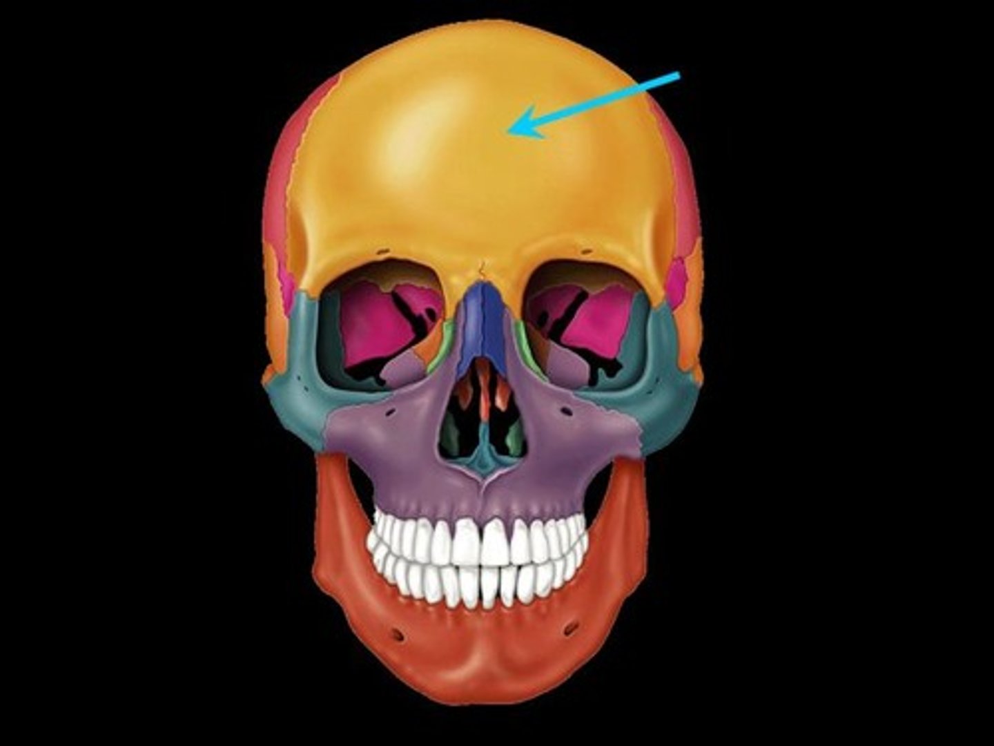
Frontal Bone
(1) Forms the forehead, superior part of the orbit, and the floor of the anterior cranial fossa
Supraorbital margin
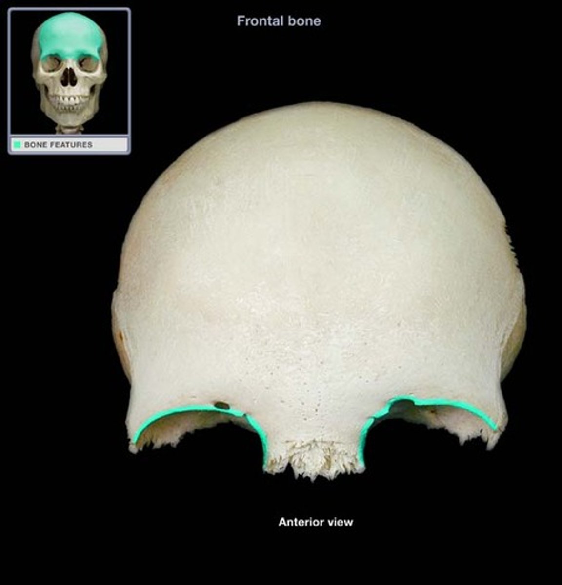
(Frontal)
Thick margin of the eye socket that lies beneath the eyebrows
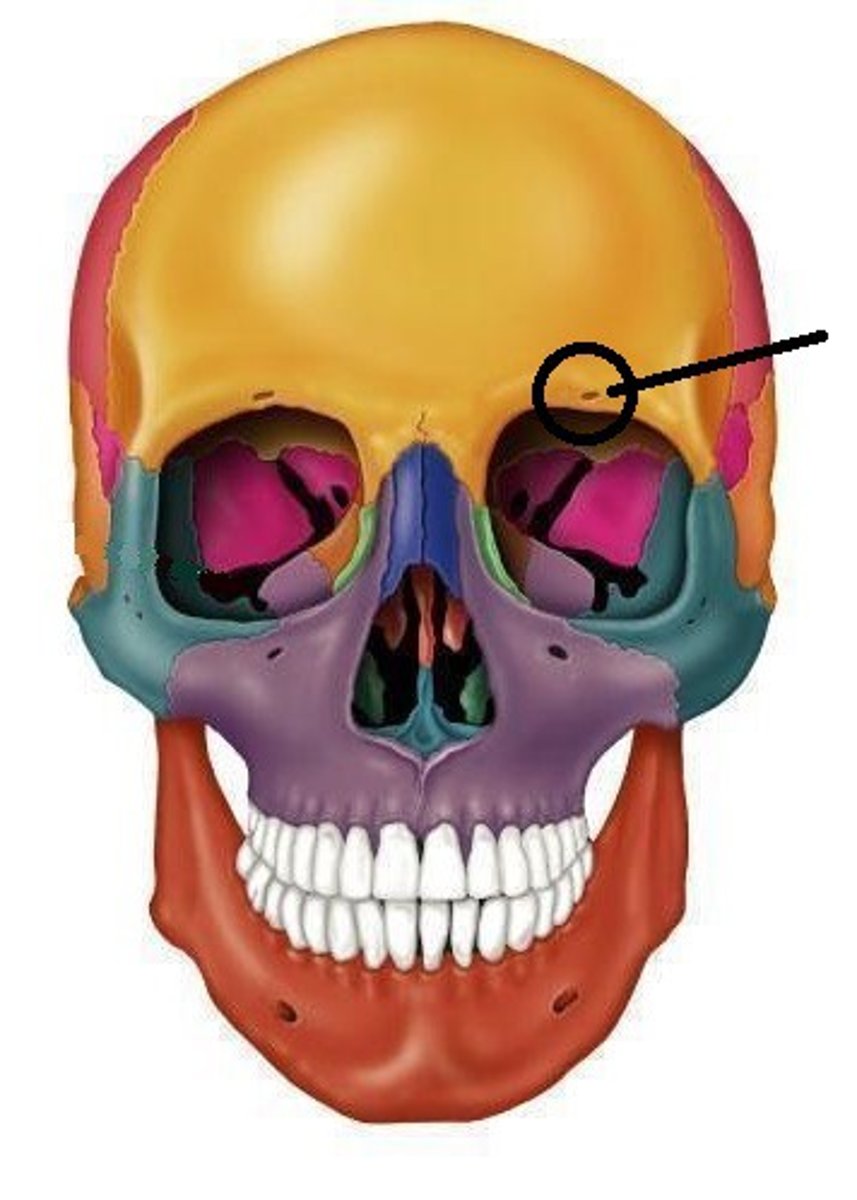
Supraorbital foramen (notch)
Opening above each orbit allowing blood vessels and nerves to pass
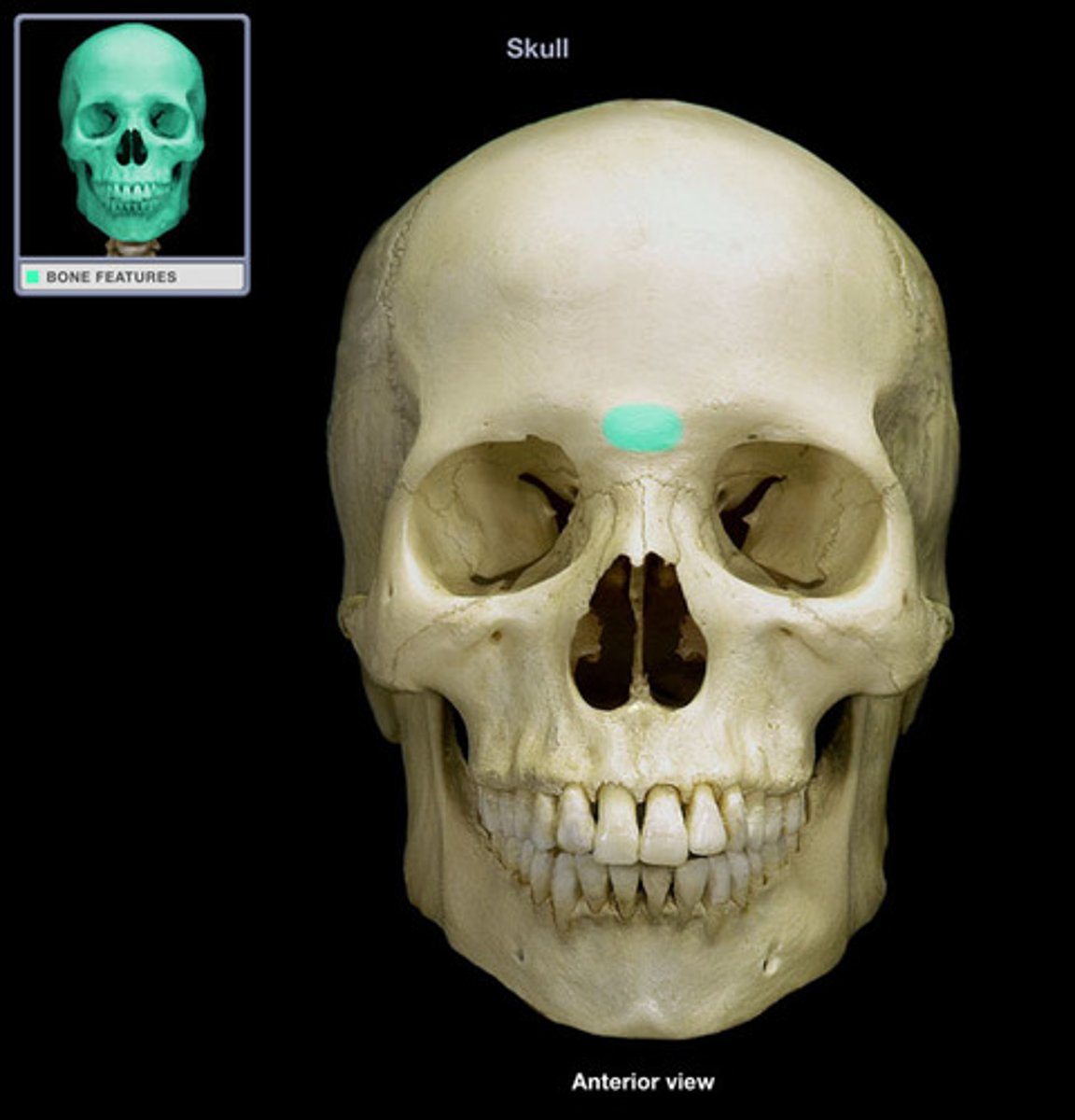
Glabella
Smooth area between the eyes
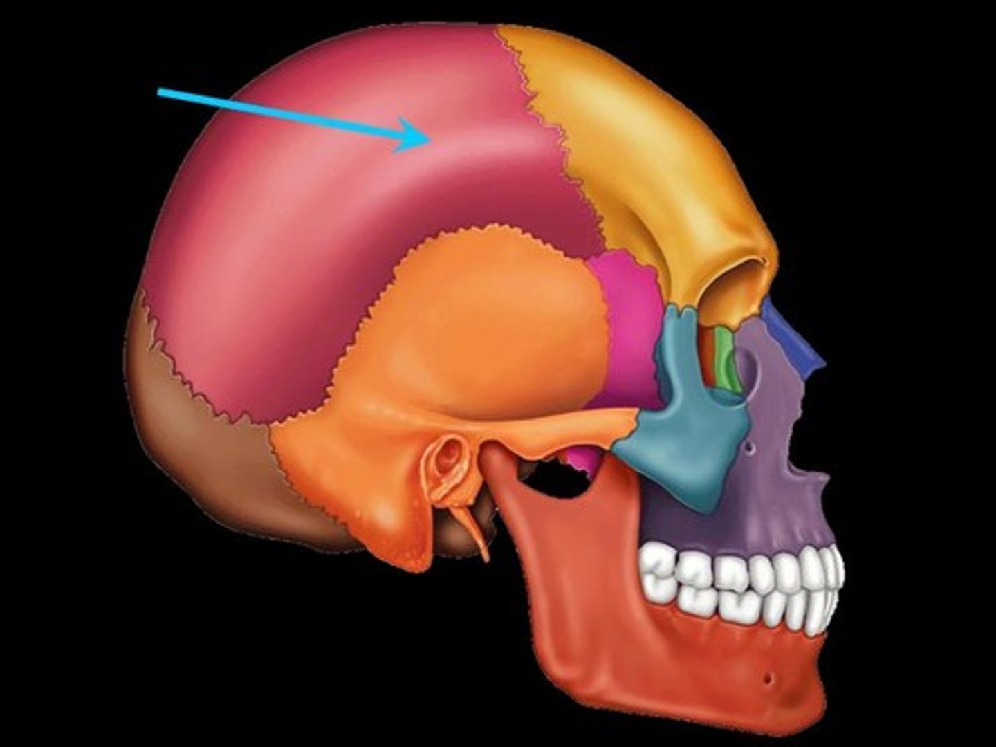
Parietal
(2)
Form the superior and lateral aspects of the skull
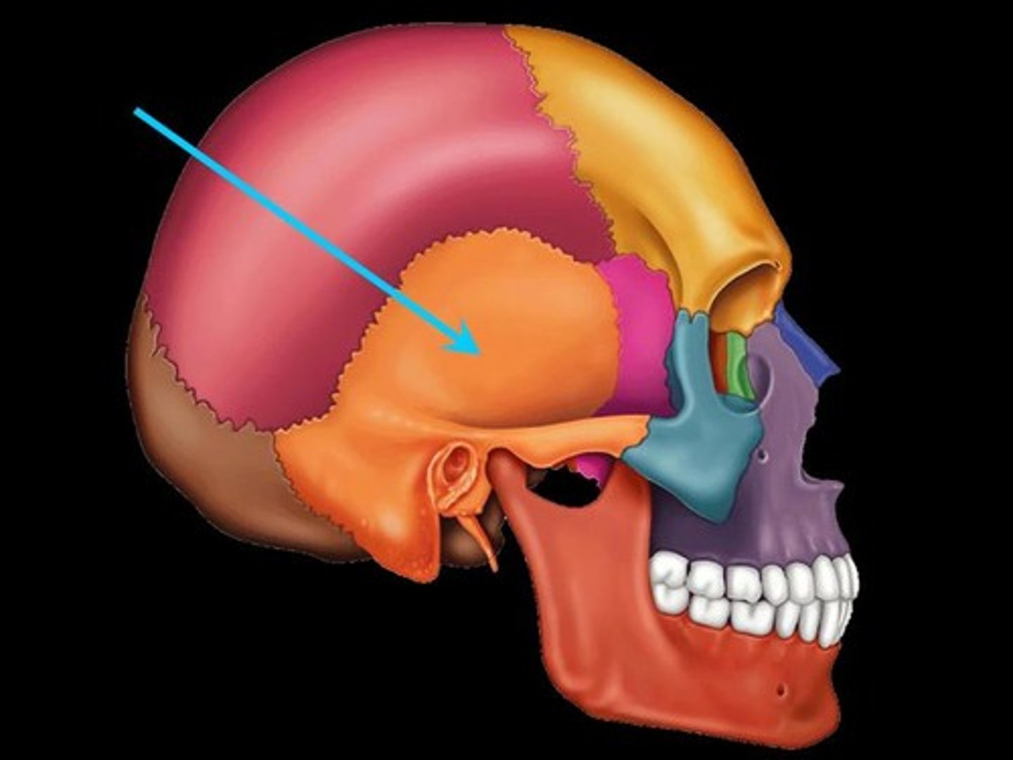
Temporal
(2)
Form the inferolateral aspects of the skull and contribute to the middle cranial fossa; each has squamous, tympanic, and petrous parts
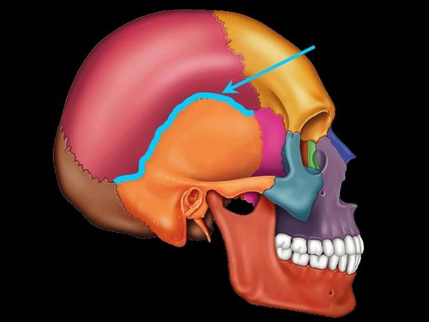
Squamous suture
(Temporal)
Located inferior to the squamous suture
Zygomatic process
(Temporal)
A bridge-like projection that articulates with the zygomatic bone to form the zygomatic arch
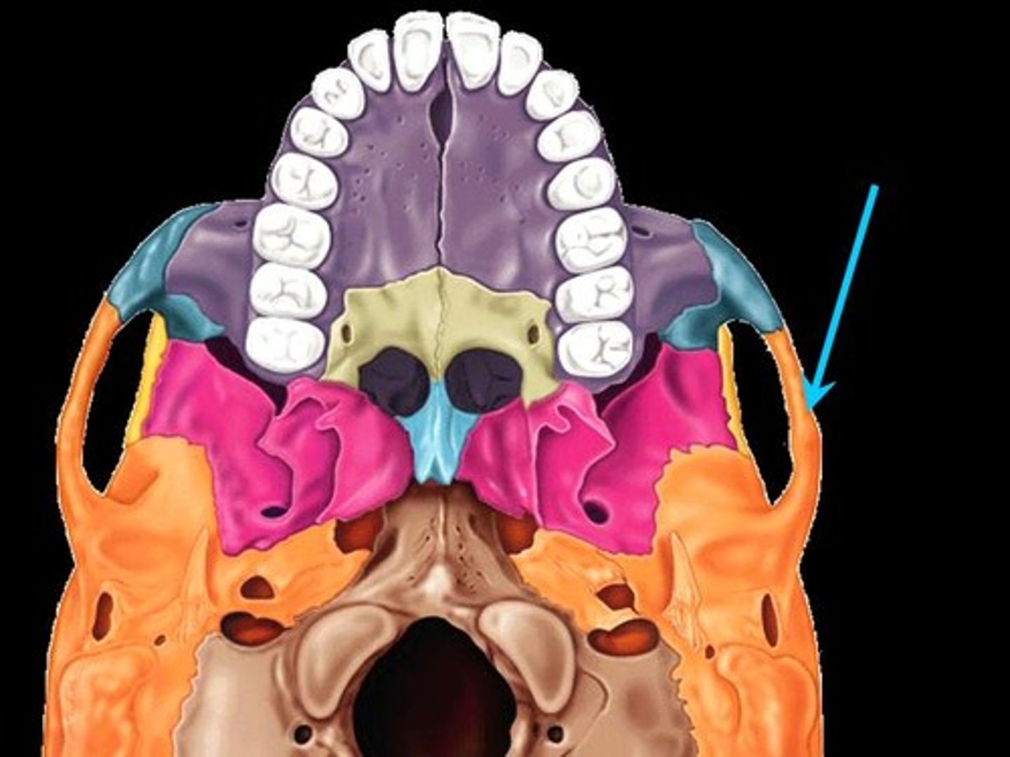
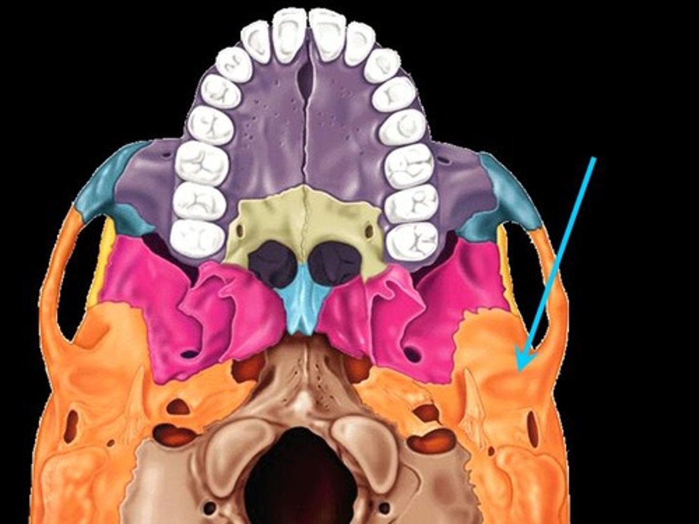
Mandibular fossa
(Temporal)
Located on the inferior surface of the zygomatic process; receives the condylar process of the mandible to form the temporomandibular joint
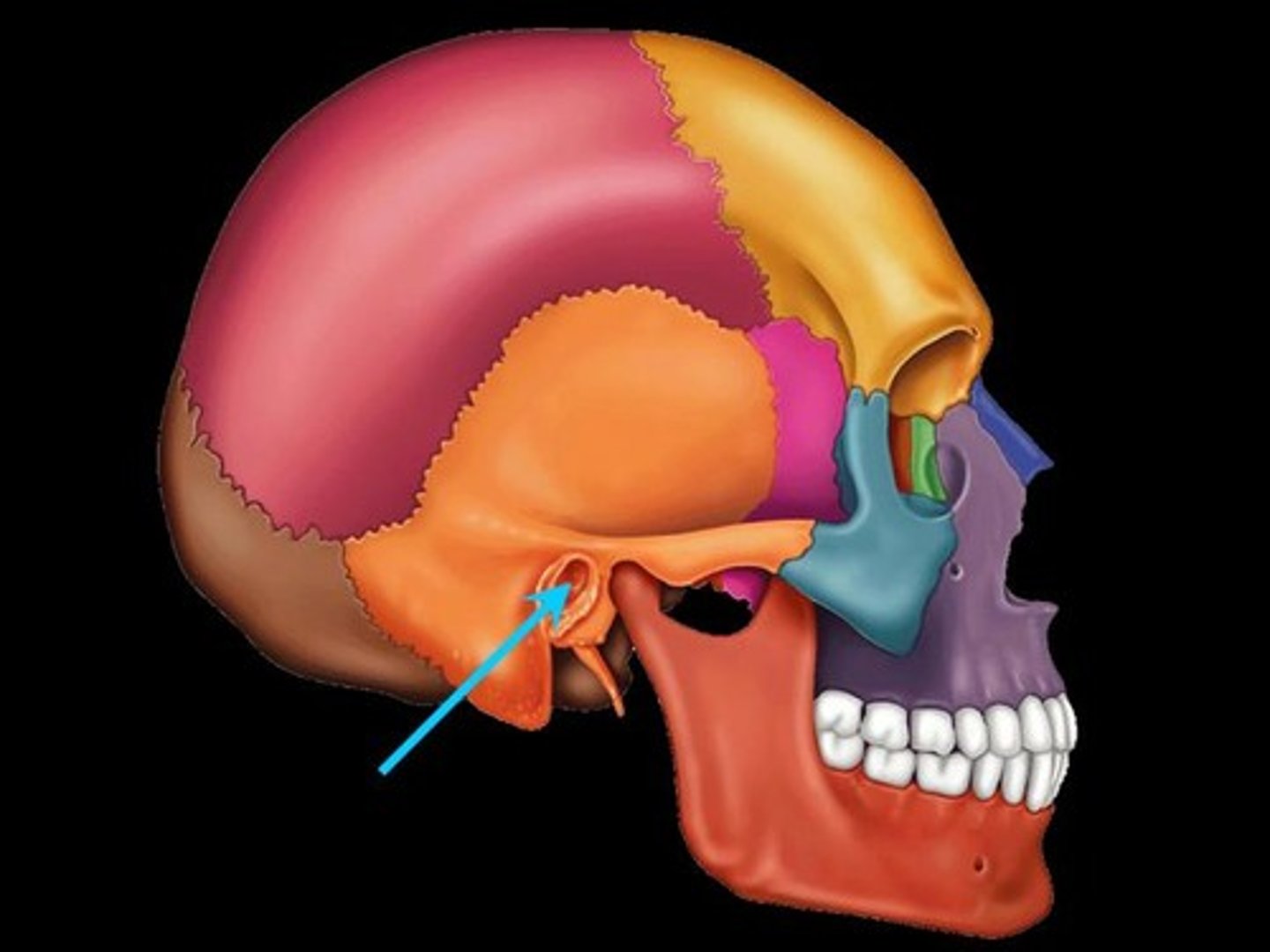
External acoustic meatus
(Temporal)
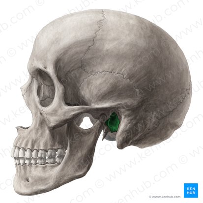
Canal leading to the middle ear and eardrum
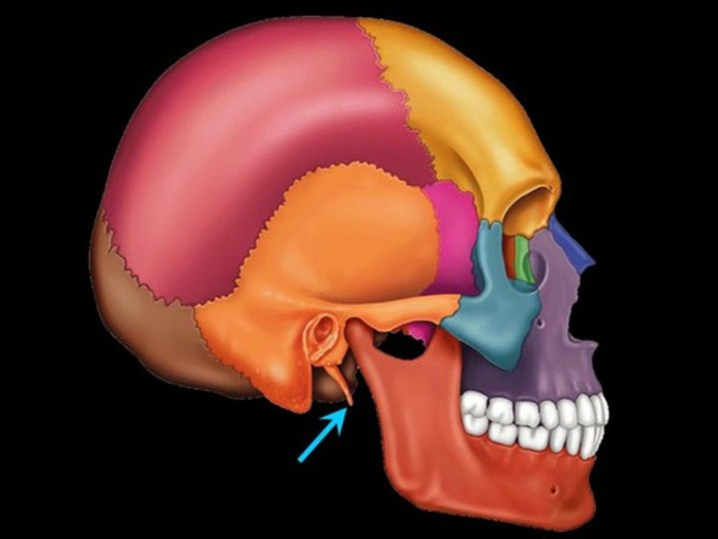
Styloid process
(Temporal)
Needlelike projection that serves as an attachment point for ligaments and muscles of the neck. (This process is often missing from demonstration skulls because it has broken off)
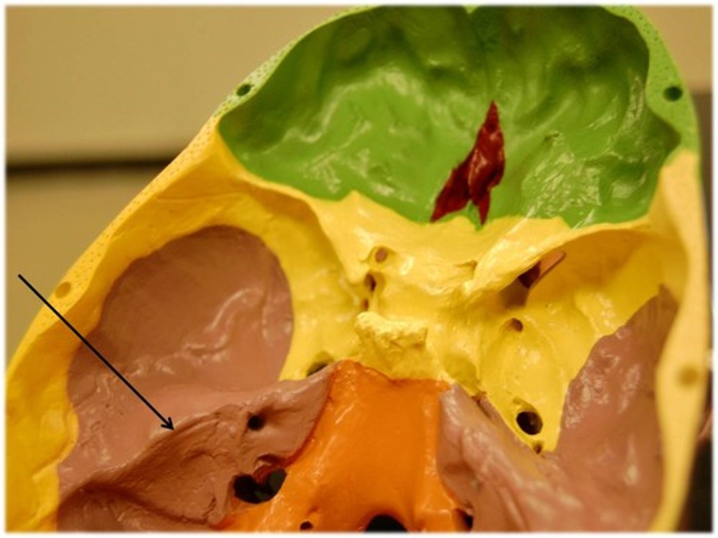
Petrous portion
(Temporal)
Forms a bony wedge between the sphenoid and occipital bones and contributes to the cranial base
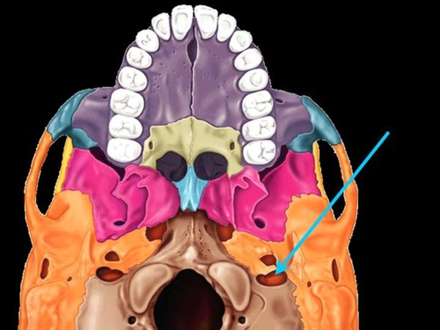
Jugular foramen
(Temporal)
Located where the petrous part of the temporal bone joins the occipital bone. Forms an opening which the internal jugular vein and cranial nerves IX, X, and XI pass
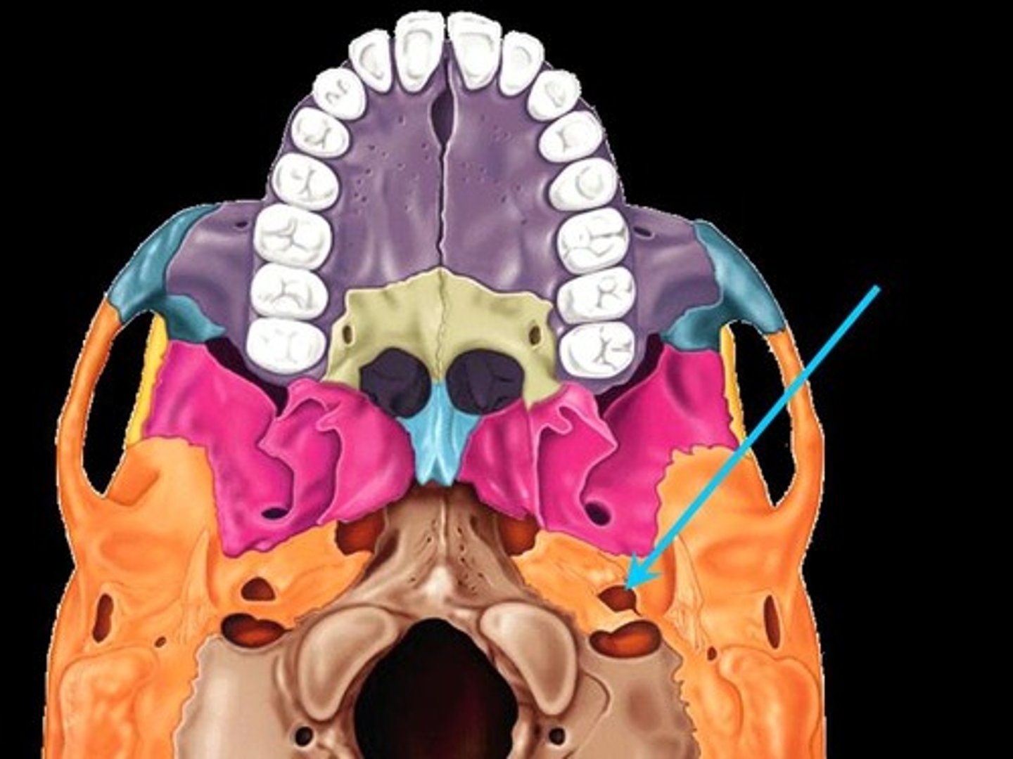
Carotid canal
(Temporal)
Opening through which the internal carotid artery passes into the cranial cavity
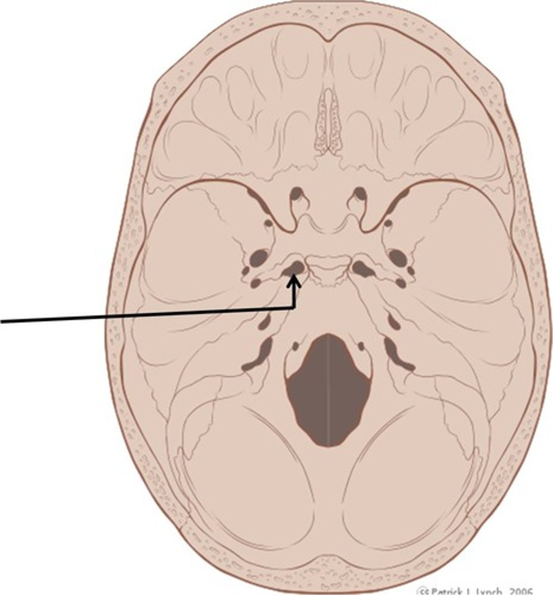
Foramen lacerum
(Temporal)
Almost completely closed by cartilage in the living person but forms a jagged opening in dried skulls
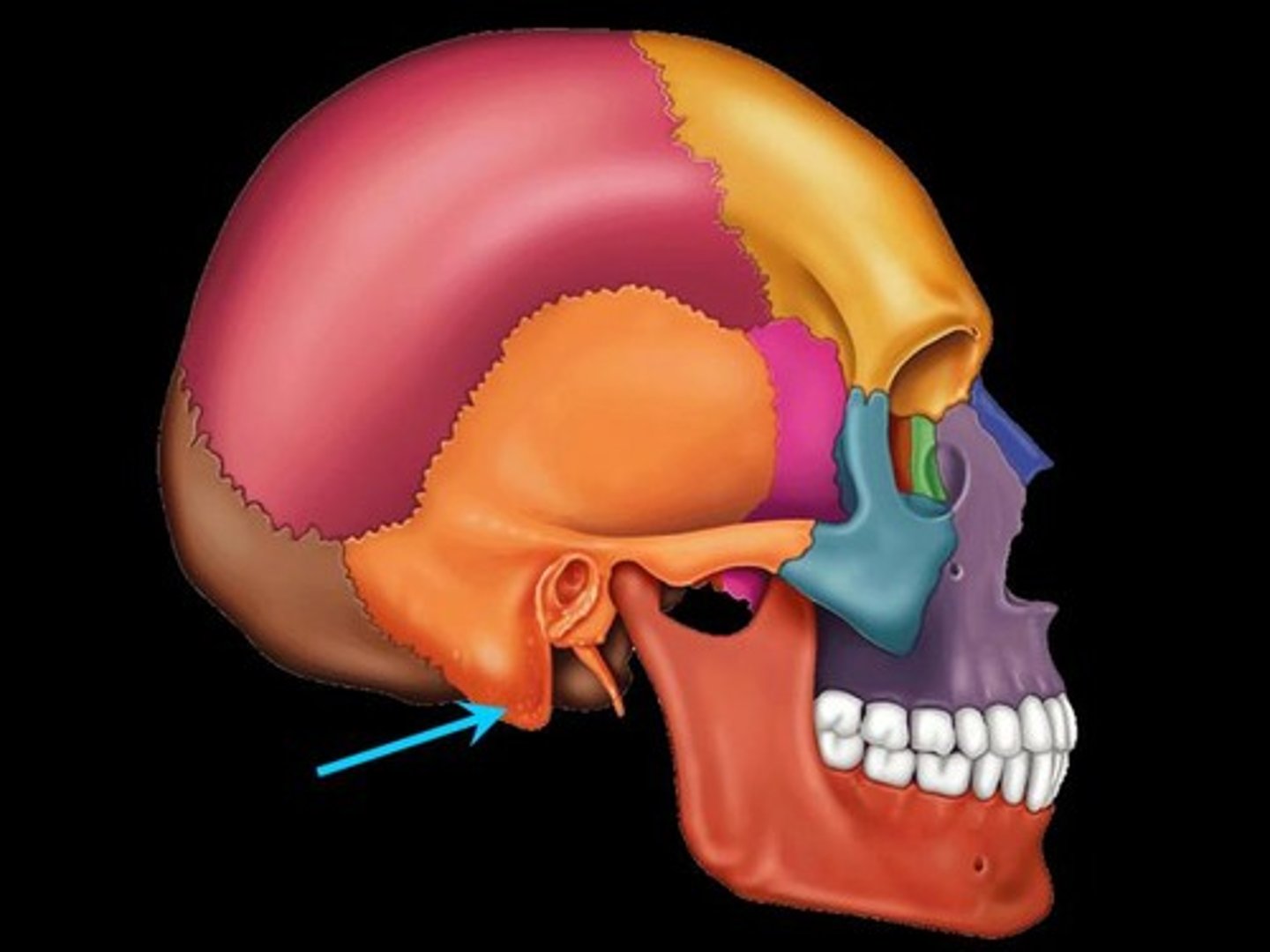
Mastoid process
(Temporal)
Located posterior to the external acoustic meatus; serves as an attachment point for neck muscles
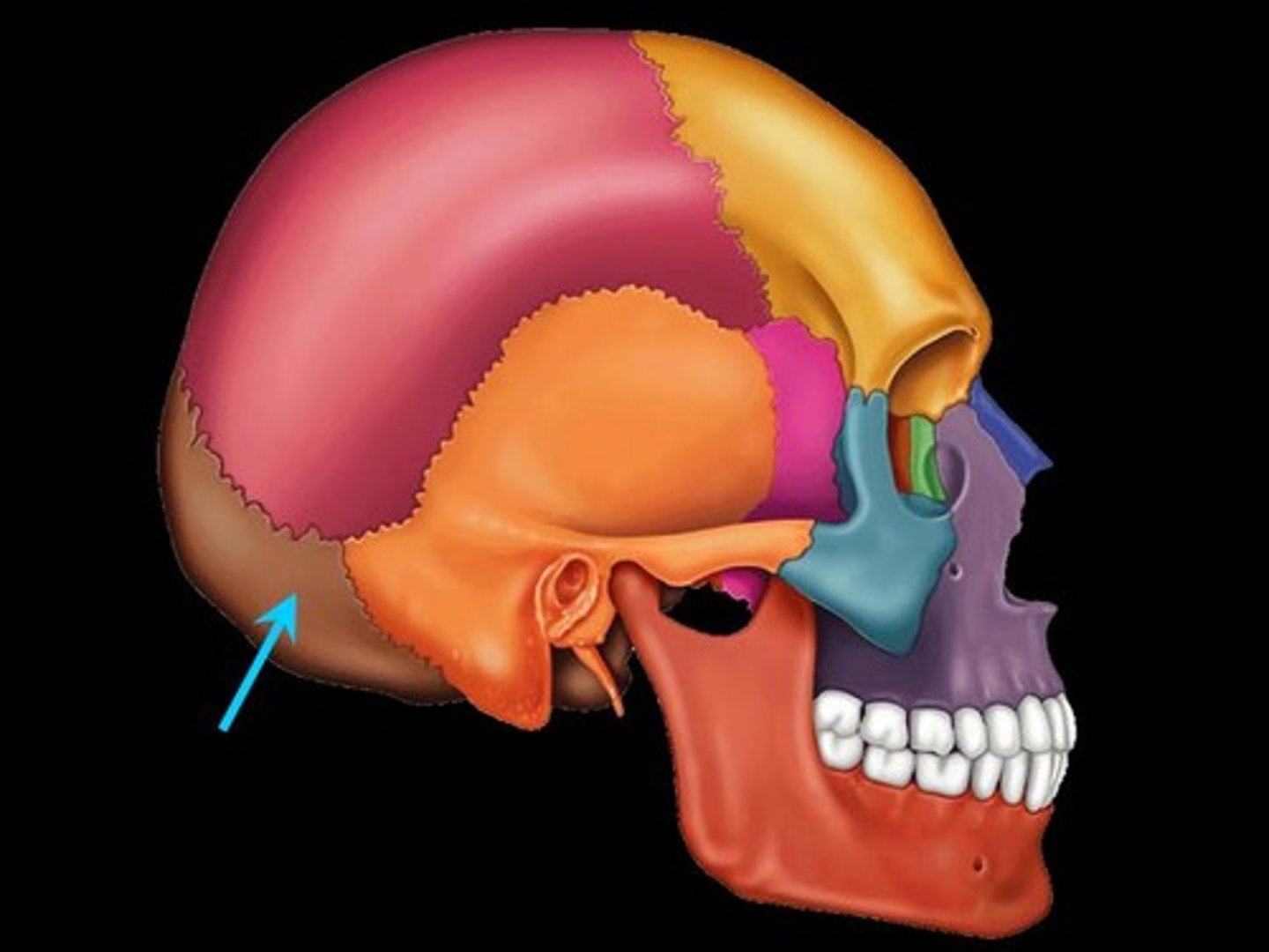
Occipital
Forms the posterior aspect and most of the base of the skull
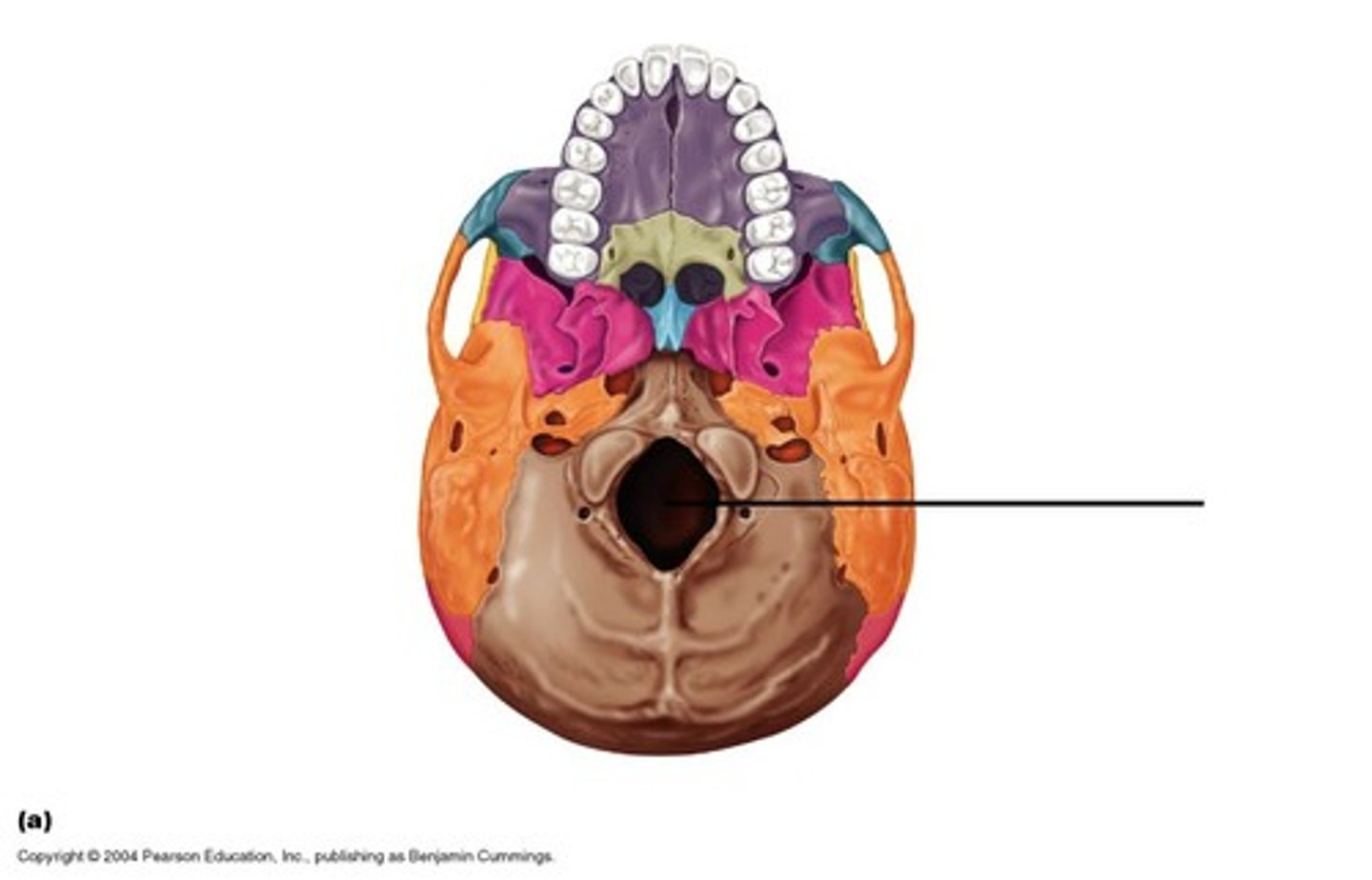
Foramen magnum
(Occipital)
Large opening in the base of the bone, which allows the spinal cord to join with the brain stem
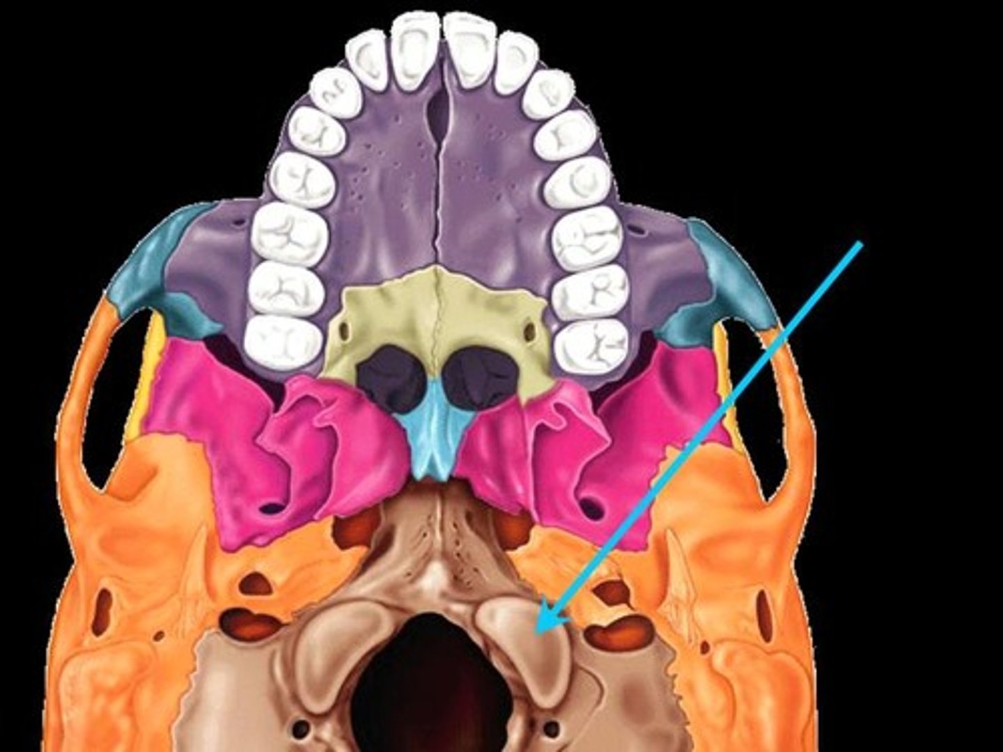
Occipital condyles
(Occipital)
Rounded projections lateral to the foramen magnum that articulate with the first cervical vertebra (atlas)
Hypoglossal canal
(Occipital)
Opening medial and superior to the occipital condyle through which cranial nerve XII passes
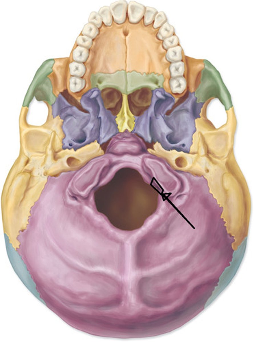
Sphenoid bone
Bat shaped bone that is described as the keystone bone of the cranium because it articulates with all other cranial bones
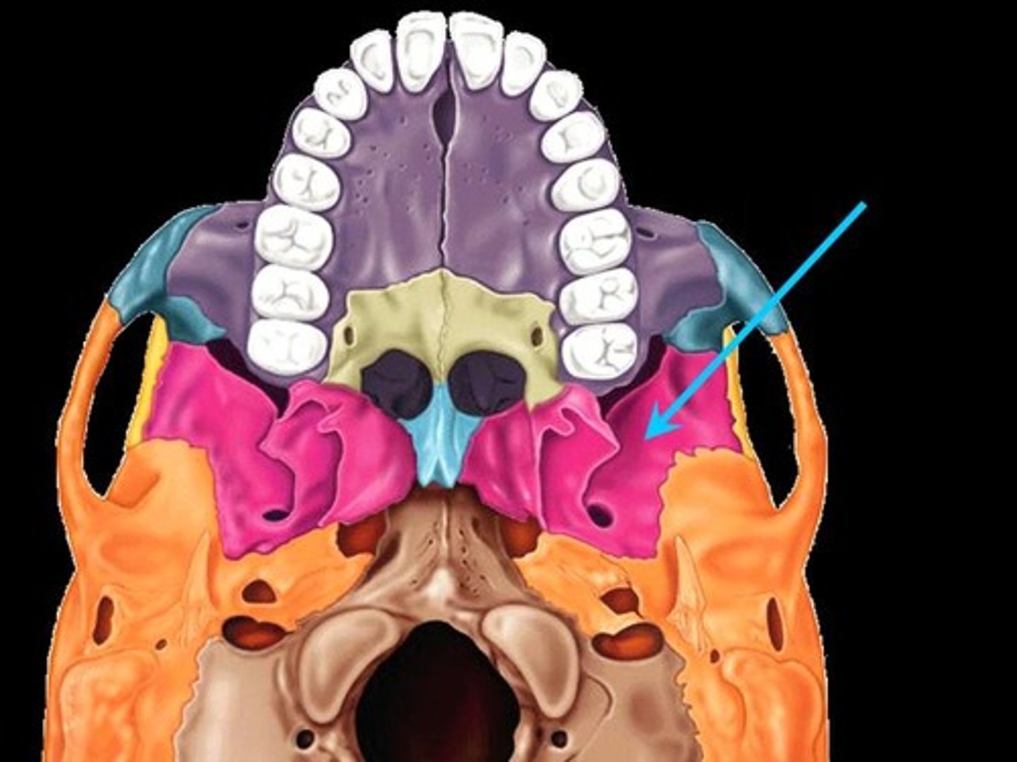
Greater wings
(Sphenoid)
Project laterally from the sphenoid body, forming parts of the middle cranial fossa and the orbits
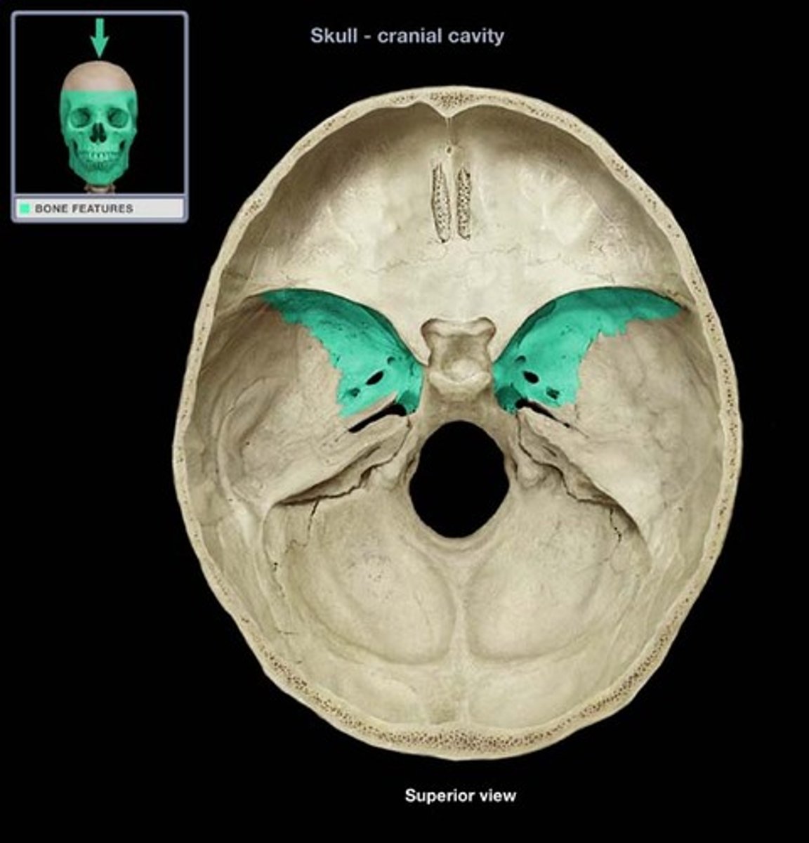
Pterygoid processes
(Sphenoid)
Project inferiorly from the greater wings; attachment site for the chewing muscles
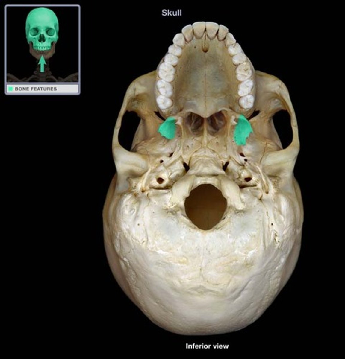
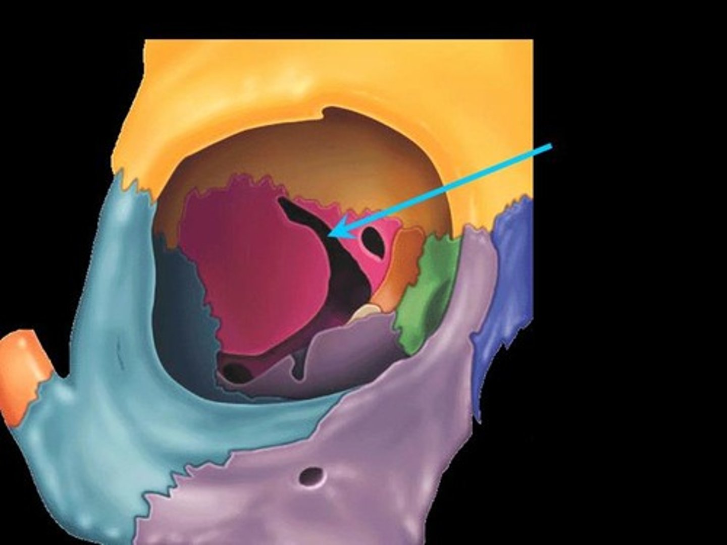
Superior orbital fissures
(Sphenoid)
Slits in the orbits providing passage of cranial nerves that control eye movements
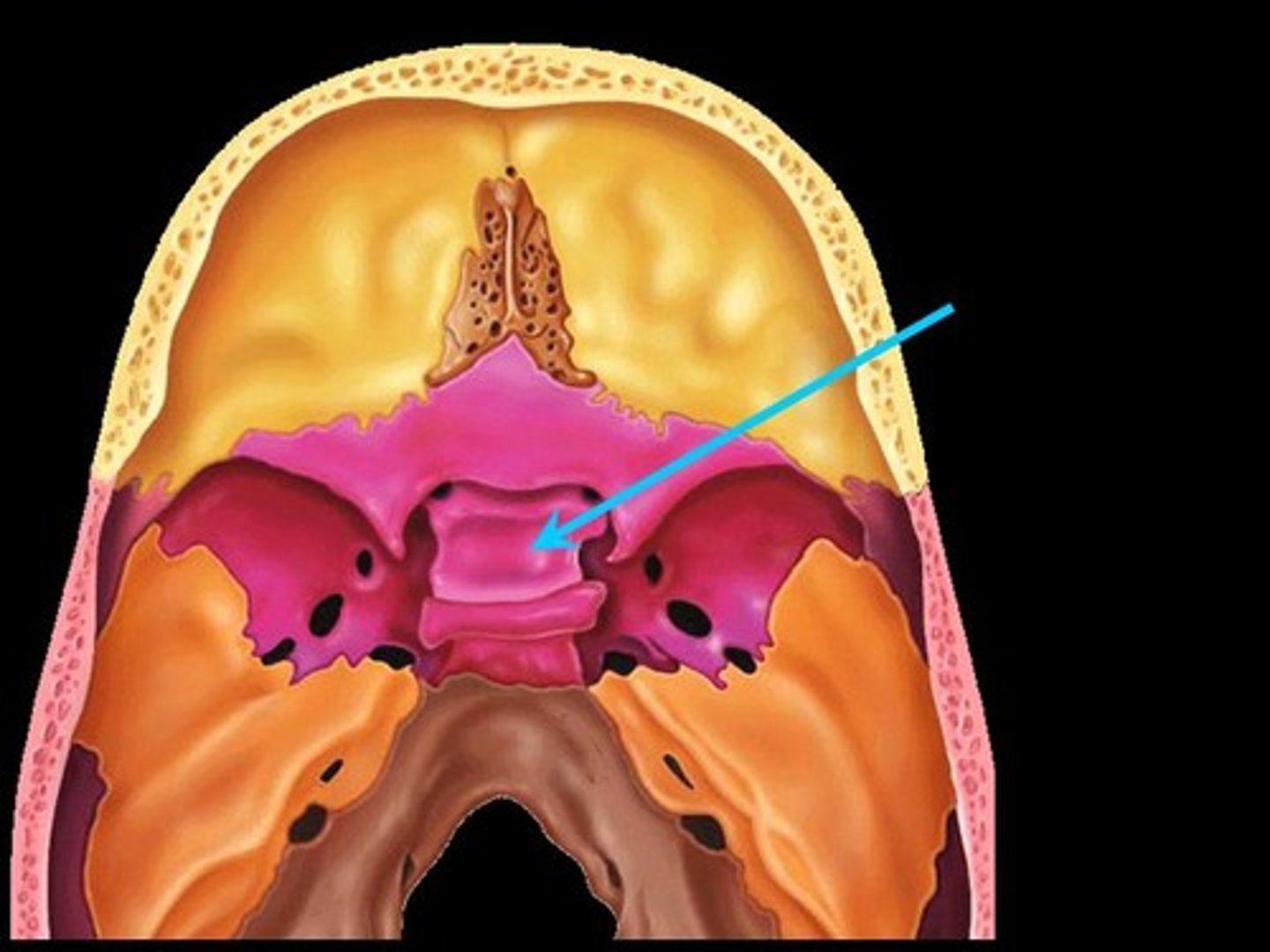
Sella turcica
(Sphenoid)
"Turkish saddle" located on the superior surface of the body; the seat of the saddle, called the hypophyseal fossa, holds the pituitary gland
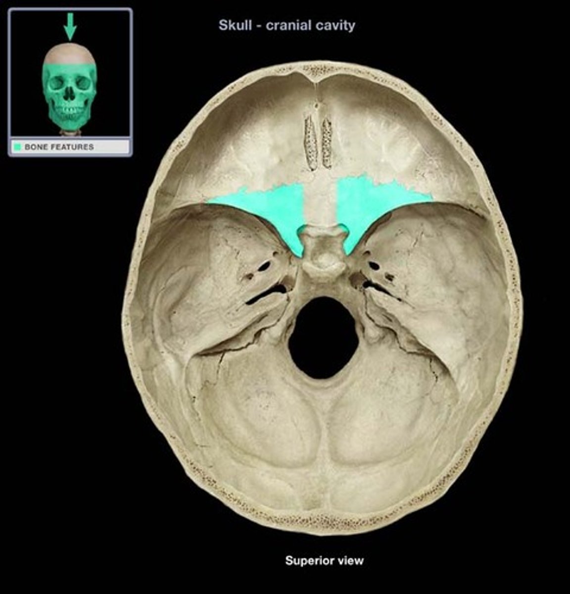
Lesser wings
(Sphenoid)
Form part of the floor of the anterior cranial fossa and part of the orbit
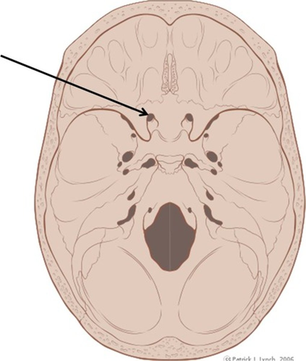
Optic canals
(Sphenoid)
Openings in the base of the lesser wings; cranial nerve II passes through to serve the eye
Foramen rotundum
(Sphenoid)
Openings located in the medial part of the greater wing; a branch of cranial nerve V passes through
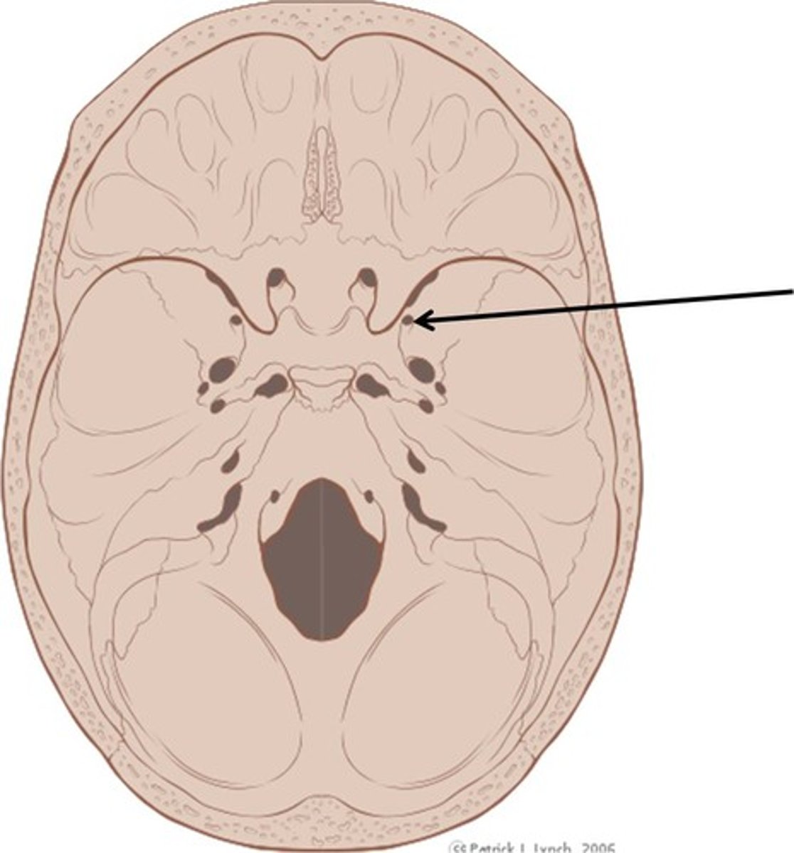
Foramen ovale
(Sphenoid)
Openings located posterolateral to the foramen rotundum; a branch of cranial nerve V passes through
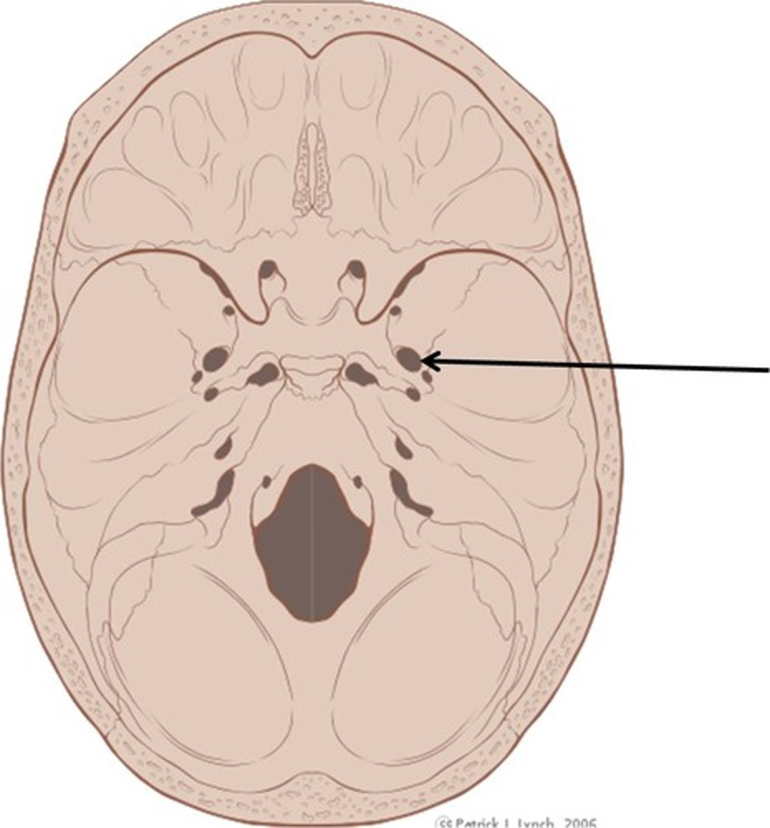
Foramen spinosum
(Sphenoid)
Openings located posterolateral to the foramen spinosum; provides passageway for the middle meningeal artery
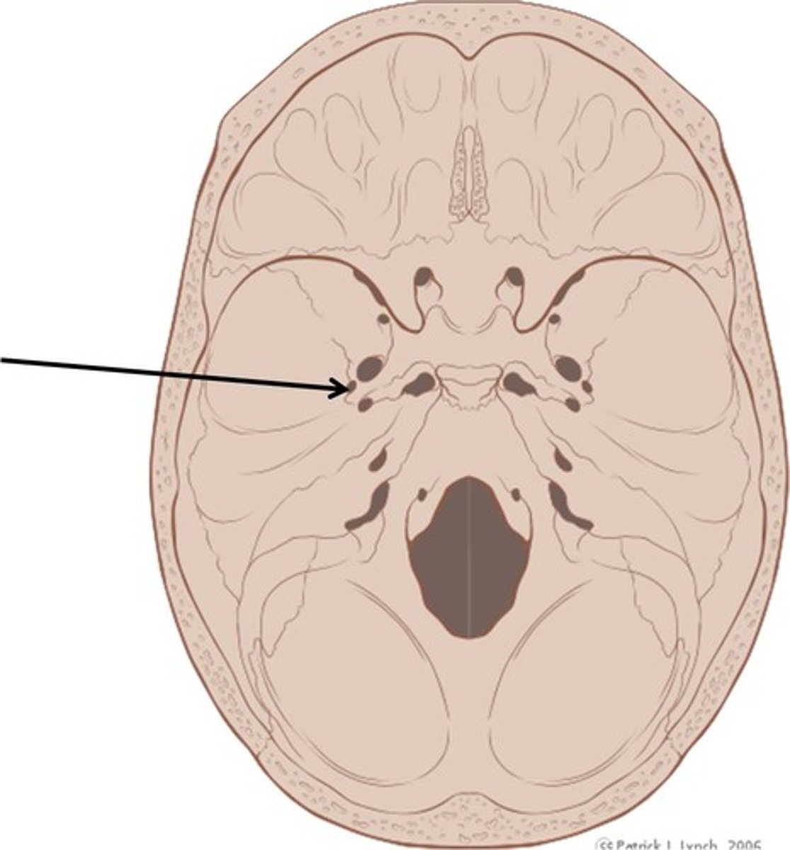
Ethmoid
Contributes to the anterior cranial fossa; forms part of the nasal septum and the nasal cavity; contributes to the medial wall of the orbit
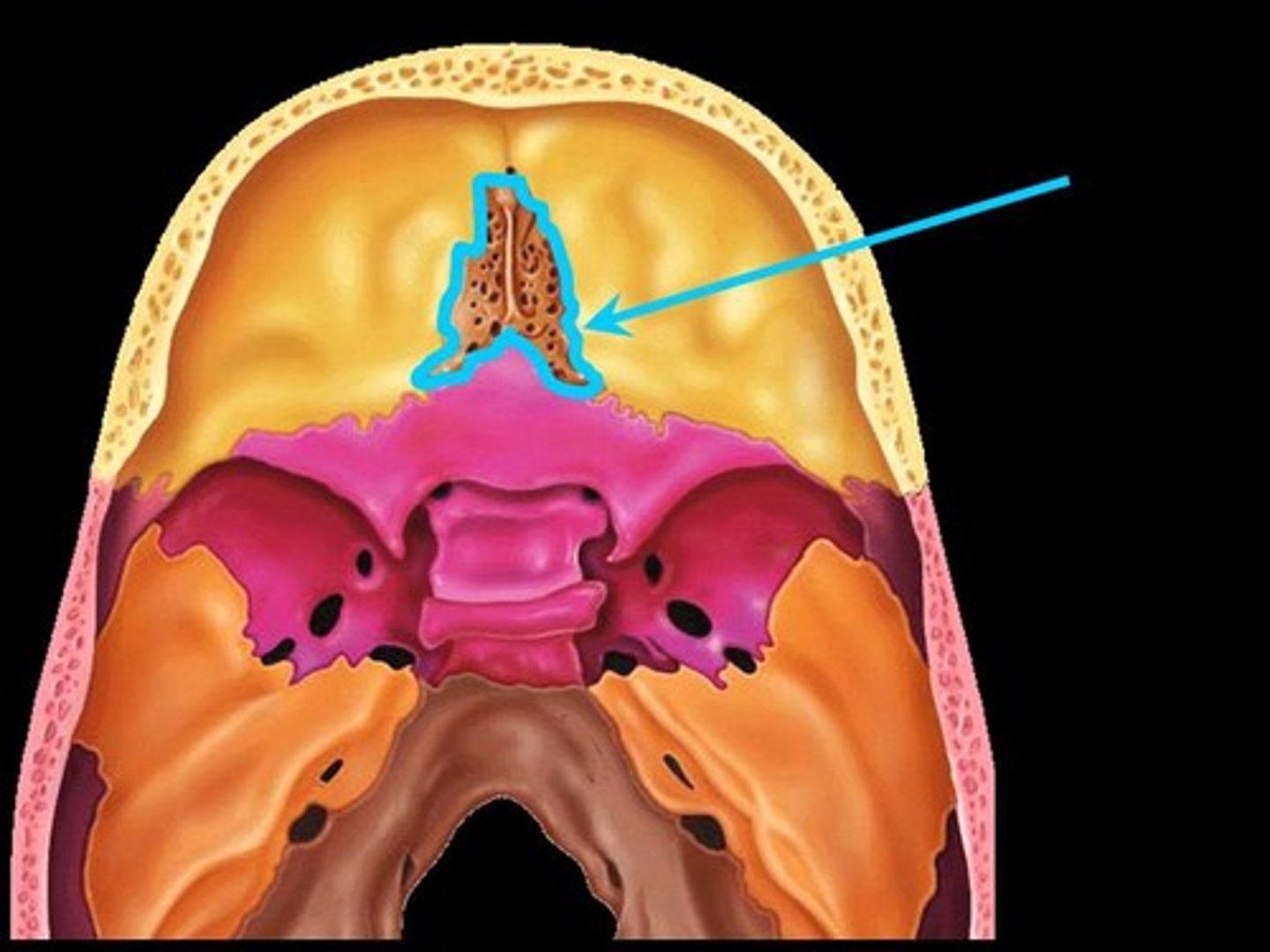
Crista galli
(Ethmoid)
"Rooster's comb"; a superior projection that attaches to the dura mater, helping to secure the brain within the skull
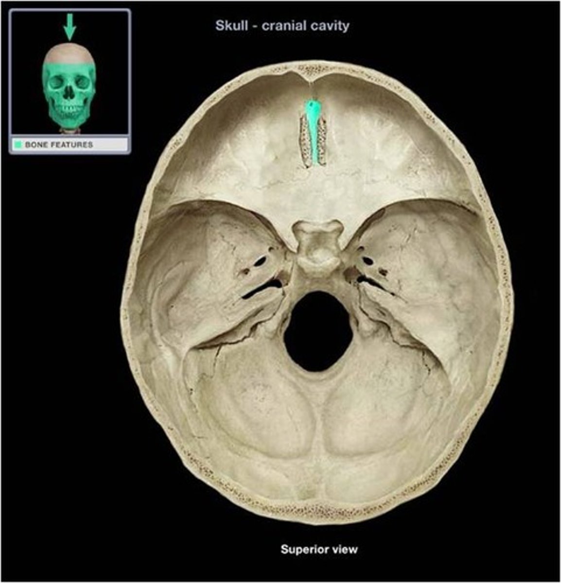
Cribriform plates
(Ethmoid)
Located lateral to the crista galli; from a portion of the roof of the nasal cavity and the floor of the anterior cranial fossa
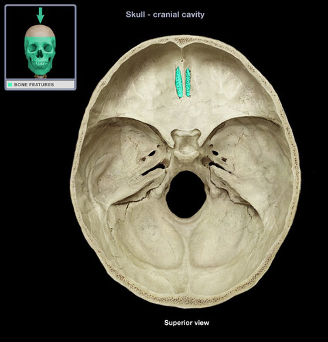
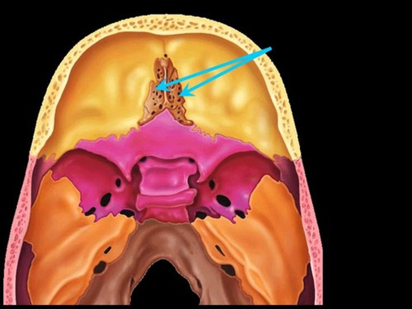
Cribriform foramina
(Ethmoid)
Tiny holes in the cribriform plates that allow for the passage of filaments of cranial nerve I
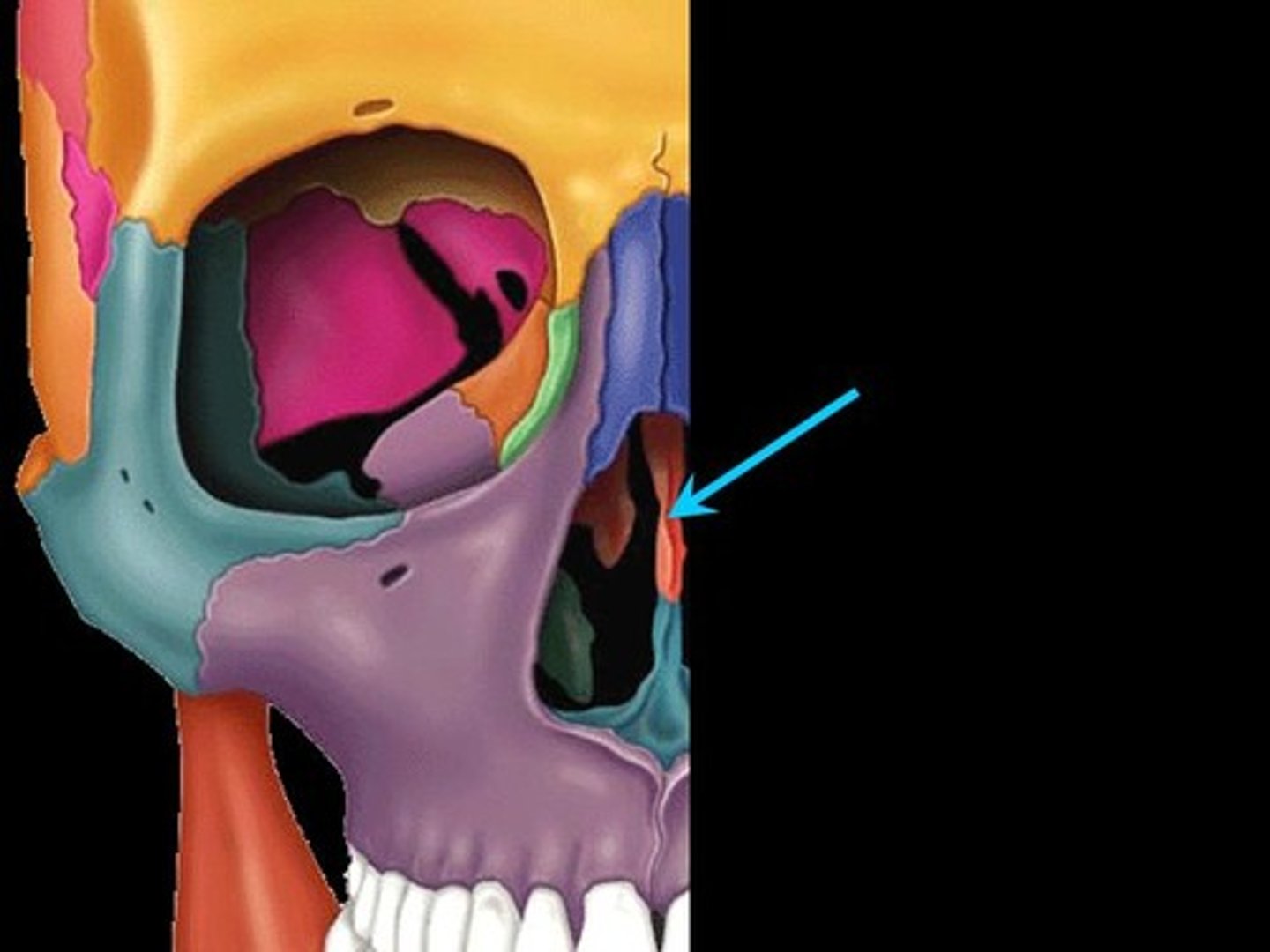
Perpendicular plate
(Ethmoid)
Inferior projection that forms the superior portion of the nasal septum
Orbital plates
(Ethmoid)
Lateral surface of the lateral masses that contribute to the medial wall of the orbits
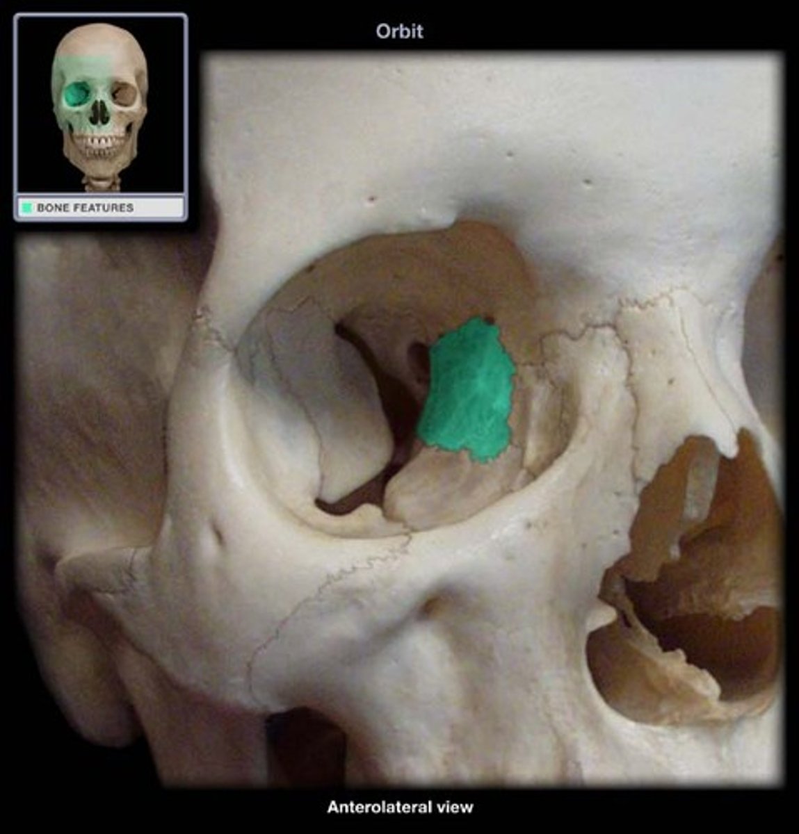
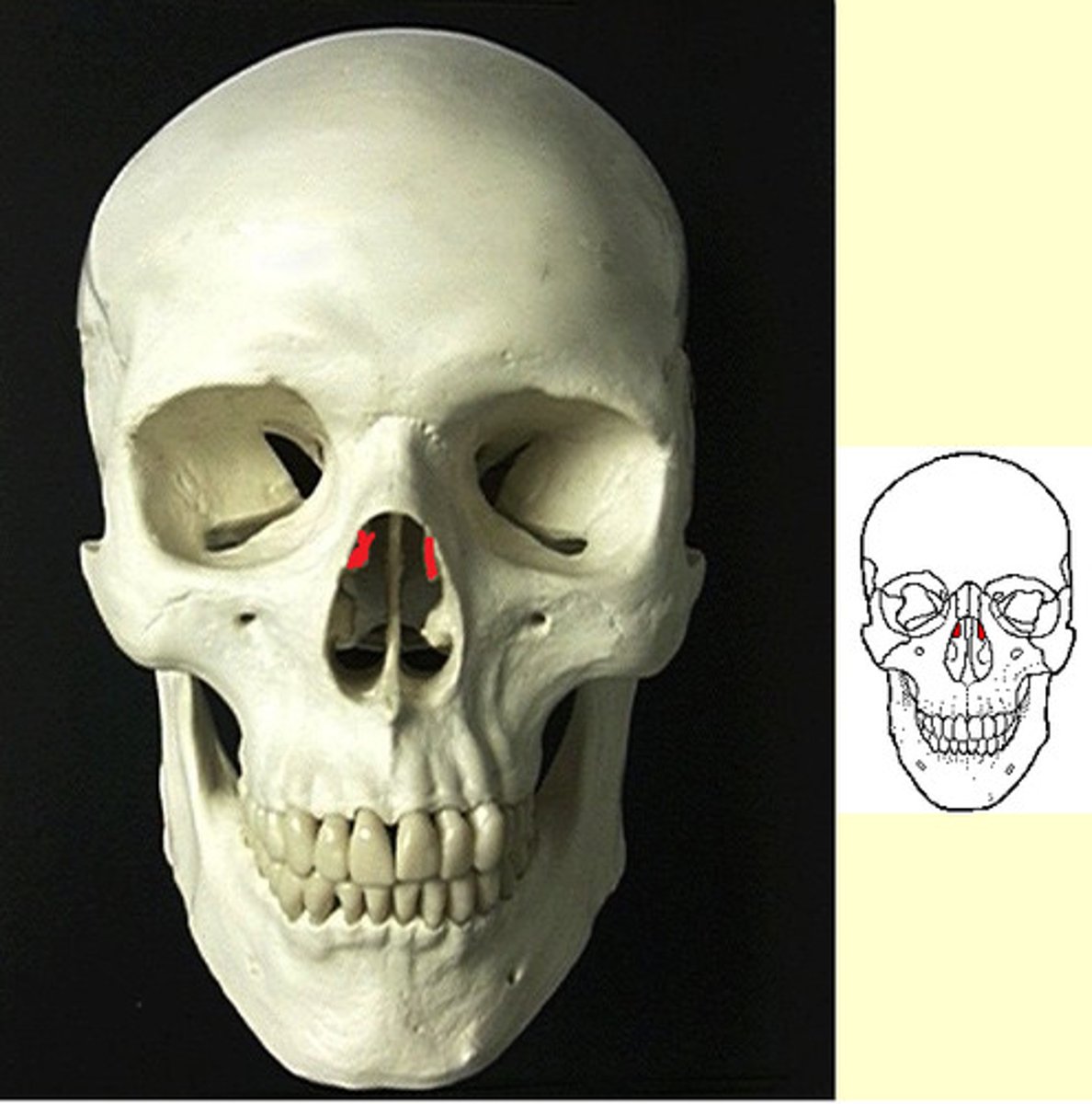
Superior and middle nasal conchae
(Ethmoid)
Extend medially from the lateral masses; act as turbinates to improve airflow through the nasal cavity
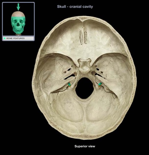
internal auditory meatus
external auditory meatus
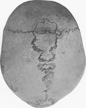
wormian bones