Week 2/3 - CT Instrumentation
 Call Kai
 Learn
Learn Practice Test
 Spaced Repetition
 Match
Match Flashcards
 Knowt Play
1/57

There's no tags or description
Looks like no tags are added yet.
Name | Mastery | Learn | Test | Matching | Spaced | Call with Kai | Chat |
|---|
No analytics yet
Send a link to your students to track their progress
58 Terms
Explain centrifugal (g) force
Acceleration of a rotating object relative to gravity, 9.81m/s2. Therefore an object rotating at accelerations of 10g means that the force is 10 x the force of gravity.
Explain the CT gantry
The large cylindrical machine that houses the x-ray tube, detectors and generators.
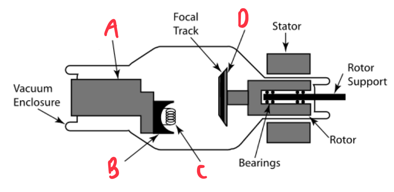
State these components of the x-ray tube
A - Cathode
B - Focussing Cup
C - Filament
D - Rotating Anode
Explain the role of the filament
The filament is heated and releases electrons via thermoionic emission, creating an electrode cloud that accelerates electrons towards the anode.
Explain why the anode is rotating
The rotation of the anode disperses heat
Explain the role of the anode
The electrons hit the anode made of tungsten metal which produces x-rays.
Explain the orientation of the x-ray tube in relation to the patient and the x-ray beam produces
The anode-cathode axis is orientated parallel to the long-axis of the patient and is perpendicular to the direction of the x-ray beam produced.
State the 2 requirements of an x-ray tube for CT
Produce high intensity x-rays
Ensure rapid heat dissipation
Differentiate between x-ray intensity and quality
Intensity - the number of x-ray photons
Quality - the energy of the x-ray photons
Explain the anode heel effect
Radiation intensity is greater at the cathode than at the anode due to the absorption of x-rays at the anode heel.
Explain how the x-ray target angle alters the anode heel effect
Small x-ray target angles will increase the amount of x-rays absorbed at the anode heel.
Explain how high x-ray intensity is achieved with the x-ray tube
High x-ray tube current (200-800 mA) increases number of electrons produced
Large focal spot size to prevent anode melting
High voltage (>120 kV) applied across the x-ray tube from the generator
State the formula to calculate power of the x-ray tube with units
Power (kW) = voltage (kV) x current (A)
State the range of power that can be produced from the latest CT scanners
20-100 kW
Explain focal spot size
The specific area of the anode bombarded by electrodes
Differentiate between the advantages and disadvantages of small vs large focal spot sizes
Smaller - better resolution however greater and more concentrated heat deposition that reduces the amount of x-ray power that can be used
Larger - poorer resolution however lower and less concentrated heat deposition that allows for higher x-ray power use
Explain why the x-ray beam intensity should not vary by more than 1%
It will form beam hardening artefacts and falsely change the value of the linear attenuation coefficient as the transmitted intensity will be higher than it should due to the absorption of lower energy photons, increasing average beam energy.
Explain how rapid heat dissipation is achieved with the x-ray tube
The anode has a large diameter and thickness that rotates at 10,000 rpm, switching the position of the focal spot as the anode rotates.
State how the use of a rotating anode alters detector width and number of projections
Halves detector width
Doubles no. of projections
Explain how older (first and second-generation) x-ray tubes achieved heat dissipation
Stationary anodes that were oil-cooled.
Explain heat units (HU)
A value that represents the amount of thermal energy the CT’s x-ray tube can safely endure.
State the range of heat capacity of a CT machine in HU
3-5 MHU (mega/million heat units)
State the formula for HU
HU = voltage (kVp) x current (mA) x time (sec)
Explain why voltage, current and time cannot be decreased to reduce heat produced
This will create a noisy image
Explain the modifications that can be made for pediatric imaging
Increased current (mA) will reduce the time required for the child to sit still during the scan
State the type of generator within the CT gantry
High-voltage, high-frequency, 3 phase generator
State and explain the components of the generator
Rectifier - converts AC to DC
Step-up transformer - amplifies voltage to be applied across the x-ray tube
Step-down transformer - lowers voltage to heat the filament
Explain why high kV generators are used in CT
To minimise photoelectric absorption and maximise Compton scattering to reduce noise, improve image quality and reduce patient dose.
State normal frequency (pulses/sec) and high frequency in CT (pulses/sec)
Normal - 50 pulses/sec
High - 30,000 pulses/sec
State whether CT scanners emit x-rays continuously or in pulses
Majority - continuously
Some, eg. Dual energy - in pulses
Explain reasons for energy loss in CT
Heat loss
Filtration
Collimation
State and explain the type of filter used in CT
A bowtie filter that removes low-energy x-rays to harden the beam, make it as mono-energetic as possible and reduce patient dose. Additionally its shape equalises the x-ray beams intensity in which it is thinner in the centre as there is more attenuation by the patient whereas it is thicker on the edges as there is less attenuation by the patient.
State and explain the types of metal that make up the filter
Copper - located towards x-ray tube in which it filters medium-energy photons and produces characteristic x-rays
Aluminium - locates towards the patient and filters low-energy photons as well as the characteristic x-rays to reduce patient dose
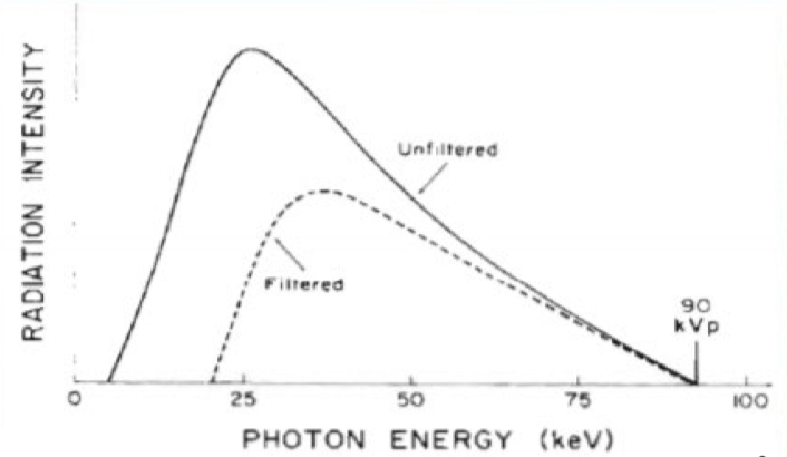
Explain filtration with reference to this graph
Filtration reduces radiation beam intensity (no. Of photons), specifically filtering lower-energy photons in which the average photon energy is shifted to the right, therefore greater.
State and explain the 2 types of collimators
Pre-patient - defines the slice thickness
Post-patient - reduces scatter reaching the detector
State the 2 types of CT detectors currently used
Solid state scintillation detectors (most common)
Gas-filled ionisation chambers (older and less common)
Explain dynamic range
The range of photon energies that the detector can pick-up/differentiate. High dynamic range means the detector can differentiate between very small differences in photon energies, therefore in attenuation.
Explain why traditional scintillation/PMT detectors are no longer used in CT
Geometric efficiency is limited as PMT’s cannot be closely packed together due to their bulkiness.
State the types of scintillation materials that were used
Na(TI)
BGO
CaF2
Briefly explain how gas-filled detectors work
Long narrow chambers filled with xenon gas and separated by metal electrodes. X-rays interact with gas and cause ionisation reactions in which ions are attracted to their respective electrodes, forming an electric pulse.
Explain why xenon gas is used in gas-filled detectors
Gas is low density which decreases the chance of interaction, therefore xenon gas with a high atomic number increases the chance of interaction.
Why are gas-filled detectors also considered intrinsically directional
The metal electrodes converge towards the x-ray tube, increases scatter rejection.
Explain why solid-state scintillation detectors are preferred over traditional scintillation detectors
Scintillation crystal is couples to thin photodiodes that convert light into electric signal rather than bulky PMTs, increasing geometric efficiency.
State the type of scintillation materials typically used in solid-state detectors
CsI
CdWO4
Yttrium
Gadolinium
Explain cross-talk between detectors
When visible light is scattered and detected by neighbouring detectors.
Explain how cross-talk is reduced
By placing a light absorbing material between detectors.
Briefly explain how photon counting detectors work
Contain a semiconductor material that directly converts photons in electric signals rather than light, then electric signals. Therefore the detectors counts each individual photon.
Briefly explain first-generation CT scanners
A rotate-translate type systems in which a pencil x-ray beam was used with one detector that were fixed in a relative position. To acquire a projection, the tube and detector were translated laterally to acquire raysums over a series of positions. The gantry then rotates by a small angle and another projection is acquired.
Explain the trade-offs of first-generation CT scanners
Very time consuming (4-5 mins per projection) therefore could only image static areas of the body, eg. Brain
Low efficiency due to only a very small fraction of the generated x-ray power being used after filtration and collimation to produce a pencil beam
Excellent scatter rejection due to use of single-detector
Briefly explain second-generation CT scanners
This involves the same rotate-translate system as first-generation scanners however a transition to the use of a small fan beam and an arc of 30 detectors.
Explain the trade-offs of second-generation CT scanners
Fan beam means x-ray tube output is more efficient
Reduced scan time due to fan beam and multiple detectors reducing the number of rotations required
Greater number of detectors increases susceptibility to scatter, artefacts and reduced image quality.
Briefly explain third-generation CT scanners
Transition to rotate-rotate type systems in which the fan beam was widened to cover the entire cross-section of the patient with an arc of 500-900 detectors. Therefore translation no longer required and only rotation required to acquire each projection.
Explain the trade-offs of third-generation CT scanners
Reference detection in which the detectors on the ends of the arc detect rays that haven’t passed through the patient, therefore continuously measure I0 for calibration.
The elimination of translational motion has dramatically reduced scan times
X-ray tube and detector are still fixed in a relative position, therefore an anti-scatter grid can be used to reduce side scatter
Due to increased number of detectors, complexities with the number of cables required and their requirement to extend and retract during gantry rotation.
Briefly explain fourth-generation CT scanners
Transition to rotate-stationary type system in which the x-ray tube is mounted and rotates between the patient and a ring of thousands of stationary detectors that completely encircle the patient.
Explain the trade-offs of fourth-generation CT scanners
Stationary detectors eliminates cabling issues
Each detector can measure I0, not a few of them.
Ring of detectors means that anti-scatter grids cannot be used
Nutation occurs in which the detectors behind the x-ray tube must wobble/tilt out of the way for the rotating source.
Image reconstruction can only occur after entire scan is complete, not after each projection as detectors are continuously being exposed.
Briefly explain fifth-generation CT scanners
Transition to stationary-stationary type systems in which all motion is eliminated. An electron beam is magnetically steered around the tungsten anode ring below the patient, producing fan beams that are detected by an arc of detectors above the patient.
Explain the trade-offs of fifth-generation CT scanners
Extremely fast acquisition times that allow for cardiac imaging without motion blurring
No rotation within the gantry
Extremely expensive
State what sixth and seventh-generation CT scanners are also named
6th - helical CT
7th - multi-slice CT