Eye Anatomy 8- 24 - 26
 Call Kai
 Learn
Learn Practice Test
 Spaced Repetition
 Match
Match Flashcards
 Knowt Play
1/44

There's no tags or description
Looks like no tags are added yet.
Name | Mastery | Learn | Test | Matching | Spaced | Call with Kai | Chat |
|---|
No analytics yet
Send a link to your students to track their progress
45 Terms
Exophthalmos
Protrusion of the eye
Cornea
Covers the iris and the pupil.
Conjunctiva
Three different types:
1.Palpebral
2.Bulbar
3.Fornix
Palpebral conjunctiva
Lines the inner surface of the eyelids

Bulbar conjucriva
Covers anterior portion of the sclera

Fornix conjunctiva
Flexible fold connecting Palpebral and bulbar region.

Limbus
Junction between cornea and sclera. Conjunctiva does not cross cornea because that could impair vision.
Osteogenesis imperfecta
Causes blue color of sclera

Arcus senile
Can come w/old age - this can happen younger if diet is bad (fatty)

Wilsons diagnosis
Too much copper in the body

Sphincter pupillae
Circular muscle around the pupil that causes miosis (construction). Involved in ANS of parasympathetic region.
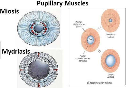
Dilator pupillae
Muscle causing pupillary mydriasis ( dilation). Crucial in the ANS of sympathetic region,

Perrla
Pupils equal round and reactive to light and accommodations
Canthus
There is the medial and lateral - they are the angles of the eye
Superior and inferior lacrimal papillae
Raised structure that contains the puncta, which is where tears drain.
Palpebrae - what are the 5 layers?
Eyelids
Skin
Subcutaneous tissue
Muscles
Tarsal plate
Conjunctiva
Hordeolom
Style - bacterial
Two types:
Internal hordeolum- infection of tarsal gland
External hordeolum- infection of ciliary gland
Chalazion
Inflammation of tarsal (melbomian) gland - not an infection- due to a build up in ails.
Eyelid muscle
Orbiculars oculi - help with pinching eye shut. Blinking. Innervated by CN VII
Levator palpebrae superioris- pulls back upper eyelid. Innervated by CN III. If damaged it can cause ptosis.
Superior tarsal muscle- works with the levator palpebrae, especially during stress or surprise.
Orbital cellulitis
Infection that occurs in post orbital septum.
Symptoms include erythema, edema, purulent, and mild entrapment or bulging
Usually developed from the ethmoid sinus

Trachoma
A severe form of bacterial conjunctivitis. Specifically due to chlymidia trachomatis.
Dx progression includes:
Cicatrization- corneal scarring
Entropion- eyelid inversion
Trichiasis - eyelashes brushing cornea leads to corneal blindness
Lacrimal gland
For secretion of tears into sclera.
Route: tears are secreted by the lacrimal gland into the sclera. Tears go to the lacrimal papillae to be drained by the punctum, the puncture leads into the lacrimal canalcoli to the sac holding place for tears, until it is drained out of nasal each mal duct to the nasal meatus.

Dacryocystits.vs dacryodenothis
Inflammation of the lacrimal sac.
Inflammation of lacrimal gland
Eye muscles:
Superior rectus: for elevation of the eye, but also adduction ( move eye towards nose )
Lateral rectus: abduction (move eye away fromnose). Innervated by CN VI
Medial rector: adduction
Inferior rector: depression /lowering the eye
Superior oblique: depresses abducts and medially rotates. Intervated by CN IV
Inferior oblique: elevates, abducts, and laterally rotates. Intervated by CN III

Responsible for eyelid movement and which for eye movement
Eye : recti and oblique
Eyelid: levator papillae superions -elevates upper eyelid

Someone presents to the Ed and their pupils do not react to light. What is happening?
Parasympathetic problem win CN 3 (could be intracranial pressure )
Edinger westphal nucleus: plays a key role in autonomic eye control such as pupil constriction /miosis
Homers syndrome characteristics
miosis
Anhidrosis
Ptosis
Homer syndrome could arise from pancoast tumor, lateral brain stem infarction, carotid dissection.
What is the fibrous, neural, and vascular layer of the eye?
Fibrous is sclera and cornea
Vascular is the uvea which is the choroid, ciliary body, and iris
Optic nerve critical vessels
Central retinal artery and vein.
Posterior compartment of the eye
Goes from the lens to the retina
Anterior compartment of the eye
The iris divides the anterior from posterior - this chamber contains aqueous fluid
Where does aqueous humor come from?
The ciliary body
Aqueous humor comes from the ciliary body, goes around the iris and into the antenor chamber where the trabecular meshwork is, which goes into schlamm’s canal into I the vein.
Open angle vs closed angle glaucoma
Closed angle is when the iridocorneal angle, where the trabecular meshwork is, is pinched 10ff which doesn't allow aqueous humor to drain.
This is a medical emergency!
Open angle is when the iridocorneal angle is open but the trabecular meshwork is not draining or drains too slow.
How does the lens work for near farsighted vision?
The lens has an important ligament called the zonules of zinn.
One end of zonules of zinn connects to the ciliary bony the other to the lens
Two things can happen :
Circular fibers of ciliary body contract, zonules loosen, lens become bulbous = near vision
Circular fibers relax, zonules tighten, the lens flatten,= distant vision
Myopia
Nearsighted → the globe is too long, but can be fixed w/ concave lens
Hyperopia
Farsighted → globe is too short for lens, fixed w/convex lens.
Vessels of the eye. Which is darker? Lighter?
The arteries are lighter on a fundoscopic exam while the veins are darker.

Diabetic retinopathy
Hard exudates: proteins and lipids that have exudate out of blood vessel
Cotton wool spots: indicative of ischemia. Yellow/orange and larger than n the hard exudates.
Hemorrhages: where blood has leaked into the retina ( shadow looking)
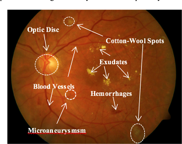

Proliferative diabetic retinopathy → extra smaller blood vessels grow. Usually a response to ischemia.

Papilledema: bright contrast between optic disc and retina reduced, this can signify high intercranial pressure (meaning don't do a spinal tap)
This term specifically means smelling at the optic disc

Central retinal vein occlusion → Blood and thunder finding
Commonly occurs due to a thrombotic event. Retina suffers from blood flow in and out
Key detail: it is painless
High contrast cup to disc ratio is what condition?
Glaucoma
This is entirely opposite of papilledema
Hypertensive retinopathy
Due to high BP (210/140)
To true be diagnosed this it has to have stage 4 papilledema
Macular degeneration -dry type
Drusen: cholesterol droplets + protein + calcium phosphate → this collects in the retina
The macula thins over time and drusen deposits under the retina

Macular degeneration-wet type
Endothelial injury →hemorraging/scarring
