Fascial Layers of the Neck
1/34
There's no tags or description
Looks like no tags are added yet.
Name | Mastery | Learn | Test | Matching | Spaced |
|---|
No study sessions yet.
35 Terms
Fascial layers
They create anatomical spaces that influence the spread of infections
Superficial cervical fascia
A layer of fatty connective tissue located between the skin and the first deep fascial layer
Contains:
Cutaneous nerves
Blood and lymphatic vessels
Superficial lymph nodes
Variable amounts of fat
Superficial cervical fascia
In which layer is the platysma muscle embedded in?
Facial expression (depresses lower jaw, draws down lip & mouth corner)
Tenses the skin (shaving and easing a tight collar)
Functions of the superficial cervical fascia
Cervical branch of the facial nerve (CN VII)
Innervation of the superficial cervical fascia
Main cutaneous nerves of neck
External jugular vein
Deep to the platysma, we find _____ (innervation)
Superficial cervical fascia
Attachments:
Superior: Deep fascia covering pectoralis major & deltoid
Inferior:
Mandible
Skin
Subcutaneous tissues of lower face
Deep cervical fascia
Supports and compartmentalizes cervical structures
Three main layers:
Investing fascia
Pretracheal fascia
Prevertebral fsascia
*Carotid sheath is also part
Investing layer
Layer of Deep Fascia
Encircles the entire neck, deep to skin & superficial fascia
Encloses trapezius and SCM muscle
Contains:
Inferior ends of anterior jugular veins
Jugular venous arch
Fat and deep lymph nodes
Investing layer
Layer of Deep Fascia
Forms a fibrous capsule around:
Submandibular gland (splitting below mandible)
Parotid gland (splitting behind mandible)
Investing layer
Which layer of the deep cervical fascia forms the suprasternal space?
Investing layer
Attachments:
Superior:
Superior nuchal line
Mastoid process
Zygomatic arch
Mandible, hyoid bone, cervical vertebrae
Inferior:
Manubrium of sternum
Clavicles
Scapulae
Suprasternal space
Space located between the sternal heads of the SCM
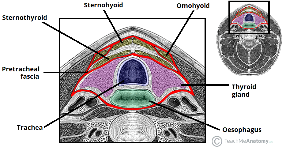
Pretracheal layer (Anterior deep fascia)
Layer of Deep Fascia
Located only in the anterior neck
Extends from hyoid bone to thorax (merging w/ fibrous pericardium)
2 Parts:
Muscular part — encloses infrahyoid muscles
Visceral part — encloses thyroid gland, trachea, esophagus
Muscular part
Which part of the pretracheal layer encloses infrahyoid muscles?
Visceral part
Which part of the pretracheal layer encloses thyroid gland, trachea, and esophagus?
Buccopharyngeal fascia
Superiorly, the pretracheal layer blends with the ______
Carotid sheath
Laterally, the pretracheal layer blends with the _____
Mediastinum
Inferiorly, the pretracheal layer communicates with the _____
Pretracheal layer
Specialization: Forms pully (trochlea) for the intermediate tendon of the digastric muscle
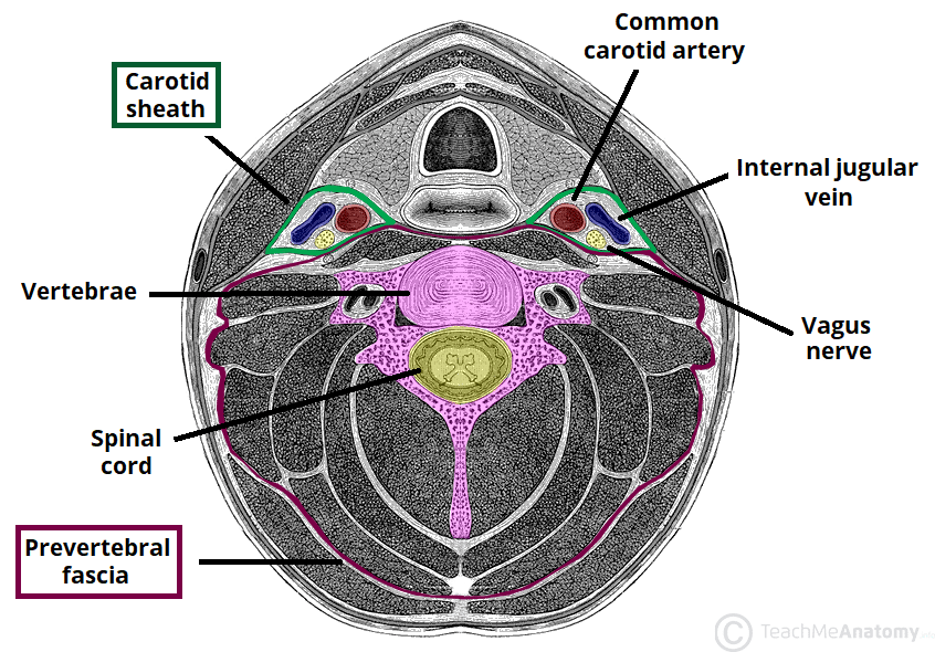
Prevertebral layer (Deepest deep fascia)
Layer of Deep Fascia
Surrounds vertebral column and associated muscles:
Longus colli
Longus capitis
Scalene muscles
Deep cervical muscles
Encloses the cervical part of the sympathetic trunk
Prevertebral layer
Attachments:
Superiorly: Cranial base
Inferiorly: Blends with endothoracic fascia at T3 vertebra
Laterally: Extends as axillary sheath (encloses axillary vessels & brachial plexus)
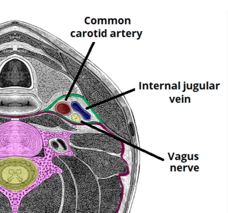
Carotid sheath
Tubular fascial structure that extends from the cranial base to the root of the neck
Carotid sheath
Contents:
Common and internal carotid arteries
Internal jugular vein
Vagus nerve (CN X)
Deep cervical lymph nodes
Carotid sinus nerve & sympathetic fibers
Carotid sheath
Fascial connections:
Anterior: Investing & pretracheal fascia
Posterior: Prevertebral fascia
Inferior: Mediastinum
Retropharyngeal space
Largest potential space in the neck
Between prevertebral fascia (posteriorly) and buccopharyngeal fascia (anteriorly)
Retropharyngeal space
Allows pharynx, esophagus, larynx, and trachea to move freely during swallowing
Retropharyngeal space
Boundaries:
Superior: Cranial base
Lateral: Carotid sheath
Inferior: Communicates w/ mediastinum
True retropharyngeal space, Danger space
What are the 2 divisions of the retropharyngeal space?
True retropharyngeal space
Division of RP space
Anterior to alar fascia
Extends from skull base to C7
Danger space
Division of RP space
Between alar fascia and prevertebral fascia
Extends from skull base to diaphragm
Infections here can spread to the thoracic cavity
Superficial infections
Spread of infections is limited to the superficial fascia
Pretracheal space infections
Infections here may spread to the anterior mediastinum
Retropharyngeal space infections
Infections here may extend to the superior mediastinum
Danger space infections
Infections here can reach the posterior mediastinum and diaphragm, leading to serious complications