IMAGES for Pathological Conditions of the Liver
 Call Kai
 Learn
Learn Practice Test
 Spaced Repetition
 Match
Match Flashcards
 Knowt Play
1/61

There's no tags or description
Looks like no tags are added yet.
Name | Mastery | Learn | Test | Matching | Spaced | Call with Kai | Chat |
|---|
No analytics yet
Send a link to your students to track their progress
62 Terms
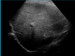
what can be said about the parenchyma of this liver
it is fatty

how would you grade this liver for fatty infiltration
normal
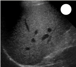
how would you grade this liver for fatty infiltration
grade 1 (mild)
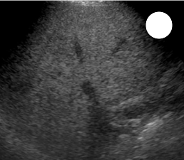
how would you grade this liver for fatty infiltration
grade 2 (moderate)
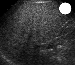
how would you grade this liver for fatty infiltration
grade 3 (severe)

PT has a fatty liver, what does the white arrow indicate
focal fatty sparing

PT has a fatty liver, what does the white arrow indicate
focal fatty sparing

what does this image show about the liver parenchyma
focal fatty infiltration
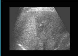
what does this image show about the liver parenchyma
focal fatty infiltration

What is the name of this sonographic sign and which disease is it linked to
starry sky, acute hepatits
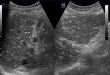
which type of hepatitis is this showing (acute/chronic)
acute
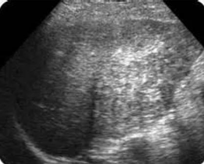
which type of hepatitis is this showing (acute/chronic)
chronic

which type of hepatitis is this showing (acute/chronic)
chronic

what is the dx
cirrhosis
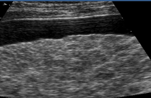
describe this image and what is the dx
pt has cirrhosis, ascites is seen anteriorly, heterogenous, coarse echotexture, micronodular contour
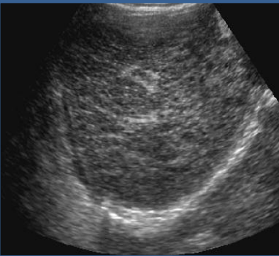
describe the echogenicity and echotexture of this liver
echogenic from fibrotic changes and fatty infiltration. coarse, heterogenous, and micronodular contour

describe the echogenicity and echotexture of this liver
echogenic due to fibrotic changes and fatty infiltration. small heterogenous liver, coarse echotexture, macronodular contour

glycogen storage disease (von gierkes disease), hepatic adenoma within caudate lobe

glycogen storage disease
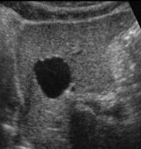
PT is asymptomatic, what is the dx and describe the sonography
simple/congenital cyst (non parasitic), anechoic, regular walls, through transmission
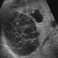
PT is asymptomatic, what is the dx and describe the sonography
hemorrhagic cyst, spider web appearance with through transmisson

what is this showing
peribiliary cysts (clustered tubular cysts within septae parallel to bile ducts)

what dx is this and describe the sonography
polycystic liver disease, multiple cysts of variable size within liver parenchyma
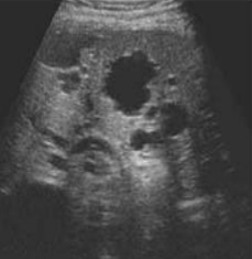
what dx is this and describe the sonography
polycystic liver disease, multiple cysts of variable size within liver parenchyma
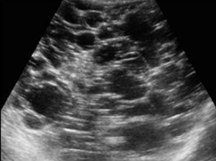
what dx is this and describe the sonography
polycystic liver disease, multiple cysts of variable size within liver parenchyma
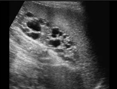
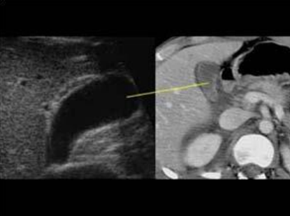
what does this image demonstrate the relationship of
left lobe of the liver at the splenic hilum

PT presents with fever, high WBC count, and RUQ pain, what is the dx
pyogenic abscess

PT presents with fever, high WBC count, and RUQ pain, what is the dx
pyogenic abscess

PT presents with fever, high WBC count, and RUQ pain, what is the dx and describe the sonography
pyogenic abscess, hypoechoic with irregular walls and acoutic enhancement

pt is immunocompromised, what is the dx
hepatic candidiasis

pt is immunocompromised, what is the dx and what is the sonography
candidiasis abscess, multiple small hypoechoic masses with echogenic central cores (bull’s eye lesion)

pt is a young girl, what is the dx
chronic granulomatous disease

pt is a young girl, what is the dx
chronic granulomatous disease

hydatid/ echinococcal cyst

hydatid/ echinococcal cyst

hydatid/ echinococcal cyst

hydatid/ echinococcal cyst

PT is undergoing chemotherapy, what dz is this
pneumocystis carinii

pt is undergoing organ transplantation, what is the dz
pneumocystis carinii

PT has entamoeba histolytica parasite, what is dx and sonography
amebic abscess, poorly defined walls, hypoechoic with internal echoes along posterior margin, posterior enhancement

PT has entamoeba histolytica parasite, what is dx and describe the sonography
amebic abscess, poorly defined wall echoes, hypoechoic, low level echoes internally along posterior margin secondary to debris

what is this showing
cavernous hemangioma

what is this showing
cavernous hemangioma

what is this showing
cavernous hemangioma
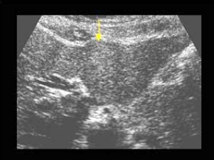
PT is on BCPs, and presents with RUQ pain and palpable mass what is the diagnosis
liver cell adenoma
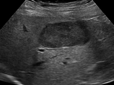
PT is on BCPs, and presents with RUQ pain and palpable mass what is the diagnosis
liver cell adenoma
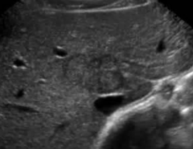
what is this showing
focal nodular hyperplasia (FNH)
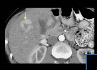
what is this showing
focal nodular hyperplasia (FNH) with arrow to the central scar

what is this showing
focal nodular hyperplasia (FNH)
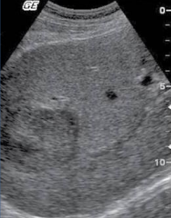
what is being shown in this and its sonography
hepatocellular carcinoma (HCC), discrete hypoechoic lesion in the right lobe
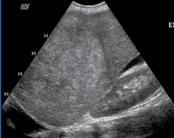
what is being shown in this and its sonography
hepatocellular carcinoma (HCC) large hyperechoic lesion in the rt lobe of the liver

what is this
HCC

what is this and what is the sonography
metastatic disease, multiple well defined hypoechoic lesions throughout the liver

what is this
diffuse metastatic liver disease

what is this
diffuse metastatic liver disease
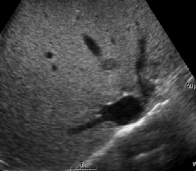
what is this showing
focal metastatic liver disease
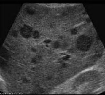
pt presents with enlarged lymph nodes and night sweats, what is the dz and sonography
lymphoma, diffuse markedly hypoechoic lesions

Pt was in an automobile accident, what is the diagnosis and describe the sonography
hematoma, large complex subcapsular hematoma
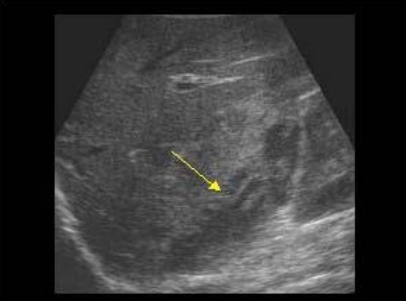
is the blood in this hematoma fresh or older
fresh
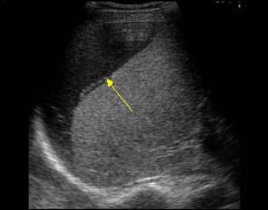
is the blood in this hematoma fresh or older
older

what is this showing
liver rupture
what is this showing delete or like wtf
liver rupture