Spinal Cord and Meninges HK*3401
 Call Kai
 Learn
Learn Practice Test
 Spaced Repetition
 Match
Match Flashcards
 Knowt Play
1/13

There's no tags or description
Looks like no tags are added yet.
Name | Mastery | Learn | Test | Matching | Spaced | Call with Kai | Chat |
|---|
No analytics yet
Send a link to your students to track their progress
14 Terms
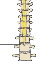
What structure is this and what vertebrae does it occur at
Medullary cone, at L2
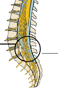
Name these 2 structures
Dural sac (extends to proximal sacrum)
Cauda Equine (circled)
Bundle of root nerves

Name these 3 sections of spinal nerve
Medullary cone
terminal filament
Cauda Equine
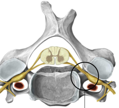
Where does this nerve exit?
Intervertebral foramen
How are spinal nerves formed branching out from spinal cord?
Rootlets (ant post) → Roots (ant post) → Spinal Nerve → Rami (ant post) divides to form rami
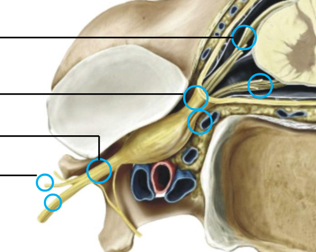
Name all the labels
Rootlets
Roots
Spinal nerve
Rami
Order of Motor information flowing away from spinal cord
Spinal Cord – Anterior Rootlets – Anterior root – Spinal Nerve -- Rami
Order of sensory information flowing towards spinal cord
From Periphery – Rami – Spinal Nerve – Posterior Root – Posterior Rootlets – Spinal Cord
What information do Anterior Rootlets/Roots carry
Spinal Nerves
Contain Efferent/Motor Fibers to
Skeletal Muscle
What information do Posterior Rootlets/Roots carry
Contain Afferent/Sensory Fibers from
Skin; Subcutaneous and Deep Tissues; Viscera
Do rootlets and roots carry sensory info motor info or both?
One or the other, never both. Spinal nerves and Ramus can carry both
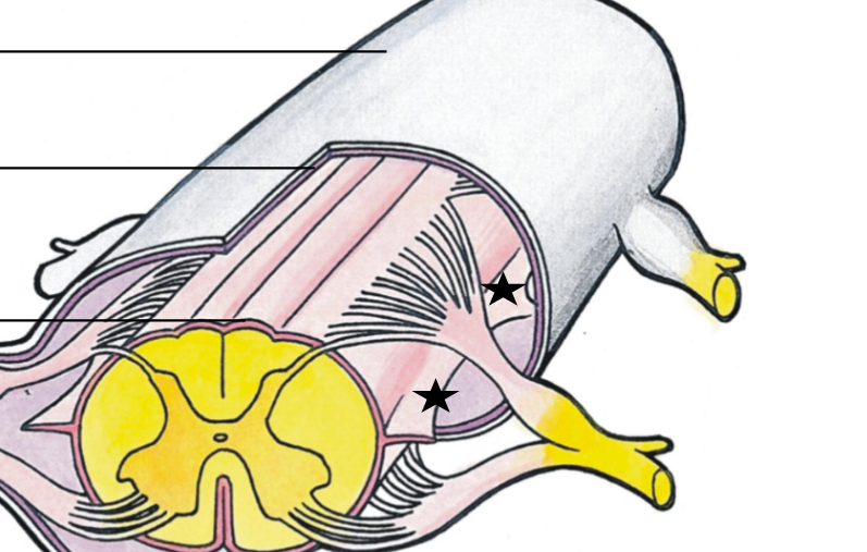
name the three layers and the starred ligament
Dura mater
Arachnoid mater
Pia mater
Denticulate Ligament (Derived from pia mater)
Name the spaces between the Mater
Epidural space
Sub Dural
Subarachnoid space (contains cerebral fluid)

Name every part of this diagram
Left side:
Dura
Arachnoid
Pia'
Denticulate ligament
Terminal filament
Right side:
Rootlets
Roots