Organisms exchange substances with their environment
1/109
There's no tags or description
Looks like no tags are added yet.
Name | Mastery | Learn | Test | Matching | Spaced |
|---|
No study sessions yet.
110 Terms
Suggest an advantage of calculating SA: mass for organisms instead of SA: V
Easier/quicker to find/more accurate because irregular shapes.
Do amoeba have high SA:V ratio?
Yes, because they are small organisms.
This means that there’s a big surface for exchange of substance but also there is a smaller distance from the outside of the organisms to the middle of it.
As a a result, small organisms can simply exchange substances across their surface.
Larger organisms
The larger an organism the lower it’s SA:V ratio.
Larger organisms will also have higher metabolic rate too, which demands efficient transport of waste out of cells and reactants into the cells.
As a result, they have adaptations that help make exchange across surfaces more efficient.
Adaptions to increase SA:V ratio
Villi and microvilli- Absorption of digested food
Alveoli and bronchioles- Gas exchange
Spiracles and tracheoles- Gas exchange
Gill filaments and lamellae- Gas exchange
Thin wide leaves- Gas exchange
Many capillaries- Capillary network
What is metabolic rate?
Amount of energy used up by an organism within a given period of time.
Often measured by oxygen uptake → as used in aerobic respiration to make ATP for energy release.
General information about terrestrial insects
Insects have an exoskeleton made of hard fibrous material for protection and a lipid layer to prevent water loss.
Don’t have lungs but instead a tracheal system.
Insect adaptations to prevent water loss:
Small SA:V ratio where water can evaporate from.
Insects have a waterproof exoskeleton- thick waxy.
Spiracles that can open and close where gases enter and water can evaporate from (closing reduces water loss).
Hairs around spiracles, to trap moist air, reducing water potential gradient so less water less/evaporation.
Insect tracheal system (spiracles, trachea and tracheoles)
Spiracles are pores on surface that can open / close to allow diffusion. Oxygen and carbon dioxide enter and leave and water evaporates via the spiracles. The trachea attach to these openings.
Trachea is a network of internal tubes with rings within them to strengthen the tubes and keep them open.
Tracheoles are the smaller tubes that the trachea branch into, deeper into the abdomen of the insects, These extend throughout all the tissues in the insect to deliver oxygen to all respiring cells.
Permeable to allow gas exchange with cells.
Trachea and tracheoles are supported by spirals of chitin.
Tracheal fluid at end of tracheoles- oxygen dissolves into this and then diffuses into muscle tissue.
3 Methods of moving gases in the tracheal system
Gas exchange by simple diffusion, as when cells respire they use up oxygen and produce carbon dioxide which creates a concentration gradient for CO2 to leave and O2 to enter.
Mass transport- An insect contracts and relaxes their abdominal muscles to move gases on mass.
When the insect is in flight, the muscle cells start to respire anaerobically to produce lactate. This lowers the water potential of the cells and therefore water moves from the tracheoles into the cells by osmosis. This decreases the volume in the tracheoles and as a result more air from the atmosphere is drawn in.
Adaptations for efficient diffusion in insects
Large number of fine tracheoles- large SA and reduces diffusion pathway.
Epithelial cell layer of tracheoles are thin and short distance between spiracles and tracheoles- short diffusion pathway
Use of oxygen and production of carbon dioxide sets up steep diffusion gradients.
Abdominal muscles contract to change pressures in body, forcing air to move in and out.
Fluid in end of tracheoles drawn into tissues by osmosis during exercise (lactate produced in anaerobic respiration lowers ψ of cells)- gases cross ends of tracheoles more quickly.
General information about fish🐠
Fish are waterproof and they have a small SA:V ratio so they require a gas exchange surface (gills).
Fish obtain oxygen from water but there is 30 times less oxygen in water than air, so they have a special adaptation to maintain the concentration gradient to enable diffusion to occur.
Fish gill anatomy
Four layers of gills on both sides of the head.
The gills are made up of stacks of gill filaments.
Each gill filament is covered in gill lamellae, position at right angles to the filament.
When fish open their mouth, water rushes in and over the gills and then out through a hole in the sides of their head.
Lamellae is where diffusion of gases occur.
Adaptations of gills for efficient gas exchange
Many gill filaments covered in many gill lamellae increase surface area.
Short diffusion distance due to a capillary network in every lamellae and
very thin gill lamellae.
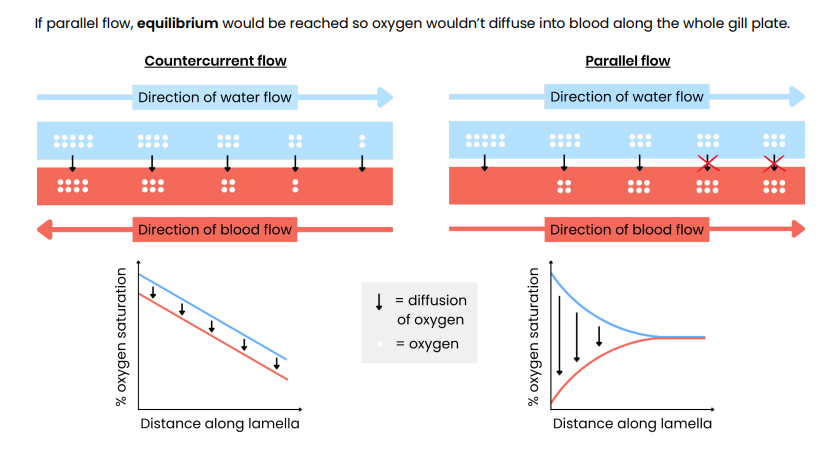
Maintaining concentration gradient through countercurrent flow mechanism.
Countercurrent Flow Mechanism
Water flows over the gills in opposite directions to the flow of blood in the capillaries.
Countercurrent flow ensures that equilibrium is not reached which ensures that a diffusion gradient is maintained across the entire length of the lamellae.
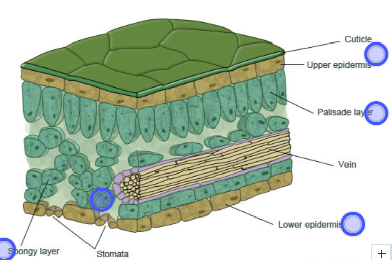
Structure of a leaf
Gas exchange at a stomata
O2 diffuses out of the stomata.
CO2 diffuses in through the stomata.
To reduce water loss by evaporation, stomata close at night when photosynthesis wouldn’t be occurring.
Plants like xerophytic plants are adapted to survive in environments with limited water and have structural features to enable efficient gas exchange to occur whilst also limiting water loss.
Explain how the leaves of dicotyledonous plants are adapted for gas exchange.
Many stomata → large surface area for gas exchange
Spongy mesophyll contains air spaces → large surface area for gases to diffuse through.
Thin → Short diffusion distance
Further adaption of xerophytes
Leaf is curled to trap moisture that evaporates to increase local humidity and therefore reduce further evaporations/transpiration as it reduces water potential gradient.
Add stuff from synoptic
Hairs to trap moisture to increase local humidity and reduce transpiration by reducing water potential gradient.
Sunken stomata to trap moisture to increase local humidity by reducing water potential gradient.
Thicker cuticle to reduce evaporation by reducing water potential gradient.
Longer network of roots to reach more water.
Spines/needles- Reduces surface area to volume ratio
What is breathing?
Movement of air into and out of lungs.
What is ventilation?
The scientific word for breathing.
What is respiration?
Chemical reaction to release energy from the form of ATP.
What is gaseous exchange?
Diffusion of oxygen from the air into the blood and carbon dioxide from the blood into the air in the alveoli.
What does the trachea have that strengthens it?
C-shaped cartilage rings.
Ventilation: inhalation and exhalation
The diaphragm muscles contract so it flattens and external intercostal muscles contract, while internal intercostal muscles relax, pulling the ribcage upwards and outwards, providing a much bigger volume in the thoracic cavity. The air pressure in the lungs then drops so air moves into the lungs down the pressure gradient during inhalation (internal relaxing because there’s antagonistic interaction between them so it'll to the opposite).
The internal intercostal muscles’ contracts while external relaxes so diaphragm relaxes, pulling the ribcage back down and inwards, decreasing volume in thoracic cavity. This increases air pressure inside the lungs to air moves out of the lungs down the pressure gradient (exhalation).
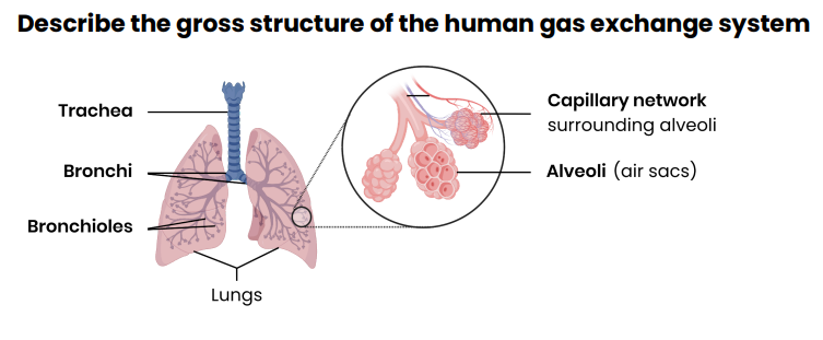
Gas Exchange in the alveoli
Alveoli are surrounded by capillaries.
Both are one cell thick.
Blood in capillaries contains deoxygenated blood and alveoli contains oxygenated air so O2 diffuses simply into capillaries from the alveolar air space and CO2 diffuses simply from the blood to the alveolar air space where it can be exhaled.
Adaptations of alveoli: most important are thin, large, SA and rich blood supply
There are 300 million in each human lung which creates a large surface area for gas exchange.
Short diffusion distance as it’s made up of one layer of epithelial cells (and capillaries are made up of a layer of endothelial cells).
Rich Blood Supply: Each alveolus is surrounded by a network of capillaries that maintain a strong concentration gradient by constantly bringing in deoxygenated blood and carrying away oxygenated blood.
Moist walls - gases dissolve in the moisture helping them to pass across the alveoli.
Elastic Walls: Alveoli are elastic, allowing them to expand and recoil during breathing, helping to ventilate the lungs efficiently.
A large diffusion gradient - breathing ensures that the oxygen concentration in the alveoli is higher than in the capillaries so oxygen moves from the alveoli to the blood. Carbon dioxide diffuses in the opposite direction.
Label human gas exchange system parts
Suggest why expiration is normally passive at rest
Internal intercostal muscles do not normally need to contract.
Expiration aided by elastic recoil in alveoli
Suggest how different lung diseases reduce the rate of gas exchange
Thickened alveolar tissue → increases diffusion distance.
Alveolar wall breakdown → reduces surface area.
Reduce lung elasticity → Lungs expand/ recoil less → reduces concentration gradients of O2/ CO2.
Suggest how different lung diseases affect ventilation
Reduces lung elasticity → lungs expand/recoil less.
Reduces volume of air in each breath (tidal volume).
Reducing maximum volume of air breathed out in one breath (forced vital capacity).
Narrow airways/ reduced airflow in and out of lungs.
Reducing maximum volume of air breathed out in 1 second (forced expiratory volume).
Reduced rate of gas exchange → Increased ventilation rate to compensate for reduced oxygen in blood.
What is a spirometer used for?
To measure lung capacity.
Graph stuff for spirometer
Tidal volume - Volume of air that enters and leaves the lungs at normal resting breath (0.5 dm³).
Vital capacity- Maximum volume of air we can inhale and exhale.
Residual volume- Volume of air left in lungs after the strongest exhalation.
Total lung capacity- Vital capacity and residual capacity (normally 5-6 dm³).
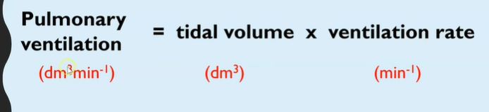
Pulmonary ventilation formula
Refers to total volume of air that is moved into the lungs during one minute.
What happens during digestion?
Large biological molecules are hydrolysed into smaller molecules that can be absorbed across cell membranes and used by body cells.
Digestion of carbohydrates
Mouth – Salivary amylase hydrolyses starch into maltose by hydrolysing glycosidic bond.
Stomach – No carbohydrate digestion (amylase is denatured by HCl).
Small Intestine:
Duodenum – Pancreatic amylase hydrolyses starch into maltose by hydrolysing glycosidic bonds.
Ileum – Membrane-bound maltase hydrolyses
Maltose → Glucose
Absorption (Ileum) – Monosaccharides absorbed by epithelial cells lining the ileum and then co-transport with Na⁺ transports it into the bloodstream.
Digestion of proteins
3 key sets of enzymes to hydrolyse the polymers/proteins.
Endopeptidases- Hydrolyse peptide bonds between amino acids in the middle of a polymer chain.
Exopeptidases- Hydrolyse peptide bonds between amino acids at the end of a polymer chain.
Stomach:
HCl denatures proteins and provides an acidic environment.
Pepsin hydrolyses proteins into polypeptides.
Small Intestine:
Duodenum –
Pancreatic endopeptidases (e.g., trypsin) hydrolyse polypeptides into shorter peptides by hydrolysing peptide bonds between 2 amino acids in middle of the polypeptide chain.
Ileum –
Exopeptidases remove terminal amino acids.
Dipeptidases (membrane-bound) hydrolyse dipeptides into amino acids.
Absorption (Ileum) – Amino acids are absorbed via co-transport with Na⁺ into the bloodstream.
Digestion of lipids: 1
Physical: Emulsification and micelle formation
Chemical: Lipase
Physical: Lipids are coated in bile salts during emulsification, forming micelles and many droplets of lipids provide a larger surface area to enable the faster hydrolysis action by lipase.
Chemical: Lipase hydrolyses the ester bonds in lipids, breaking it down into monoglycerides, glycerol and fatty acids.
Digestion of lipids: 2
When the micelles encounter the ileum epithelial cells, due to the non-polar nature of the fatty acids and monoglycerides, they can simply diffuse across the cell surface membrane to enter the epithelial cells.
Once in the cell, these will be modified back into triglycerides inside of the ER and the Golgi apparatus, which aggregate into globules.
Globules are coated with proteins, forming chylomicrons which are then packaged into vesicles.
Chylomicrons are extruded from the epithelial cell via exocytosis and enter a lacteal (lymph capillary).
It eventually enters the bloodstream through the capillary system for distribution around the body.
What is a micelle?
A water soluble vesicle formed of the fatty acids, glycerol and bile salts.
They deliver the fatty acids, glycerol and monoglycerides to the epithelial cells lining the ileum for absorption and release them so they can move into the epithelial cells by diffusion.
Absorption
In mammals, the products of digestion are absorbed across the cells lining the ileum.
The ileum wall is covered in villi, which have thin walls surrounded by a network of capillaries and epithelial cells have even smaller microvilli.
These features maximise absorption by increasing the surface area, decreasing the diffusion distance and maintaining a concentration gradient.
How are monosaccharides and amino acids absorbed?
Via co-transport
Suggest why membrane-bound enzymes are important in digestion
Membrane-bound enzymes are located on cell membrane of epithelial cells lining ileum.
By hydrolysing molecules at the site of absorption, they maintain concentration gradients for absorption.
Red blood cells: Structure and function
Biconcave shape with no nucleus.
Function: Contain haemoglobin so oxygen can be transported in the blood.
Hb associates with O2 at high pO2 and dissociates from O2 at low pO2.
Haemoglobin
Groups of proteins that are found in different organisms.
Quaternary structure proteins, made of 4 polypeptide chains- 2 alpha and 2 beta.
Each polypeptide chain contains a haem group and this contains iron and this is where O2 would bind.
What does affinity of haemoglobin mean?
The ability of haemoglobin to attract or bind oxygen.
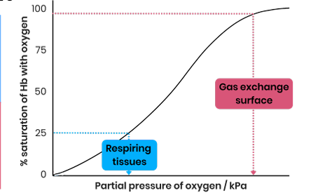
Name area with low and high pO2
Low= respiring tissues
High= gas exchange surfaces
Saturation shown by graph
What does saturation of haemoglobin with oxygen mean?
When haemoglobin is holding the maximum amount of oxygen it can bind.
What does loading/association of haemoglobin mean?
The binding of oxygen to haemoglobin.
Unloading/dissociation refers to the unbinding/detachment of oxygen from haemoglobin.
Oxyhaemoglobin dissociation curve
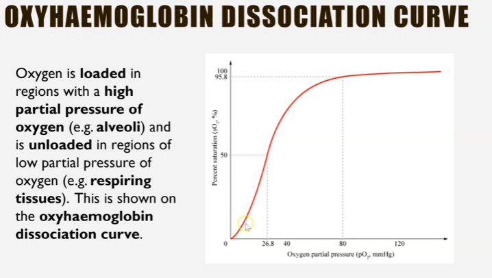
High partial pressure= high affinity, more association e.g. alveoli.
Lower partial pressure= lower affinity, more unloading where the oxygen is needed e.g. respiring tissues.
Cooperative binding
The cooperative nature of oxygen binding to haemoglobin is due to the haemoglobin changing shape when the first oxygen binds.
This makes it easier for 2 and 3 to bind as haem group binding sites are uncovered.
4 has difficulty binding due to saturation of haemoglobin.
Describe evidence for the cooperative nature of oxygen binding.
● A low pO2 as oxygen increases there is little/slow increase in % saturation of Hb with oxygen
○ When first oxygen is binding
● At higher pO2 ,as oxygen increases there is a big/ rapid increase in % saturation of Hb with oxygen ○ Showing it has got easier for oxygens to bind
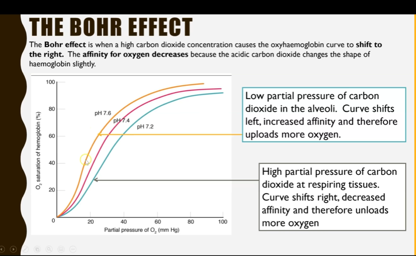
The Bohr Effect
When a high carbon dioxide concentration causes oxyhaemoglobin to shift to the right (less saturation).
The affinity for oxygen decreases because the acidic carbon dioxide (which reacts with water to form carbonic acid) changes the shape of haemoglobin slightly.
Lower affinity= more unloading e.g. at respiring tissues which is advantageous as it allows for faster aerobic respiration → more ATP produced.
Do different animals have different types of haemoglobin?
Yes, these have different affinities for oxygen, which is an adaption to their environments e.g. llama, earthworm, dove, foetus.
Foetal haemoglobin
Further to the left= higher affinity for oxygen= more association → can load even at lower partial pressures so more saturated in oxygen at lower partial pressures.
Advantageous as foetus can’t exhale or inhale so this allows foetus to survive by receiving sufficient amounts of oxygen.
Llamas haemoglobin
Live at high altitudes where there is a lower partial pressure of oxygen.
To the left= higher affinity for oxygen= loading at low partial pressures.
Dove haemoglobin
Shift to the the right= lower affinity= more unloading oxygen to respiring tissues.
Advantageous as it has a faster metabolism so needs more oxygen for respiration to provide energy for contracting muscles.
Earthworms haemoglobin
Shifts to left= higher affinity for oxygen= loading even at low partial pressures e.g. underground.
Blood vessels overview
Arteries are connected to arterioles which are connected to capillaries which are connected to venules which are connected to veins.
Veins have valves.
Suggest the importance of a double circulatory system
Prevents mixing of oxygenated and deoxygenated blood.
So blood pumped to the body is fully saturated with oxygen for aerobic respiration.
Blood can be pumped to body at higher pressure.
Substances taken to/removed from body cells more efficiently.
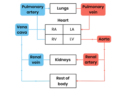
Pattern of blood circulation in a mammal
Capillaries
Form capillary beds as exchange surfaces.
These are many branched capillaries.
They all have a narrow diameter to slow blood flow.
Red blood cells can only just fit through and are squashed against the wall.
This maximises diffusion as there’s more time for diffusion.
Blood vessels: Muscle layer (controls diameter)
Arteries muscle layer is thicker than veins so that constriction (increase blood flow to certain areas e.g. vasoconstriction) and dilation (reducing blood flow to certain areas e.g. vasodilation) can occur to control the volume of blood.
Relatively thin in veins so it cannot control the blood flow.
Arteriole muscle layer is thicker than in arteries to help restrict blood flow into the capillaries so it doesn’t burst because pressure is lower. It’s also involved in vasoconstriction and vasodilation so it needs to be able to control the flow of blood.
Capillaries do not have a muscle layer.
Blood vessels: Elastic Layer
Arteries elastic layer is thicker than veins to help maintain blood pressure.
The walls can stretch and recoil in response to the heartbeat.
Stretch when blood surges through (systole)
Recoil to maintain smooth pressure and keep blood moving (diastole)
Relatively thin in veins as the pressure is much lower and doesn’t fluctuate.
Thinner in arterioles than arteries as the pressure is lower and their main role is to control flow using muscle, not recoil.
Capillaries do not have an elastic layer because blood pressure is low and steady and an elastic layer would increase diffusion distance.
Blood vessels: wall thickness
Arteries have thicker walls than veins to help prevent the vessels bursting due to the high pressure.
Thin in veins as the pressure is much lower so there is a low risk of bursting.
The thinness means the vessels are easily flattened, which helps the flow of blood up to the heart.
Arterioles= thinner as the pressure is slightly lower but thicker than capillaries as the pressure is slightly higher and needs to be reduced before it reaches capillaries.
Capillaries are one cells thick because blood pressure is low→ this provides a short diffusion distance for exchanging materials between the blood and cells.
What is cardiac muscle?
The muscle layer in the walls of the heart.
Thick but thickness varies between different chambers.
Unique properties:
It’s myogenic
It never fatigues, as long as it has a supply of oxygen and glucose.
What does it mean by the cardiac muscle being myogenic?
It can contract or relax without nervous or hormonal stimulation.
What are coronary arteries?
Blood vessels surrounding the heart that branch off from the aorta and supply the cardiac muscle with oxygenated blood.
If they become blocked, cardiac muscle won’t receive oxygen, therefore will not be able to respire and the cells will die.
This results in myocardial infarction (heart attack).
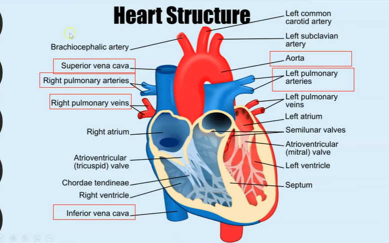
Heart structure: Four chambers
2 atria: left and right
2 ventricles: left and right
Atria:
Thinner muscular walls as they do not need to contract as hard as they are not pumping blood far but only to the ventricles and it goes along gravity.
They have elastic walls which stretch when blood enters.
Ventricles:
They have thicker muscular walls to enable bigger contraction, creating a higher blood pressure to enable blood to flow longer distances (to the lungs and the rest of the body) and against gravity.
Right ventricle has thinner muscular wall than left because the right ventricle pumps blood to the lungs and blood needs to be at slightly lower pressure to prevent damage to capillaries in the lungs and also so that blood flows slowly to allow time for gas exchange.
The left ventricle has thicker muscular wall to enable larger contractions to of the muscle to create higher pressure as it pumps blood to the body, so higher pressie is needed to ensure blood reaches all the cells in the body.
Structure of the heart
Veins:
Vena cava- Carries deoxygenated blood from the body into the right atrium.
Pulmonary vein- Carries deoxygenated blood from the lungs to the left atrium.
Arteries:
Pulmonary artery- Carries deoxygenated blood from the right ventricle to the lungs to become oxygenated.
Aorta- Carries oxygenated blood from the left ventricle to the rest of the body.
Valves:
Semi-lunar valves between aorta and left ventricle and right ventricle and pulmonary artery.
Atrioventricular valves between atria and ventricles.
Bicuspid (mitral) on left side and Tricuspid on right side (related to the number of flaps which make it up).
Septum:
Separates the deoxygenated blood from the oxygenated blood which maintains a high concentration of oxygen to maintain concentration gradient to enable diffusion to respiring cells.
How to valves prevent backflow of blood?
Open when pressure is higher behind valve and close when pressure is lower behind the valve.
The Cardiac Cycle: Diastole
The atria and ventricular muscles are relaxed.
This is when blood will enter the atria via the vena cava and pulmonary veins and this flows passively to ventricles as the blood flowing into the atria increases the pressure within the atria.
Atrioventricular valves open when pressure in atria exceeds pressure in ventricles.
The Cardiac Cycle: Atrial Systole
The atria muscular walls contract, increasing the pressure further as the volume of the atria decreases.
This causes, the atrioventricular valves to open and blood to flow into the ventricles.
The ventricular muscular walls are relaxed (ventricular diastole).
(SL remain shut because pressure in arteries exceeds pressure in ventricles).
The Cardiac Cycle: Ventricular Systole
After short delay, the ventricle muscular walls contract, increasing the pressure beyond that of the atria.
This causes the atrioventricular valves to close when ventricular pressure is greater than atria pressure and the semi-lunar valves to open when ventricular pressure is greater than pressure in arteries.
Blood is pushed out of the ventricles into the arteries where it then leaves the heart.
ECG
Measures the electrical activity of the heart.
Spike= ventricular contraction.
Cardiac output
This refers to the volume of the blood which leaves the heart in one minute. (dm³min^-1
Cardiac Output= Heartbeat x Stroke volume
It can be calculated by multiplying the heart rate by the stroke volume.
Heart Rate= Beats of the heart per minute (minute ^-1).
Stroke volume= The volume of blood that leaves the heart each beet (dm³).
Valves in the cardiac cycle
Controlling the blood and preventing backflow.
Open when pressure is higher behind the valve.
Close when pressure is higher in front of valve.
Atrioventricular valves open when the pressure is higher in the atria compared to the ventricles.
They close when the pressure is higher in the ventricles compared to the atria.
Semi-lunar valves open when the pressure is higher in the ventricles compared to the arteries (aorta and pulmonary artery).
They close when the pressure is higher in the arteries than the ventricles.
Interpretation of pressure changes
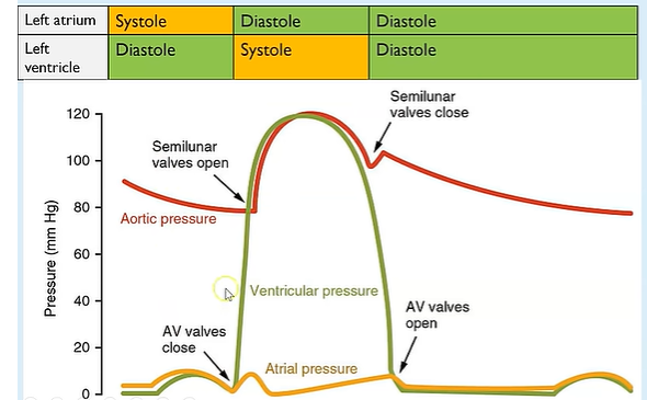
🔶 Starting Point – Far Left of the Graph
At this point:
🟠 Atrial Pressure is a bit higher — because the atrium is full of blood from the lungs.
🟢 Ventricular Pressure is low — the ventricle is relaxed and filling.
🔴 Aortic Pressure is steady — it's always relatively high because it's full of blood and elastic.
🔸 First Small Rise in Orange (🟠 Atrial Pressure Rises Slightly)
Why?
The left atrium contracts — this is atrial systole.
This little squeeze adds more blood to the ventricle, causing a small increase in atrial pressure.
🟢 Ventricular pressure also bumps up a little as it fills.
🔸 Sharp Rise in Green (🟢 Ventricular Pressure Increases Fast)
Why?
The ventricle starts contracting — this is ventricular systole.
Because it's now squeezing, pressure inside rises rapidly.
This pressure soon gets higher than atrial pressure, so the AV valves close (to stop backflow).
This sharp upward slope in green is the start of the powerful pump phase.
🔸 Red Line (🔴 Aortic Pressure) Starts to Rise
Why?
As the ventricle continues to contract, its pressure becomes higher than the pressure in the aorta.
This forces open the semilunar valves, and blood is ejected into the aorta.
So, aortic pressure also rises.
🧠 Both the green and red lines are high here because the ventricle is pumping strongly and the aorta is filling fast.
🔸 Green Line Peaks and Starts to Fall (🟢 Ventricular Pressure Drops)
Why?
The ventricle stops contracting and begins to relax — this is ventricular diastole.
Pressure inside it drops quickly.
Once the pressure in the ventricle is lower than in the aorta, the semilunar valves close (to stop blood flowing back in).
This drop is quite sharp because the ventricle is no longer squeezing.
🔸 Red Line Falls Slowly (🔴 Aortic Pressure Decreases)
Why?
No more blood is being pumped into the aorta (semilunar valve is now closed).
But blood is still flowing away to the rest of the body, so pressure slowly drops.
It’s a gradual fall because the aorta is elastic and maintains pressure.
🔸 Green Line Falls Below Orange (🟢 Ventricular Pressure < 🟠 Atrial Pressure)
Why?
As the ventricle relaxes further, its pressure becomes lower than the atrium’s.
This causes the AV valves to open again.
Blood starts to flow passively from the atrium to the ventricle.
This is a quiet, filling stage — no big pressure spikes, everything is relaxing and preparing for the next beat.
🔸 Final Small Rise in Orange (🟠 Atrial Pressure Increases Again)
Why?
The atrium is filling with blood returning from the lungs.
Pressure slowly increases until it’s ready to contract again.
The Cardiac Cycle- Summary
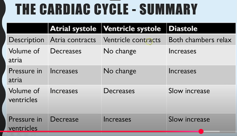
The reason for the slight increase in atrial pressure when volume increases is because blood begins to enter due to increased volume/
The reason for the slight increase in ventricular pressure is due to the blood from atria beginning to trickle into the ventricles slowly.
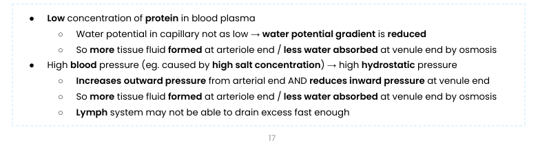
What is tissue fluid?
The fluid containing water, glucose. amino acids, fatty acids, ions and oxygen which bathes the tissues.
Allows for essential molecules to diffuse in.
How is tissue fluid formed? (arteriole side of capillaries)
Capillaries have small gaps in the walls which are small enough for liquid and small molecules can be forces out.
As blood enters the capillaries from arterioles, the smaller diameter results in a high hydrostatic pressure (pressure when water isn’t moving e.g. water coming out a bucket with holes), so water, glucose, small proteins, amino acids, fatty acids, ions and oxygen are forced out in a process known as ultrafiltration.
This tissue fluid then surrounds the nearby tissue before reabsorption.
Red blood cells, platelets and large proteins remain.
How is tissue fluid reabsorbed?
Large molecules remain in the capillaries and therefore create a lowered water potential.
Towards the venule end of the capillaries, the hydrostatic pressure is lowered due to the loss of liquid, but the water potential is very low.
Water re-enters the capillaries by osmosis with waste products dissolved in it: CO2 and urea.
Waste products can then be transported away for removal.
Not all the liquid will be reabsorbed by osmosis, as equilibrium will be reached.
The rest of the tissue fluid that the capillaries couldn’t reabsorb is absorbed into the lymphatic system and eventually drains back into the bloodstream near the heart.
The lymphatic system surrounds blood vessels.
The lymphatic system will eventually bring the fluid back into the blood through lymph vessels near the heart (similar to veins as they have valves).
Suggest and explain causes of excess tissue fluid accumulation
Explain the rate of transpiration between 5am and midday shown in Figure 9
1. (Rate of) transpiration increases due to increased temperature
2.(So) increased kinetic energy
3.Stomata open (at sunrise/after 5 am) allowing gas exchange
4. (Some) stomata close at midday
To observe the fish gills with an optical microscope, the scientists used two different stains.
The first stain binds to DNA; the second binds to red blood cells.
Explain why a second stain would be needed and which molecule in RBCs the stain could bind to.
Second stain is needed because red blood cells do not have DNA.
Haemoglobin.
Other than surface area: volume ratio, describe one way in which this uncontrolled cell division changes the gills.
Uncontrollable cell division could thicken gill filament
Increasing diffusion distance for gas exchange
Which slows the rate of gas exchange.
What is transpiration?
The loss of water vapour from the stomata, down a water potential gradient by evaporation.
What 4 key conditions affect the rate of transpiration?
Light intensity- Positive correlation as more light causes more stomata to open → larger surface area for evaporation.
Temperature- Positive correlation as more heat means more kinetic energy of molecules so they therefore evaporation is quicker.
Humidity- Negative correlation as more water vapour in the air will make the water potential outside of the leaf higher/more positive and this reduces the water potential gradient, reducing the rate of evaporation.
Wind- Positive correlation as more wind will blow away humid air containing water vapour, therefore maintaining a high water potential gradient.
Why can water travel several metres against gravity from roots to leaves?
Due to cohesion-tension theory
Suggest how xylem tissue is adapted for its function
Cells joined with no end walls, forming a long continuous tube -> water flows as a continuous column.
Cells contain no cytoplasm / nucleus → easier water flow/ no obstructions.
Thick cell walls with lignin → Provides support/ withstand tension/ prevents water loss.
Pits in side walls → allow lateral water movements.
Cohesion-tension theory: Cohesion
Water is a dipolar molecule.
This enables hydrogen bonds to form between the partially positive hydrogen and partially negative oxygen of different water molecules.
This creates cohesion between water molecules- they stick together. Therefore, water travels up the xylem as a continuous water column.
Cohesion-tension theory: Capillarity
Adhesion of water is when water sticks to other molecules.
Water adheres to xylem walls.
The narrower the xylem, the bigger the impact of capillarity as a larger proportion of the water is in contact with the walls of the straw so it moves further up the xylem.
Cohesion-tension Theory: Root Pressure
As water moves into the roots by osmosis, it increases the volume of liquid inside the root and therefore the pressure inside the root increases. This is known as root pressure.
This increase in pressure in the roots forces water above it to move upwards (positive pressure).
Movement of water up the xylem
Water vapour evaporates out of stomata on leaves. This loss in water volume creates a lower pressure.
As a result of this water lost by transpiration, more water is pulled up the xylem to replace it (moves due to negative pressure).
Due to the hydrogen bonds between water molecules, they are cohesive, creating a continuous column of water within the xylem.
Water molecules also adhere to the walls of the water which helps to pull the water column upwards.
As this column of water is pulled up the xylem, it creates tension, pulling the xylem in to become narrower, increasing the impact of capillarity.
Suggest how light intensity affect transpiration rate
Increases rate of transpiration
Stomata open in light to let in CO2 for photosynthesis.
Allowing more water to evaporate faster.
Stomata closes when it’s dark so there is a low transpiration rate.
Suggest how temperature affect transpiration rate
Water molecules gain kinetic energy as temperature increases.
So water evaporates faster.
Suggest how wind intensity affect transpiration rate
Wind blows away water molecules from around stomata.
Decreasing water potential of air around stomata.
Increasing water potential gradient so water evaporates faster.
Suggest how humidity affect transpiration rate
Decreases rate of transpiration.
More water in air so the area around stomata has a high water potential.
Decreasing water potential gradient from leaf to air.
Water evaporates slower.
What’s the function of the leaves?
Photosynthesis
All cells require the sugar produced in photosynthesis in order to respire so there has to be a method of transporting it- translocation.
What 2 key cells make up phloem tissue?
Sieve tube elements
Companion cells on both sides.
Function is to transport organic substances e.g. sucrose in plants.
Sieve tube elements
Living cells
Contains no nucleus
Contain few organelles
These 2 features maximise space for organic substances to flow through, easier flow.
End walls are perforated