radiology 4.1 - ABCs of heart disease
 Call Kai
 Learn
Learn Practice Test
 Spaced Repetition
 Match
Match Flashcards
 Knowt Play
1/119

There's no tags or description
Looks like no tags are added yet.
Name | Mastery | Learn | Test | Matching | Spaced | Call with Kai | Chat |
|---|
No analytics yet
Send a link to your students to track their progress
120 Terms
What can cause the ascending aorta to go pass the right ventricle?
🔵A lot of pressure, such as hypertension.
True/false- The aortic knob can be too large.
True.

What forms the first cardiac contour on the patient's right on a PA CXR?
The ascending aorta.
How should the normal ascending aorta look on a PA CXR?
Relatively low density and mostly straight; it should not bulge beyond the right atrial border.
The ascending aorta protruding farther right than the right atrium is normal or abnormal?
Abnormal; the normal ascending aorta should not project beyond the right heart border.
What normal contour change occurs where the ascending aorta meets the heart?
A normal indentation.
What is the "double-density sign" on a PA CXR?
A second density projected through/inside the right heart border, indicating left atrial enlargement.

Which chamber is enlarged when a double-density sign is present?
The left atrium.
From superior to inferior, what are the main right-sided contours on a PA CXR?
Ascending aorta, then right atrium.
From superior to inferior, what are the 3 main left-sided cardiac contours on a PA CXR?
Aortic knob → main pulmonary artery → left ventricle.
Which pulmonary artery hilum is normally higher?
The left pulmonary artery/hilum is usually higher than the right.
What is the first contour on the patient's left side of the mediastinum/heart?
The aortic knob.
What structures create the aortic knob?
The aortic arch and proximal descending aorta.
What is the second contour on the patient's left side?
The main pulmonary artery.
What vessels branch from the main pulmonary artery contour on CXR?
The right and left pulmonary arteries, creating branching/squiggly hilar vascular markings.
What is the third and lowest major contour on the patient's left side?
The left ventricle.
Which chamber normally contributes most of the left lower cardiac border?
The left ventricle.
Image recognition: a second curved density is visible inside the right heart border. What is the finding?
Double-density sign from left atrial enlargement.
How is cardiothoracic ratio calculated on an upright PA CXR?
Maximum transverse cardiac width ÷ maximum internal thoracic width.
What cardiothoracic ratio suggests cardiomegaly on an upright PA CXR?
Greater than 0.50, meaning the heart is more than 50% of the internal chest width.
If you have an example of an enlarged heart, blunted costophrenic angles, etc, what would be the next step?
Echocardiogram.
This image is not caused by high pressure/hypertension.

Why should the 50% cardiothoracic-ratio rule not be trusted on an AP portable film?
AP projection magnifies the heart and can falsely make it look enlarged.
How does the heart usually respond to chronic valvular stenosis?
Pressure overload causes myocardial hypertrophy—thicker muscle walls.
How does the heart usually respond to chronic valvular regurgitation?
Volume overload causes chamber dilation/enlargement.
If cardiomegaly may be due to fluid overload/CHF, what other CXR findings should you seek?
Kerley B lines, thickened/prominent fissures, peribronchial cuffing, and pleural effusions.
Define cardiomegaly radiographically on an adequate upright PA chest radiograph.
Cardiac silhouette greater than 50% of the internal thoracic diameter.
What view is required for the most reliable cardiothoracic-ratio assessment?
An upright PA chest radiograph with adequate inspiration.
What lateral-CXR clue suggests cardiac enlargement?
Diminished/obliterated clear space posterior to the heart.
Why does the retrocardiac clear space decrease with cardiomegaly?
An enlarged heart extends farther posteriorly on the lateral view.
A large cardiac silhouette does not always mean true cardiomegaly. What 3 categories can mimic it?
Pericardial effusion, extracardiac factors, and poor radiographic technique—especially an AP view, which magnifies the heart because it is farther from the detector.
What is the key distinction between cardiomegaly and an enlarged cardiac silhouette?
Cardiomegaly means enlarged heart chambers; an enlarged silhouette may instead be fluid, projection, body habitus, or poor technique.
Name 6 common causes of pericardial effusion from this lecture.
CHF, infection, metastasis, lupus, trauma, and postsurgical causes—all can cause fluid to accumulate in the pericardial sac.
What is the study of choice for suspected pericardial effusion?
Echocardiography.
How can CT help when the cardiac silhouette is enlarged?
It can distinguish soft-tissue density of the heart from fluid density in the pericardial space.
Can CXR reliably distinguish cardiomegaly from pericardial effusion?
No. CXR may show a large silhouette, but echocardiography is needed to identify and assess the effusion.
What is the most common technical cause of apparent cardiomegaly?
An AP chest radiograph, especially a portable AP film.
Why does an AP CXR exaggerate heart size?
The heart is farther from the detector and is magnified.
Besides AP projection, what technical factors can falsely enlarge the cardiac silhouette?
Poor inspiration and patient rotation can make the heart look larger.
On an AP film, a borderline-large heart is interpreted how?
It may be normal because AP magnification commonly exaggerates size.
On an AP film, a markedly enlarged heart is interpreted how?
True enlargement is likely despite AP magnification.
On an AP film, what strongly supports definite cardiomegaly?
The cardiac silhouette touches or nearly touches the left lateral chest wall.
What is the basic mechanism behind pulmonary findings in CHF?
Elevated hydrostatic pressure causes 🔵 fluid overload🔵 to back up from the heart into the pulmonary interstitium and then alveoli.
What are the 2 radiographic types/stages of pulmonary edema in CHF?
🔵 Interstitial edema and alveolar edema.
Which generally occurs first: interstitial or alveolar edema?
Interstitial edema usually occurs first; worsening fluid then spills into alveolar airspaces.
🔵 List the 4 classic CXR signs of interstitial pulmonary edema in this lecture.
-Kerley B lines
-Peribronchial cuffing
-Fluid/thickening in the fissures
-Pleural effusions.
What anatomic structure is thickened to create Kerley B lines?
Interlobular septa.
Where should you look for Kerley B lines?
At the lung periphery, especially the lower lateral lungs near the costophrenic angles.
What do Kerley B lines look like?
Short, straight, horizontal peripheral lines that contact the pleura.
What dimensions were given for Kerley B lines?
About 1-2 cm long and less than 1 mm thick.
Kerley B lines indicate what process?
Interstitial pulmonary edema, often from CHF.
Image recognition: short horizontal lines at the lateral lung bases extending to the pleura indicate what?
Kerley B lines from thickened interlobular septa.
What is peribronchial cuffing?
Fluid/thickening around a bronchial wall, making the bronchus look like a ring or doughnut when seen end-on.
Peribronchial cuffing is a sign of which type of pulmonary edema?
Interstitial pulmonary edema.
Why does peribronchial cuffing look like a doughnut?
The air-filled bronchial lumen is dark, while the edematous thickened wall around it is white.
What does "fluid in the fissures" mean in CHF?
Edema/fluid accumulates between lobes along the major or minor fissures, making the fissures abnormally thick or conspicuous.
How do normal and CHF-associated fissures differ?
A normal fissure may be faintly visible; CHF makes it thicker and more prominent because of fluid.
Where should you first look for a pleural effusion on upright CXR?
At the costophrenic angles for blunting/meniscus.
What distribution of pleural effusions is typical in CHF?
Usually bilateral, although one side can be larger.
What physical-exam findings may occur over a pleural effusion?
Decreased breath sounds and dullness to percussion at the lung base; diaphragmatic excursion may also be reduced.
So make sure to always listen!

On the lecture CT image, what 3 findings supported interstitial edema/CHF?
Pleural effusion, thickened fissure, and interlobular septal thickening/Kerley B equivalents.
What is alveolar edema?
Fluid has progressed out of the interstitium and into the alveolar airspaces.
How does alveolar edema look on CXR?
Fluffy, ill-defined airspace opacities.
What is the most common cause of alveolar pulmonary edema in this lecture?
CHF/cardiogenic pulmonary edema.
Why are alveolar edema margins indistinct?
Fluid fills airspaces and merges across adjacent alveoli rather than following sharp anatomic borders.
What is the classic central distribution of cardiogenic alveolar edema called?
Bat-wing pattern.
(or Butterfly/angel-wing pattern)
Describe the bat-wing pattern on CXR.
Bilateral fluffy, ill-defined, centrally/perihilarly located airspace opacities, often with relative peripheral sparing.

Image recognition: bilateral central fluffy perihilar opacities in a patient with dyspnea suggest what?
Alveolar pulmonary edema, classically cardiogenic CHF.
Which associated findings make a bat-wing pattern more likely cardiogenic?
Cardiomegaly, pleural effusions, Kerley B lines, vascular congestion, and fissural fluid.
What is the basic mechanism of noncardiogenic pulmonary edema?
Increased pulmonary capillary permeability rather than elevated left-sided hydrostatic pressure.
Besides CHF, what 2 organ problems can cause volume overload?
Renal failure and severe liver disease—both can cause fluid retention.
Which inhalational/environmental causes of 🔵noncardiogenic edema🔵 were listed? [3]
Smoke inhalation, near drowning, and high-altitude pulmonary edema.
Which neurologic/procedural causes of noncardiogenic edema were listed?
Neurogenic pulmonary edema and re-expansion pulmonary edema.
Which systemic illnesses from the lecture can produce 🔵noncardiogenic edema🔵?
Sepsis, uremia, and DIC.
Compared with cardiogenic edema, how does noncardiogenic edema usually affect heart size?
The cardiac silhouette is more often normal-sized.
Compared with cardiogenic edema, are Kerley B lines and pleural effusions more or less common in noncardiogenic edema?
🔵Less common.🔵
Compared with cardiogenic edema, where may noncardiogenic opacities be more prominent?
More peripheral, although the pattern is highly variable.
Does a normal heart size rule out CHF?
No. CHF/pulmonary edema can occur without cardiomegaly.
Quick comparison: cardiogenic vs noncardiogenic pulmonary edema.
Cardiogenic: usually cardiomegaly, Kerley B lines/effusions, and central bat-wing opacities.
Noncardiogenic: usually normal heart size, 🔵fewer Kerley B lines/effusions🔵, and more peripheral or variable opacities.
What cardiac adaptation results from chronic systemic hypertension?
Left ventricular hypertrophy from chronic pressure overload.
Why can the CXR heart size remain 🔵normal🔵 in hypertensive cardiovascular disease despite LVH?
Hypertrophy increases wall thickness and may not initially enlarge the external cardiac silhouette.
What aortic change can chronic hypertension produce on CXR?
Unfolding of the aorta with more prominent ascending and descending aortic contours.
Image recognition: prominent ascending/descending aortic contours with thick LV walls suggests what?
Hypertensive cardiovascular disease with LV hypertrophy and aortic unfolding.
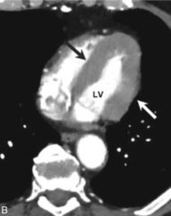
What are the 3 main types of cardiomyopathy?
Dilated, hypertrophic, and restrictive.
Define an aortic aneurysm.
🔴 Aortic enlargement classically more than 50% above the vessel's normal size.
What specific diameter values were listed for thoracic aortic enlargement - ascending aorta?
Over 3.5 cm.
What specific diameter values were listed for thoracic aortic enlargement - descending aorta?
Over 3.0 cm.
What specific diameter values were listed for thoracic aortic enlargement that is considered aneurysmal?
🔴 Over 4.0 cm.
At roughly what thoracic aortic size did the lecture say rupture risk becomes concerning?
🔴 More than 5-6 cm at risk of rupture 🔴; actual intervention thresholds depend on location, growth rate, symptoms, and patient risk factors.
What is the main diagnostic imaging study for a suspected thoracic aortic aneurysm?
🔴 CT angiography (CTA).
What may a thoracic aortic aneurysm look like on CXR?
Prominent/widened ascending or descending aortic contour and possible mediastinal widening.
Can CXR definitively diagnose or size an aortic aneurysm?
No. It may raise suspicion; CTA is needed for anatomy and measurement.
What happens in an aortic dissection?
A tear in the aortic intima 🔴 allows blood to enter and split the layers of the aortic wall.🔴
Which portion of the thoracic aorta is most often involved in the lecture?
🔴 The ascending aorta.
What pain pattern is classic for acute aortic dissection?
🔴Extremely painful.🔴
Sudden, severe chest and/or back pain, often described as tearing or ripping.
What major risk factor is most common in aortic dissection?
🔴 Chronic hypertension.
What substance exposure increases the risk of aortic dissection?
Cocaine/stimulant use.
Which connective-tissue disorders predispose to aortic dissection?
Marfan syndrome and Ehlers-Danlos syndrome.
What historical infection was listed as an aortic-disease risk factor?
Syphilis, classically associated more with thoracic aortic aneurysm/aortitis.
What is the classic CXR warning sign for aortic dissection?
🔴 Widened mediastinum.
What other concerning CXR findings can occur with dissection?
Abnormal/lost aortic knob contour, tracheal deviation, and pleural effusion—especially a new left effusion/hemothorax.