Chapter 9: Nervous & Endocrine System
1/235
There's no tags or description
Looks like no tags are added yet.
Name | Mastery | Learn | Test | Matching | Spaced | Call with Kai |
|---|
No analytics yet
Send a link to your students to track their progress
236 Terms
What are neurons specialized to do?
Specialized cells that transmit and process information from one part of the body to another.

What form does the information transmitted by neurons take?
The information takes the form of electrochemical impulses known as action potentials.
What is an action potential?
Localized area of depolarization of the plasma membrane that travels in a wave-like manner along an axon.

What happens when an action potential reaches the end of an axon at a synapse?
The signal is transformed into a chemical signal with the release of neurotransmitter into the synaptic cleft, a process called synaptic transmission.

What is the basic functional and structural unit of the nervous system?
The neuron.
What are the main structural components of a neuron?
Neurons have a central cell body (soma), axons, and dendrites.
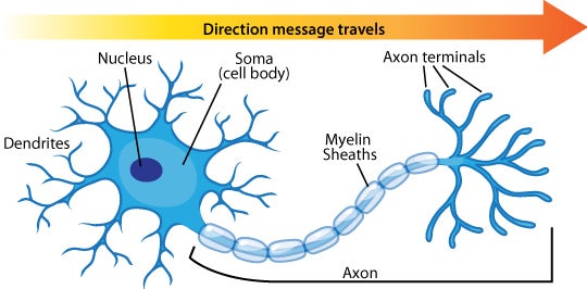
What does the soma contain, and what is its function?
Contains the nucleus and is where most of the biosynthetic activity of the cell takes place.

How many axons and dendrites do neurons typically have?
Neurons have only one axon but may have many dendrites.

What are neurons with one dendrite called?
Bipolar neurons.

What are neurons with many dendrites called?
Multipolar neurons.

In which direction do neurons generally carry action potentials?
Dendrites receive signals, and axons carry action potentials away from the cell body.

What is released when action potentials reach the synaptic knob?
Chemical messengers are released.
What motor protein drives the movement of vesicles and organelles along microtubules in axons?
Kinesin

What is the specific direction of movement driven by kinesin in neurons?
Anterograde movement (from the soma toward the axon terminus).
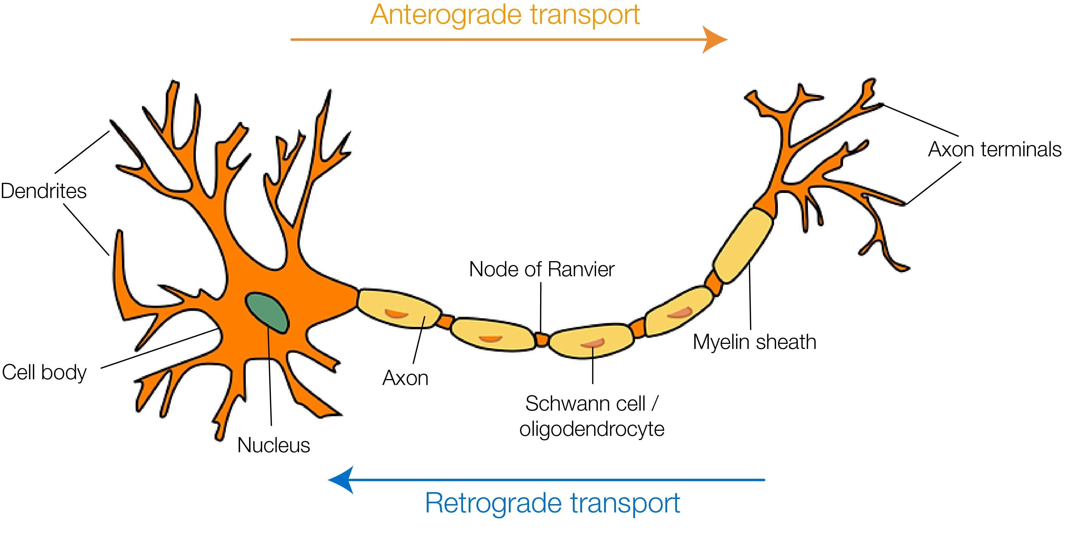
What is the likely result of adding a kinesin inhibitor to neurons in culture?
Atrophy of axons.
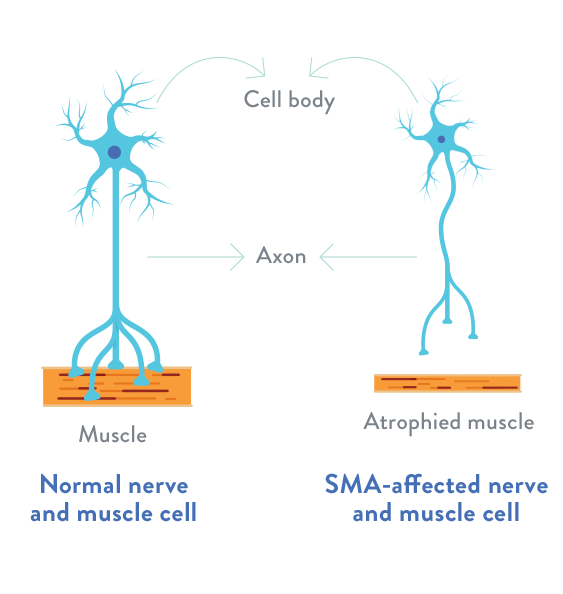
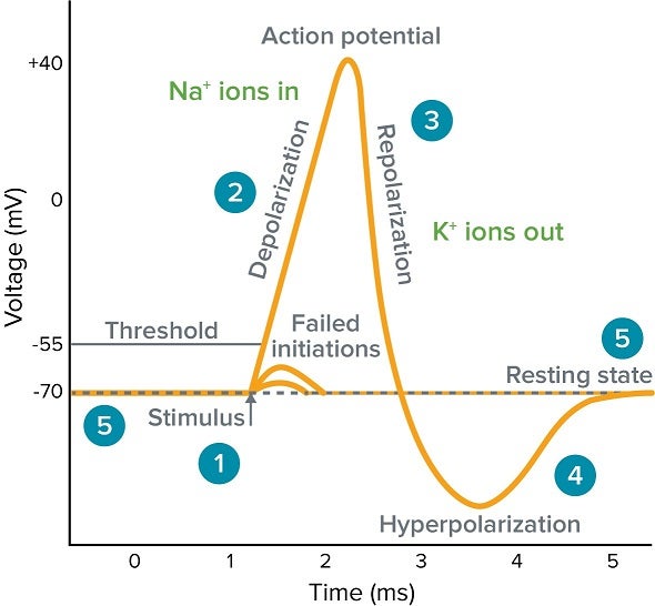
What is the resting membrane potential of a neuron?
Approximately –70 millivolts (mV).
Which two primary membrane proteins are required to establish the resting membrane potential?
The Na+/K+ ATPase and the potassium leak channels.
How many sodium and potassium ions does the Na+/K+ ATPase pump, and in which direction?
The Na+/K+ ATPase pumps three sodium ions out of the cell and two potassium ions into the cell.
What form of transport is carried out by the Na+/K+ ATPase?
Active transport.

What are potassium leak channels?
Channels that allow potassium ions to flow down their gradient out of the cell.
What is the effect of potassium leak channels on the resting membrane potential?
They contribute to the cell having a net negative charge by allowing positive potassium ions to leave the cell.
How does the interior of the cell become polarized?
The combined action of the Na+/K+ ATPase and potassium leak channels results in a negative charge inside the cell and a positive charge outside.
What is depolarization?
A change in membrane potential from the resting potential of approximately –70 mV to a less negative or even positive potential.

What proteins are key in the propagation of action potentials?
Transmembrane proteins — Voltage-gated sodium channels.
What is the effect of opening voltage-gated sodium channels on the membrane potential?
It causes depolarization of the membrane.

At what membrane potential do voltage-gated sodium channels open?
At a threshold potential of approximately –50 mV.

What is the result of opening voltage-gated sodium channels during an action potential?
Sodium flows into the cell, depolarizing that section of the membrane to about +35 mV before the channels inactivate.

Which causes the interior of the neuron to have a momentary positive charge?
Opening of voltage-gated sodium channels.
How do voltage-gated sodium channels respond to a membrane depolarization from –70 mV to –60 mV?
None of the channels open.
What happens to sodium channels shortly after they open during depolarization?
Voltage-gated sodium channels inactivate very quickly after they open, shutting off the flow of sodium into the cell.
How do voltage-gated potassium channels contribute to repolarization?
Voltage-gated potassium channels open more slowly than sodium channels and stay open longer, allowing potassium to leave the cell and return the membrane potential to negative values.
What is the effect of the Na+/K+ ATPase and potassium leak channels on membrane potential?
They continue to function to bring the membrane back to resting potential, even without the voltage-gated potassium channels, but the process would take longer.
What creates the myelin sheath around axons in the peripheral nervous system?
Schwann cells, a type of glial cell.
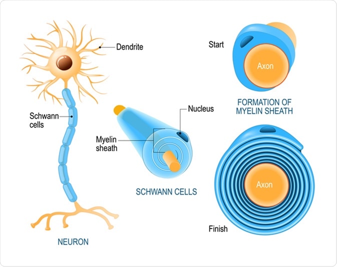
What are nodes of Ranvier and their function?
They are periodic gaps in the myelin sheath where voltage-gated sodium and potassium channels are concentrated, allowing for saltatory conduction.

How does myelin affect the conduction of action potentials?
Myelin speeds the movement of action potentials by forcing them to jump from node to node.
What is true concerning myelinated and unmyelinated axons?
The amount of energy consumed by the Na+/K+ ATPase is much less in myelinated axons than in unmyelinated axons.
What are glial cells and their general function?
Glial cells are specialized, non-neuronal cells that provide structural and metabolic support to neurons.
What is the equilibrium potential of an ion?
It is the membrane potential at which the driving force (the gradient) for an ion does not exist, resulting in no net movement of ions across the membrane.
What is the equilibrium potential for Na+ and what drives it?
The equilibrium potential for Na+ is approximately +50 mV, driven by the chemical gradient driving sodium in and the electrical gradient driving sodium out.
What is the equilibrium potential for K+ and what drives it?
The equilibrium potential for K+ is approximately –90 mV, driven by the chemical gradient driving potassium out and the electrical gradient driving potassium in.
What equation is used to predict the equilibrium potential for an ion?
The Nernst equation.
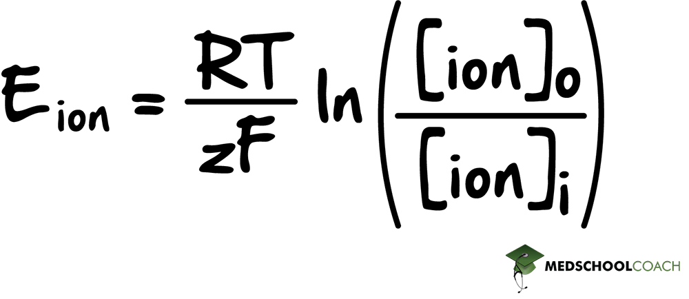
What does the resting membrane potential of –70 mV reflect?
It reflects the differences in the equilibrium potentials for Na+ and K+ and the relative numbers of leak channels for these ions.
What is the refractory period?
It is a period during which a neuron is nonresponsive to membrane depolarization and unable to transmit another action potential immediately after one has passed.

What are the two phases of the refractory period?
The absolute refractory period and the relative refractory period.

What characterizes the absolute refractory period?
During the absolute refractory period, a neuron will not fire another action potential no matter how strong the membrane depolarization is.
What characterizes the relative refractory period?
During the relative refractory period, a neuron can transmit an action potential, but it requires a greater stimulus because the membrane is hyperpolarized.
How would quicker closing of voltage-gated potassium channels after repolarization affect the refractory period in a fruit fly mutant?
The refractory period would be shorter.
What is a synapse?
A synapse is a junction between the axon terminus of a neuron and the dendrites, soma, or axon of another neuron or an organ.
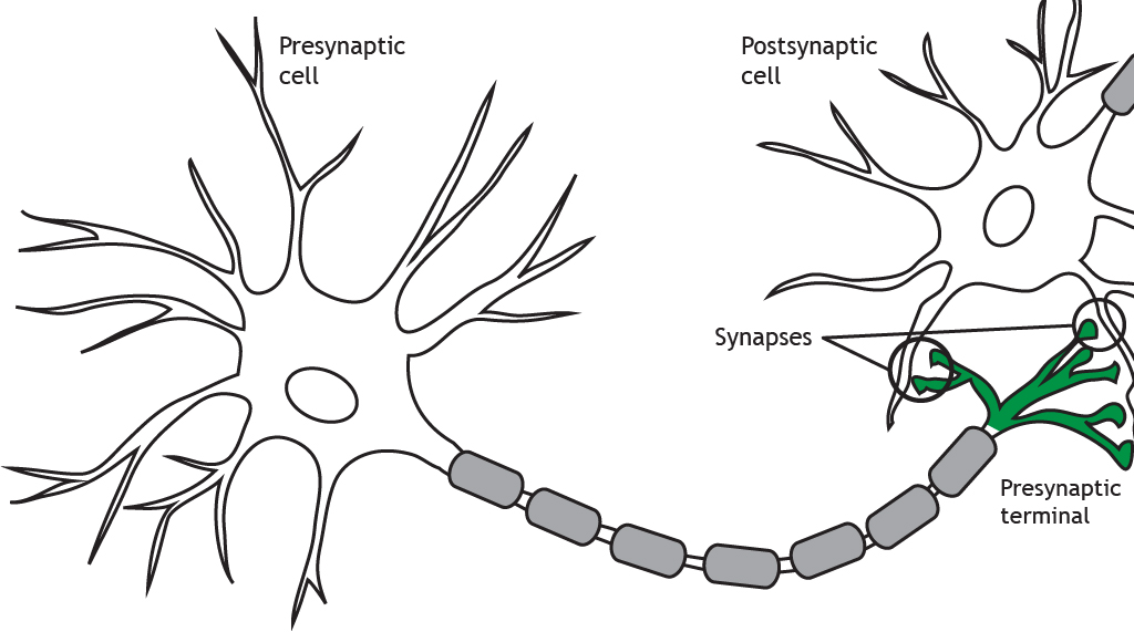
What are the two types of synapses and where are they found?
1) Electrical synapses occur where cytoplasms of two cells are joined by gap junctions. They are important in smooth and cardiac muscle.
2) Chemical synapses are found at the ends of axons where they meet their target cell and convert action potentials into chemical signals.
What are the steps involved in chemical synaptic transmission?
1) An action potential reaches the synaptic knob.
2) Depolarization opens voltage-gated calcium channels.
3) Calcium influx causes neurotransmitter release via exocytosis.
4) Neurotransmitter diffuses across the synaptic cleft.
5) Neurotransmitter binds to receptors on the postsynaptic membrane.
6) Opening of ion channels alters membrane polarization.
7) If depolarization reaches threshold, an action potential is initiated.
8) Neurotransmitter is degraded or removed to terminate the signal.
What neurotransmitter is commonly used at the neuromuscular junction?
Acetylcholine (ACh).
What determines whether a neurotransmitter is excitatory or inhibitory?
It depends on whether the neurotransmitter opens channels that depolarize (excitatory) or hyperpolarize (inhibitory) the postsynaptic membrane.
If a neurotransmitter causes chloride entry into the postsynaptic cell, is it excitatory or inhibitory?
Inhibitory
What happens if an inhibitor of acetylcholinesterase is added to a neuromuscular junction?
The postsynaptic membrane will be depolarized longer with each action potential.
What ensures signals are sent in only one direction through synapses like the neuromuscular junction?
Axons can propagate action potentials in only one direction.
What is summation in synaptic transmission?
It's the process where the effects of multiple EPSPs and IPSPs are combined to determine whether a postsynaptic neuron will fire an action potential.
What are EPSPs and IPSPs?
EPSPs are excitatory postsynaptic potentials that depolarize the postsynaptic membrane. IPSPs are inhibitory postsynaptic potentials that hyperpolarize the membrane.
What are the two forms of summation?
Temporal summation (EPSPs or IPSPs piling up due to rapid firing) and spatial summation (summing EPSPs and IPSPs from all synapses at a given moment).
How can a presynaptic neuron increase the intensity of the signal it transmits?
Increase the frequency of action potentials.
What is the threshold depolarization required for initiating an action potential?
About –50 mV.
What are the three main functions of the nervous system?
Sensory function, integrative function, and motor function.

Which part of the nervous system is responsible for receiving information?
The peripheral nervous system (PNS).
What is the integrative function of the nervous system and which part performs it?
Processing information, carried out by the central nervous system (CNS).
Which part of the nervous system is responsible for acting on information?
The motor function, carried out by the peripheral nervous system (PNS).
What is the role of motor neurons in the nervous system?
To carry information from the nervous system toward organs which can act upon that information (effectors).

What are the two types of effectors?
Muscles and glands.

What are efferent neurons?
Neurons that carry information away from the central nervous system and innervate effectors.
What are afferent neurons?
Neurons that carry information toward the central nervous system.
What is a reflex?
A direct motor response to sensory input which occurs without conscious thought, often without any involvement of the brain.
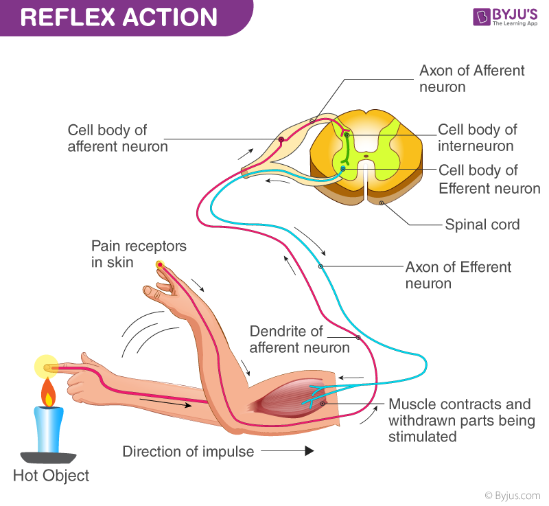
Describe a monosynaptic reflex arc.
A reflex arc involving only two neurons and one synapse, such as the muscle stretch reflex.
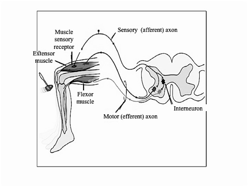
What happens during the muscle stretch reflex?
A sensory neuron detects stretching of a muscle and transmits an impulse to a motor neuron in the spinal cord, causing the muscle to contract.
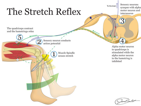
What is reciprocal inhibition?
The process where the contraction of one muscle (e.g., quadriceps) is accompanied by the relaxation of the opposing muscle (e.g., hamstring)
What is the role of an inhibitory interneuron in a reflex arc?
To form an inhibitory synapse with a motor neuron innervating the opposing muscle, facilitating reciprocal inhibition.
What are the two main subdivisions of the peripheral nervous system (PNS)?
The somatic division and the autonomic division.
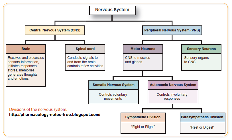
What does the somatic division of the PNS control?
Conscious sensation and deliberate, voluntary movement of skeletal muscle.
What does the autonomic division of the PNS control?
Involuntary processes like digestion, metabolism, circulation, and perspiration.
What are the two subdivisions of the autonomic division of the PNS?
The sympathetic system and the parasympathetic system.
What is the function of the sympathetic system?
To prepare the body for "fight or flight" responses.
What is the function of the parasympathetic system?
To prepare the body to "rest and digest."
What role does epinephrine play in the sympathetic system?
Many sympathetic effects result from the release of epinephrine into the bloodstream by the adrenal medulla.

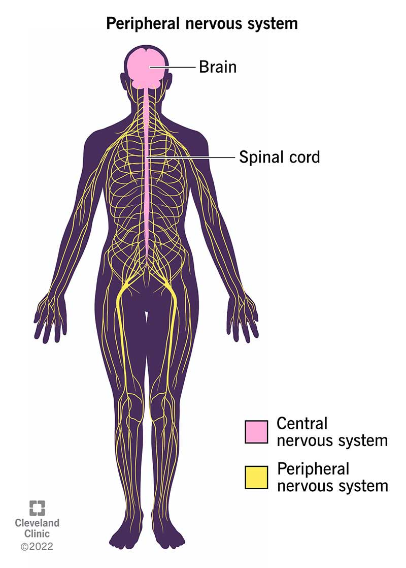
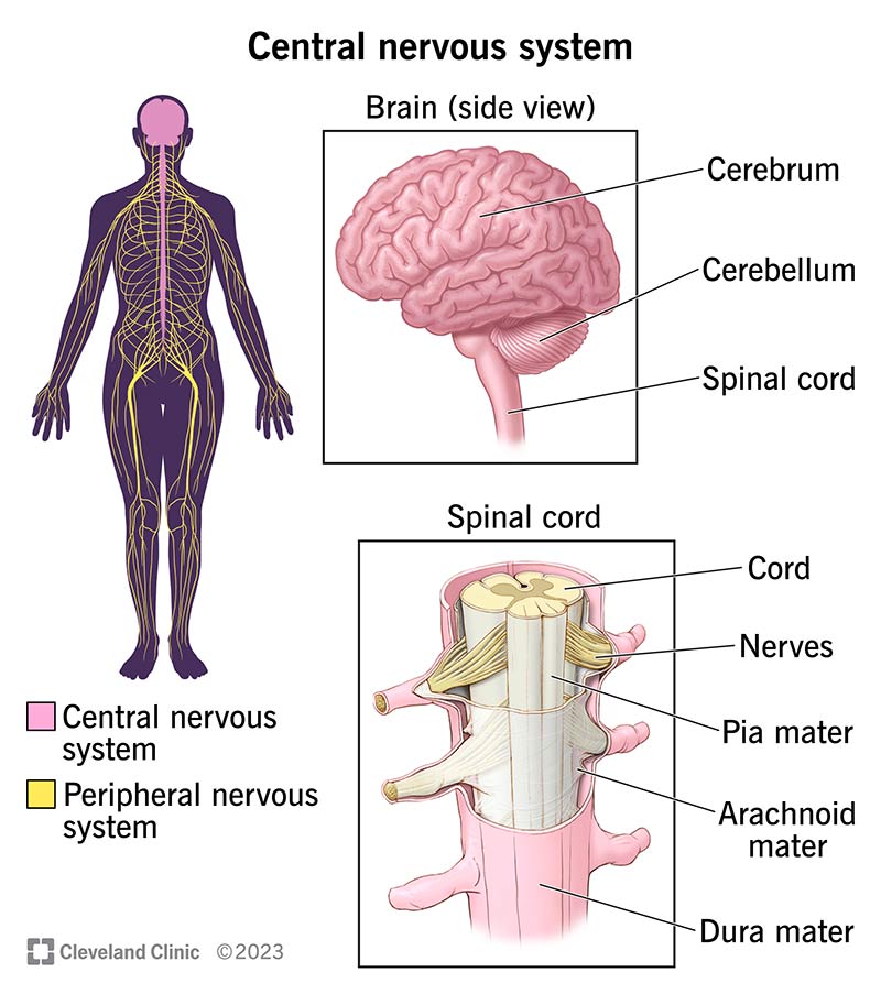
What are the two main anatomical divisions of the nervous system?
The central nervous system (CNS) and the peripheral nervous system (PNS).
What does the central nervous system (CNS) include?
The brain and spinal cord.
What does the peripheral nervous system (PNS) include?
All other axons, dendrites, and cell bodies outside the CNS.
Where are neuronal cell bodies mostly found?
Within the central nervous system (CNS).
What are clusters of cell bodies in the CNS called?
Nuclei
What are clusters of cell bodies in the PNS called?
Ganglia
What are the three subdivisions of the brain?
The hindbrain, midbrain, and forebrain.

What is the function of cerebrospinal fluid (CSF)?
It absorbs shock and facilitates nutrient and waste exchange with the CNS.
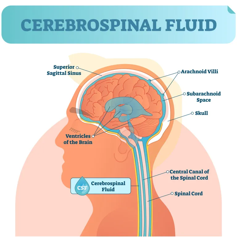
What are the functions of the spinal cord?
It connects to the brain, processes simple reflexes, and is involved in primitive processes like walking and urination.
What structures are included in the hindbrain?
The medulla, pons, and cerebellum.
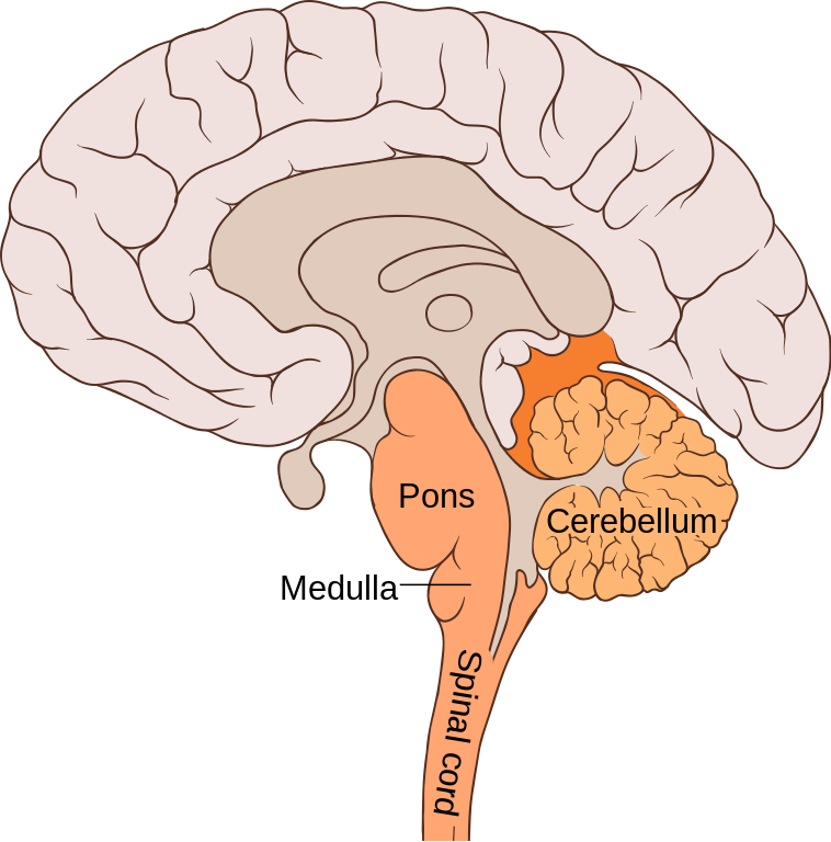
What does the medulla (or medulla oblongata) regulate?
Vital autonomic functions like blood pressure, digestion, and respiratory rhythmicity.

What is the role of the pons?
It connects the brainstem to the cerebellum and helps control autonomic functions and movement coordination.

What is the primary function of the cerebellum?
Coordinating complex movements and maintaining balance and coordination.

What is the role of the midbrain?
It relays visual and auditory information and contains the reticular activating system (RAS) for arousal and wakefulness.

What three structures make up the brainstem?
The medulla, pons, and midbrain.

What does the diencephalon include?
The thalamus and hypothalamus.

What is the function of the thalamus?
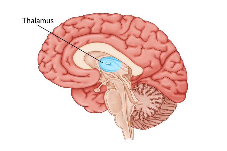
It acts as a relay and processing center for sensory information.
What does the hypothalamus control?
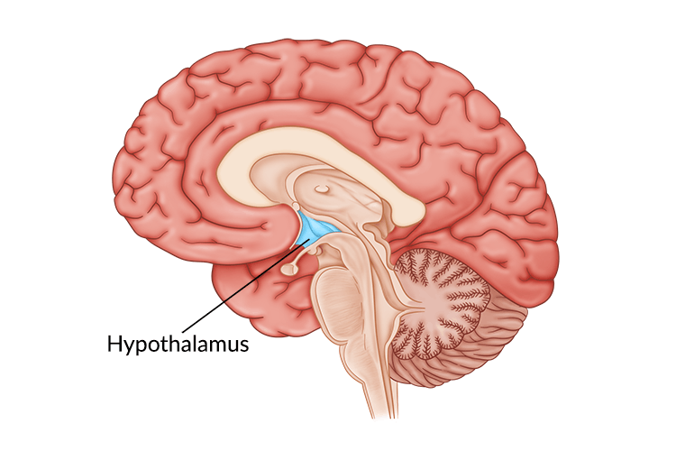
Emotions, autonomic functions, hormone production, and the pituitary gland.
What are the two main parts of the forebrain?
The diencephalon and the telencephalon.

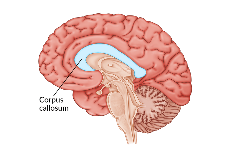
What connects the two cerebral hemispheres?
The corpus callosum.
