parasites
1/36
There's no tags or description
Looks like no tags are added yet.
Name | Mastery | Learn | Test | Matching | Spaced | Call with Kai |
|---|
No analytics yet
Send a link to your students to track their progress
37 Terms
parasite
An organism that lives in or on a host organism and benefits at the host's expense, often causing harm.
parasite direct life cycles
swithching b/w multiple hosts
asexual and sexual reporoduction
transmission of parasites in same species
indirect life cycle
transmission of parasites from 1 host to next in different species
heterogenous parasites
definitive host: parasites reach sexual maturity
intermediate host: parasite development but where parasites do not reach sexual maturity.
helminths
Nematodes (Roundworms)
Cylindrical, unsegmented
GI and tissue infections
Examples: Ascaris, Enterobius (pinworm), Strongyloides
Cestodes (Tapeworms)
Flat, segmented
Absorb nutrients through skin
Examples: Taenia, Echinococcus
Trematodes (Flukes)
Flat, leaf-shaped
Infect blood, liver, lungs
Examples: Schistosoma, Fasciola
arthropods
Characteristics: Exoskeleton, segmented body, jointed limbs
Roles in Disease:
Ectoparasites (live on host)
Direct irritation + secondary infection
Examples: Mites (Scabies), lice, fleas, ticks
Vectors (transmit disease)
Mechanical (carry pathogens): e.g., flies
Biological (pathogen develops in vector):
Mosquitoes – malaria, dengue
Ticks – Lyme disease, babesiosis
Fleas – plague
protozoa
unicellular euk organisms that can cause diseases in humans and animals, typically spreading through water or food.
asexual reproduction usually: binary fission
movement: pseudopodia, cilia, flagella. gliding
giardia
Location: small intestine
Reproduction: binary fission;
Motility: by using flagella;
Infection: Ingest cysts (contaminated water/food)
Excystation: Cysts release trophozoites in the jejunum (triggered by bile & pH ~7.8)
Encystation: Trophozoites form cysts again
Cysts excreted in feces → survive months in environment
Morphology:
Trophozoite
Size: 9–21 µm
Pear-shaped (rounded front, pointed back)
Flattened ventral surface with adhesive disc
Two nuclei
Active, feeding stage
Cyst
Size: 8–14 × 7–10 µm
Oval shape
Four nuclei, axonemes (flagella parts), disc fragments
Infective, resistant stage
Toxoplasma gondii - life cycle and stages in environment
Life Cycle
Indirect life cycle (2 hosts)
Definitive host: Cats (sexual & asexual stages in small intestine)
Intermediate host: Any warm-blooded animal (asexual stages in any nucleated cell)
Sporozoites → tachyzoites → invade & multiply → burst host cells
Tachyzoites convert to bradyzoites → form tissue cysts
Found in brain, muscles, visceral organs
Persist for life of host
Occasionally rupture
Stages in Environment
Cats shed unsporulated oocysts in feces
Sporogony occurs in environment (1–5 days)
Sporulated oocyst: 2 sporocysts, each with 4 sporozoites
Oocysts can survive 12–18 months, resist disinfectants
Toxoplasma gondii - infection routes
Ingestion of sporulated oocysts
From contaminated food/water
Main route for herbivores
Ingestion of tissue cysts
From raw/undercooked meat
Affects carnivores & omnivores
Tachyzoite transmission
Transplacental (mother to fetus)
Milk (goats)
Blood transfusion, organ transplant
Lab accidents
Toxoplasma gondii - Disease & Risks
Immunocompetent hosts: Control infection
Immunocompromised hosts (e.g. AIDS):
Reactivation of cysts → tachyzoites → severe disease (e.g., encephalitis)
Congenital toxoplasmosis:
Transmission to fetus → abortion or severe pathology
Zoonotic potential:
Affects humans, sea mammals, marsupials
Behavioral changes seen in mice, possibly humans
Toxoplasma gondii - survival and inactivation
Oocysts: survive up to 18 months
Tissue cysts in meat:
Survive up to 2 months at 4–6°C
Killed below –12°C or above 65°C
Plasmodium (Malaria) - Transmission & Hosts
Transmitted by: Female Anopheles mosquitoes
Indirect life cycle:
Definitive host: Mosquito (sexual reproduction)
Intermediate host: Human (asexual reproduction)
Plasmodium (Malaria)- Life Cycle in Humans (Asexual Stage)
Mosquito bite → injects sporozoites
Sporozoites → travel to liver → invade hepatocytes
Multiply asexually → liver cells burst → release merozoites
Merozoites → invade red blood cells (RBCs)
Multiply asexually → RBCs burst → more merozoites released
Some develop into gametocytes (male & female
Plasmodium (malaria) - Life Cycle in Mosquito (Sexual Stage)
Mosquito bites infected human → ingests gametocytes
In mosquito gut:
Gametocytes → gametes → fertilize → zygote
Zygote → ookinete → becomes oocyst
Oocyst releases sporozoites → migrate to salivary glands
Ready to infect another human during next bite
Helminths (Worms)
Phylum Platyhelminthes (flatworms)
Phylum Nematoda (roundworms)
Phylum Platyhelminthes (flatworms) - classes
Trematoda (Flukes)
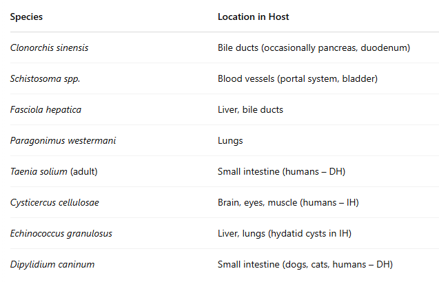
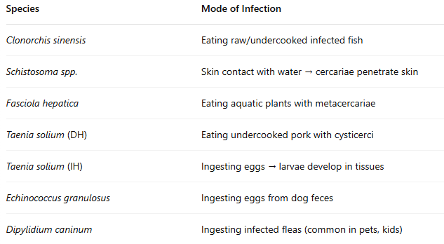
Clonorchis sinensis
Schistosoma spp.
Cestoda (Tapeworms)
Taenia solium, T. saginata
Dipylidium caninum
Phylum Platyhelminthes (flatworms) - general features
Body: flat (dorso-ventrally), bilaterally symmetrical, no body cavity
Organs in parenchyma (specialized connective tissue)
Incomplete or absent digestive tract
Suckers/hooks for attachment
Mostly hermaphroditic (except Schistosoma)
Class Trematoda (Flukes)
Short, flat (2–7.5 cm)
Have oral & ventral suckers
Incomplete digestive tract
Life cycle: indirect, needs at least 1 intermediate host
Examples:
Clonorchis sinensis (liver fluke)
Schistosoma spp. (blood fluke – dioecious)
Fasciola hepatica, Opisthorchis spp., Paragonimus westermani
Class Cestoda (Tapeworms)
Long (mm to meters), segmented body
Segments = proglottids; head = scolex (with hooks/suckers)
No digestive tract
Hermaphroditic
Life cycle: indirect, larval stage infects tissues
Examples:
Taenia solium, T. saginata, Echinococcus granulosus
Hymenolepis nana, Dipylidium caninum, Diphyllobothrium latum
Anatomic Locations in the Host
how hosts become affected
Phylum Nematoda (Roundworms) - classes
Ascaris lumbricoides
Ancylostoma caninum (hookworms)
Ascaris lumbricoides morphological features
Adults:
Large cylindrical worms, up to 35 cm in length.
Anterior end has 3 prominent lips.
Feed on intestinal contents and epithelial cells.
Eggs:
Oval, yellow-brown, and have a thick mammillated shell.
Contains one cell inside.
Can survive in the environment for 5–7 years
Ascaris lumbricoides Biology / Life Cycle
Female worms produce hundreds of thousands of eggs per day.
Eggs are passed in feces.
In the environment, embryonated eggs develop in 10–15 days.
Host becomes infected by ingesting embryonated eggs.
Inside host:
Eggs hatch in small intestine.
Larvae migrate: intestine → liver → heart → lungs → alveoli → pharynx → swallowed → small intestine.
Mature into adult worms in small intestine.
Ascaris lumbricoides anatomical location
Primary: Small intestine.
Others: Bile ducts, pancreatic ducts, stomach.
May be seen vomited, or exit through mouth/nose in heavy infections
Ascaris lumbricoides Infection Mode
Ingestion of embryonated eggs from contaminated food, water, or hands.
Ancylostoma caninum (Hookworm) - Morphological Features
Adults:
Anterior end is dorsally bent (hook-shaped).
Large buccal capsule with 3 pairs of sharp teeth.
Feed by biting the intestinal mucosa, creating wounds.
Frequent change of feeding sites (4–6 times/day).
Eggs:
Thin-shelled, morula stage (many cells inside).
About 16,000–20,000 eggs/day laid by females.
Ancylostoma caninum (Hookworm) - Biology / Life Cycle
Eggs passed in dog feces.
In environment:
Eggs develop: L1 → L2 → L3.
L3 is the infective stage.
Infection routes:
Transcutaneous: L3 penetrates skin → migrates to lungs → trachea → pharynx → swallowed → small intestine.
Oral ingestion: Direct maturation in the small intestine.
Transmammary: Arrested larvae reactivated during pregnancy → passed to puppies through milk.
Via paratenic hosts (mice, rats, birds).
Some larvae go into hypobiosis (dormant stage), reactivating later.
Ancylostoma caninum (Hookworm) - Anatomic Location
Dogs: Adult worms in small intestine – cause severe blood loss.
Humans:
Adults may cause eosinophilic enteritis.
Larvae cause cutaneous larva migrans (CLM), folliculitis, etc
Ancylostoma caninum (Hookworm) - infection mode
Skin penetration (walking barefoot on contaminated ground).
Ingestion of infective larvae or paratenic hosts.
Transmammary transmission in dogs.
Phylum Arthropoda - general morphological features
Body:
Segmented body covered by a protective exoskeleton (made of chitin).
Legs:
Jointed appendages (arthron = joint, podos = foot).
Reproduction:
Most are dioecious (separate sexes).
Development:
Molting (ecdysis) is necessary due to the rigid exoskeleton.
Metamorphosis:
Hemimetabolous: Incomplete metamorphosis (egg → nymph → adult).
Holometabolous: Complete metamorphosis (egg → larva → pupa → adult)
Pediculus humanus capitis (Head Louse) - morphology
Size: Up to 3.6 mm.
Body shape: Dorso-ventrally flattened.
Segments: Head, thorax, abdomen.
Legs: Three pairs (adapted to cling to hair shafts)
Pediculus humanus capitis (Head Louse) - biology/life cycle
Eggs (nits) are glued to hair shafts.
Eggs hatch in ~7 days.
Three nymph stages (look like small adults).
Total life cycle: 3–4 weeks (egg to egg).
Do not survive long off the host
Pediculus humanus capitis (Head Louse) - anatomical location and infection mode
Anatomic Location
Scalp hair, especially near the nape of the neck and behind ears.
Mode of Infestation
Mainly via head-to-head contact.
Also via contaminated items: combs, hats, headphones, brushes, etc.
Sarcoptes scabiei - Essential Morphological Features
Size: Up to 450 µm (microscopic).
Body: Rounded, compact, unsegmented.
Legs:
4 pairs of short legs in adults and nymphs.
Legs are stubby and suited for burrowing.
Mouthparts: Adapted for skin tunneling.
Eyes: Absent.
Cuticle: Covered with spines and bristles.
Sarcoptes scabiei - Biology & Life Cycle
Species: One species – Sarcoptes scabiei – with host-specific variants (e.g., var. hominis in humans).
Hosts: Wide range – humans, pigs, dogs, cattle, goats, sheep, etc.
Infestations in non-preferred hosts are short-lived and non-reproductive.
Egg
Larva (6 legs)
Nymph (8 legs)
Adult (8 legs)
Gravid females:
Burrow tunnels in the stratum corneum (outer layer of skin).
Lay 1–3 eggs/day.
Live for 1–2 months.
All life cycle stages occur on the host (no environmental stages required).
Transmission:
Direct contact (15–20 minutes of close skin-to-skin contact).
Indirect: Via contaminated bedding, clothing, towels – less efficient.
Immune response:
Immediate and delayed hypersensitivity to mite secretions and feces.
Leads to intense itching (pruritus), especially at night.
Sarcoptes scabiei - anatomical location and host infection
Mites live and reproduce in epidermal tunnels.
Commonly affected areas:
Webbing of fingers
Wrists
Elbows
Armpits
Waistline
Genital area
Human-to-human contact (most common route).
Close contact in families, schools, or care facilities.
Fomites (e.g., towels, bedding) may spread mites indirectly.