12. POPLITEAL FOSSA & LEG

0.0(0)
Studied by 0 people
call kaiCall Kai
Locked
learnLearn
examPractice Test
spaced repetitionSpaced Repetition
heart puzzleMatch
flashcardsFlashcards
GameKnowt Play
Card Sorting

1/39

encourage image

There's no tags or description

Looks like no tags are added yet.

Last updated 2:44 AM on 8/19/26
Name
Mastery
Learn
Test
Matching
Spaced
Call with Kai
Chat

No analytics yet

Send a link to your students to track their progress

40 Terms

1
New cards

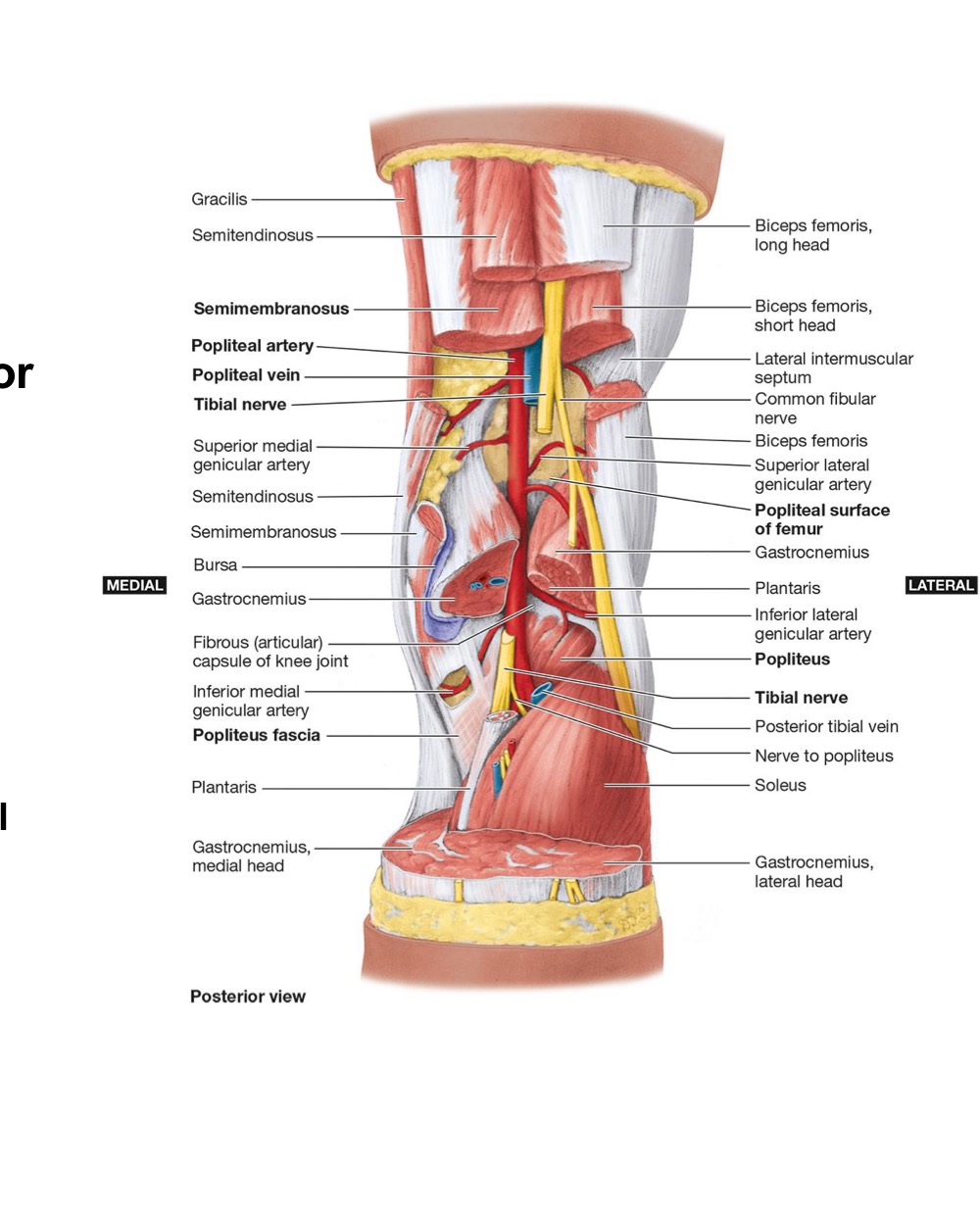
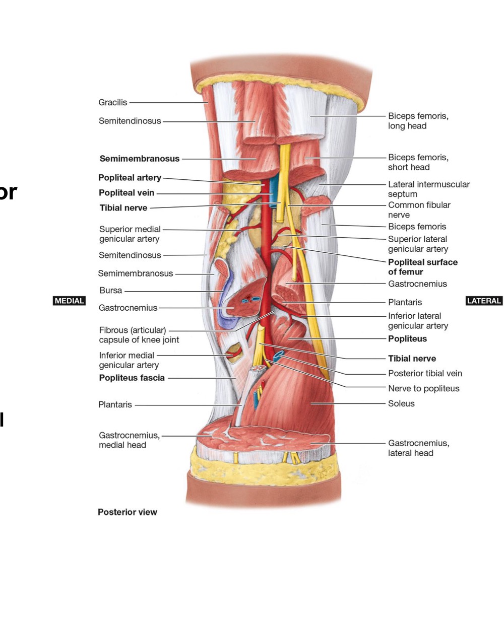
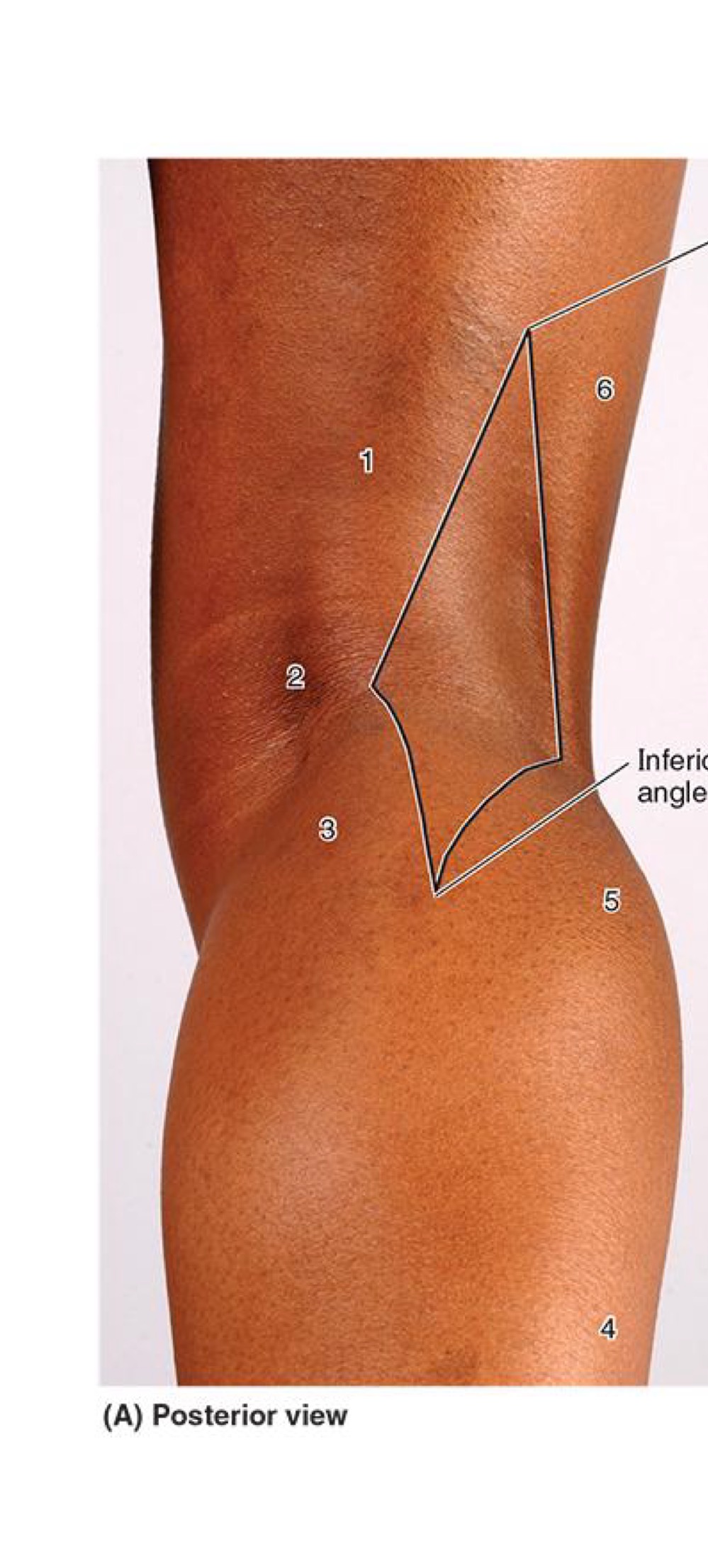
POPLITEAL FOSSA + BORDERS

  • Diamond-shaped depression POST KNEE joint


  • Borders:

    • Superomedial → Semimembranous & Semitendinous m.

    • Superolateral → Biceps Femoris m.

    • Inferomedial → Medial Head of Gastrocnemius

    • Inferolateral → Lateral head of Gastrocnemius

    • Posterior (roof) → Popliteal fascia

    • Floor → Supracondylar Lines of Femur, POST KNEE joint + Popliteus m.

<ul><li><p><strong>Diamond-shaped depression POST KNEE joint</strong></p></li></ul><p></p><ul><li><p><strong>Borders:</strong></p><ul><li><p><strong>Superomedial → Semimembranous &amp; Semitendinous m.</strong></p></li><li><p><strong>Superolateral → Biceps Femoris m.</strong></p></li><li><p><strong>Inferomedial → Medial Head of Gastrocnemius</strong></p></li><li><p><strong>Inferolateral → Lateral head of Gastrocnemius</strong></p></li><li><p><strong>Posterior (roof) → Popliteal fascia</strong></p></li><li><p><strong>Floor → Supracondylar Lines of Femur, POST KNEE joint + Popliteus m.</strong></p></li></ul></li></ul><p></p>
2
New cards

POPLITEAL FASCIA

  • Subcutaneous Fascia (of Skin) overlying popliteal fossa

    • Small saphenous vein

    • POST cutaneous Nerve of THIGH

    • Medial & Lateral Sural Cutaneous Nerves


  • Popliteal Fascia

    • Strong,deep fascia continuous w/ FASCIA LATAE ABOVE + DEEP FASCIA of LEG below


3
New cards

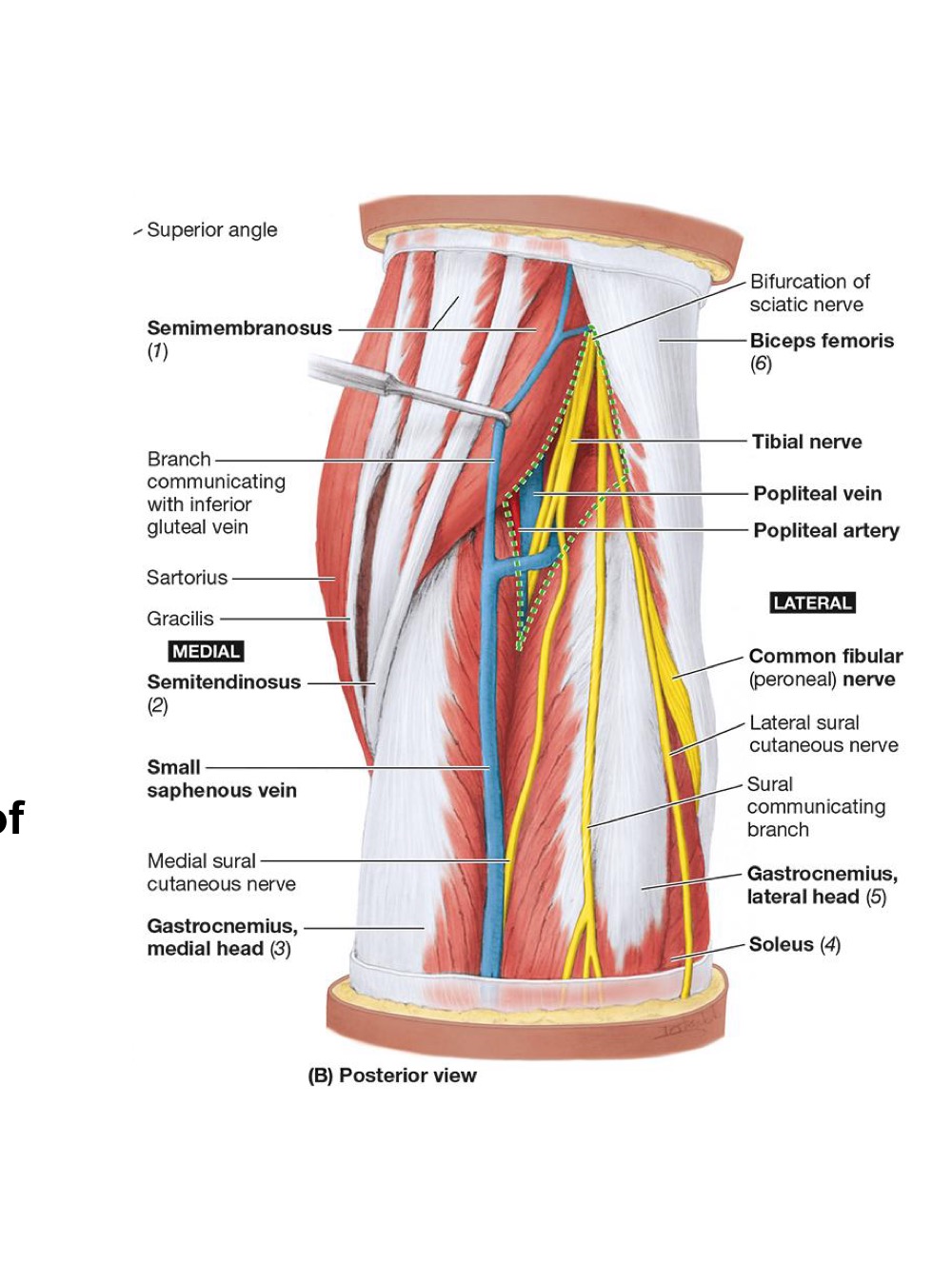
POPLITEAL FOSSA: CONTENTS

  • Popliteal A,

  • Popliteal V.

    • Small Saphaneous drains into

  • Tibial N.

  • Common Fibular (Peroneal) N.

  • POST Cutaneous N. of the THIGH

  • Popliteal Lymph Nodes + Lymphatic Vessels

<ul><li><p><strong>Popliteal A,</strong></p></li><li><p><strong>Popliteal V.</strong></p><ul><li><p><strong>Small Saphaneous drains into</strong></p></li></ul></li><li><p><strong>Tibial N.</strong></p></li><li><p><strong>Common Fibular (Peroneal) N.</strong></p></li><li><p><strong>POST Cutaneous N. of the THIGH</strong></p></li><li><p><strong>Popliteal Lymph Nodes + Lymphatic Vessels</strong></p></li></ul><p></p>
4
New cards

POPLITEAL FOSSA: NERVES

  • Sciatic N. ends + divides into:

    • Tibial N.

      • Medial Sural N. → forms Sural N.

    • Common Fibular N.

      • Sural communicating branch → forms Sural N.

      • Deep Fibular N.

      • Superficial Fibular N.


  • POSTERIOR Cutaneous N. Of the Thigh ends at Popliteal Fossa
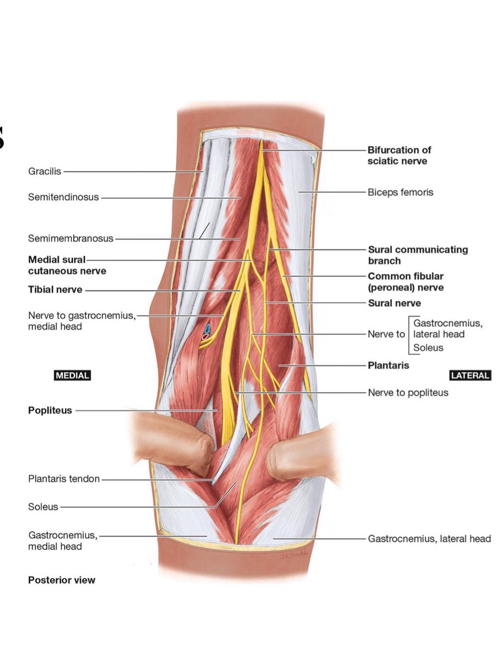

<ul><li><p><strong><mark data-color="yellow" style="background-color: yellow; color: inherit;">Sciatic N. ends + divides into:</mark></strong></p><ul><li><p><strong><mark data-color="yellow" style="background-color: yellow; color: inherit;">Tibial N.</mark></strong></p><ul><li><p><strong><mark data-color="yellow" style="background-color: yellow; color: inherit;">Medial Sural N. → forms Sural N. </mark></strong></p></li></ul></li><li><p><strong><mark data-color="yellow" style="background-color: yellow; color: inherit;">Common Fibular N.</mark></strong></p><ul><li><p><strong><mark data-color="yellow" style="background-color: yellow; color: inherit;">Sural communicating branch → forms Sural N.</mark></strong></p></li><li><p><strong><mark data-color="yellow" style="background-color: yellow; color: inherit;">Deep Fibular N.</mark></strong></p></li><li><p><strong><mark data-color="yellow" style="background-color: yellow; color: inherit;">Superficial Fibular N.</mark></strong></p></li></ul></li></ul></li></ul><p></p><ul><li><p><strong>POSTERIOR Cutaneous N. Of the Thigh ends at Popliteal Fossa</strong></p></li></ul><p></p>
5
New cards

Ignore

Ignore

<p>Ignore </p>
6
New cards

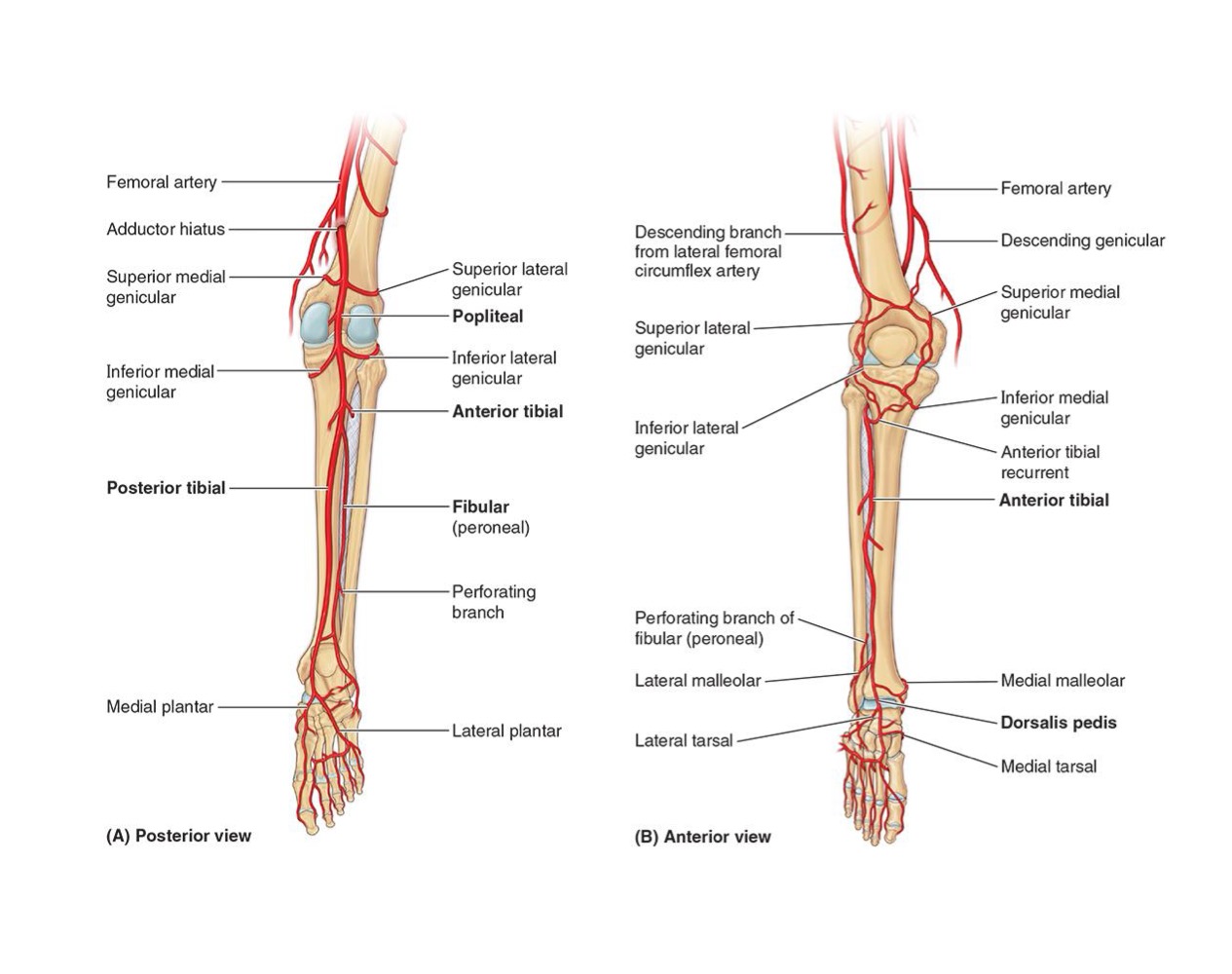
POPLITEAL FOSSA: ARTERIES

  • Popliteal Artery

    • Continuation of FEMORAL ARTERY as it passes through ADDUCTOR Hiatus

    • Ends as bifurcation → into ANT + POST Tibial Arteries
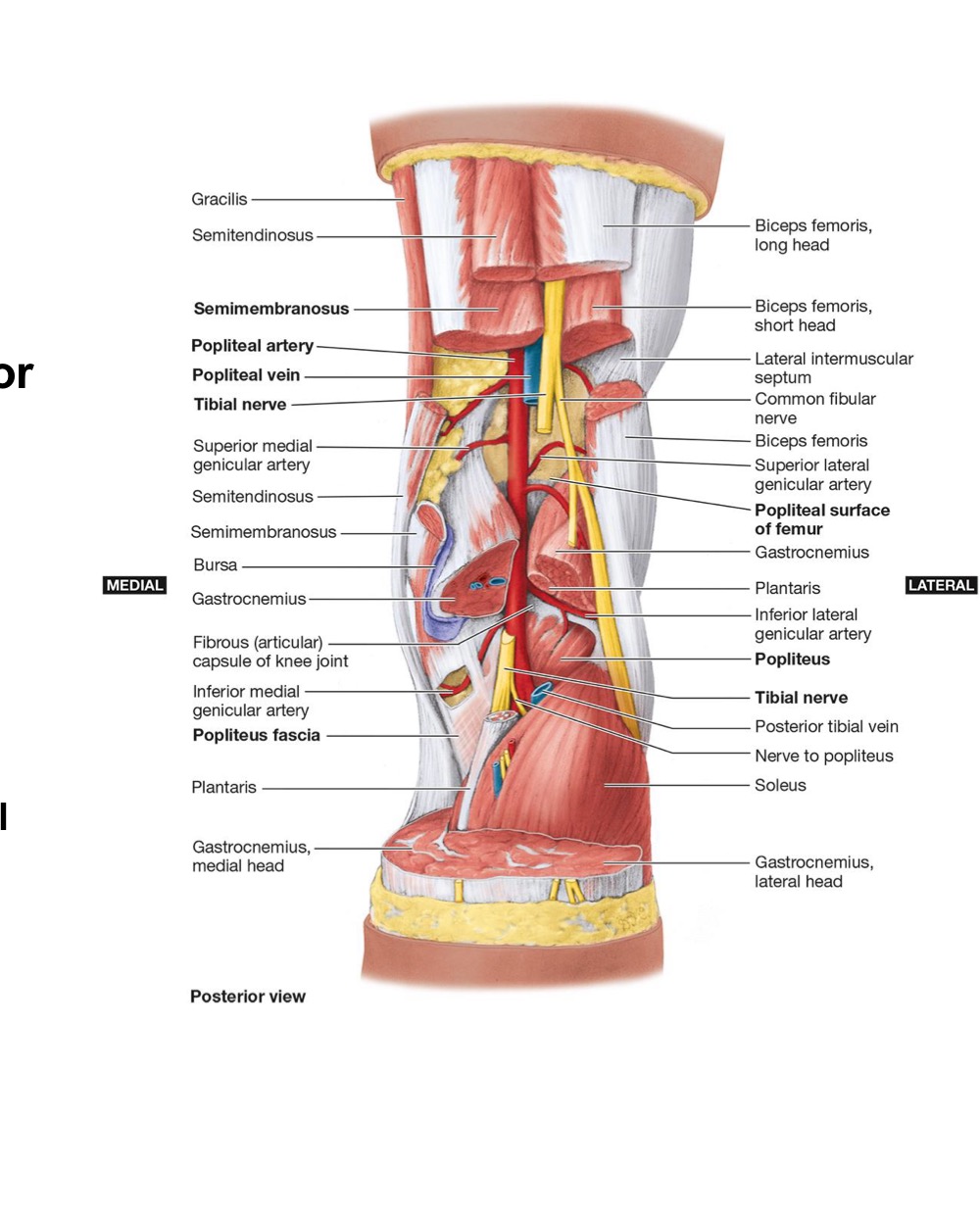


<ul><li><p>Popliteal Artery</p><ul><li><p>Continuation of FEMORAL ARTERY as it passes through ADDUCTOR Hiatus</p></li><li><p>Ends as bifurcation → into ANT + POST Tibial Arteries</p></li></ul></li></ul><p></p><p></p><p></p>
7
New cards

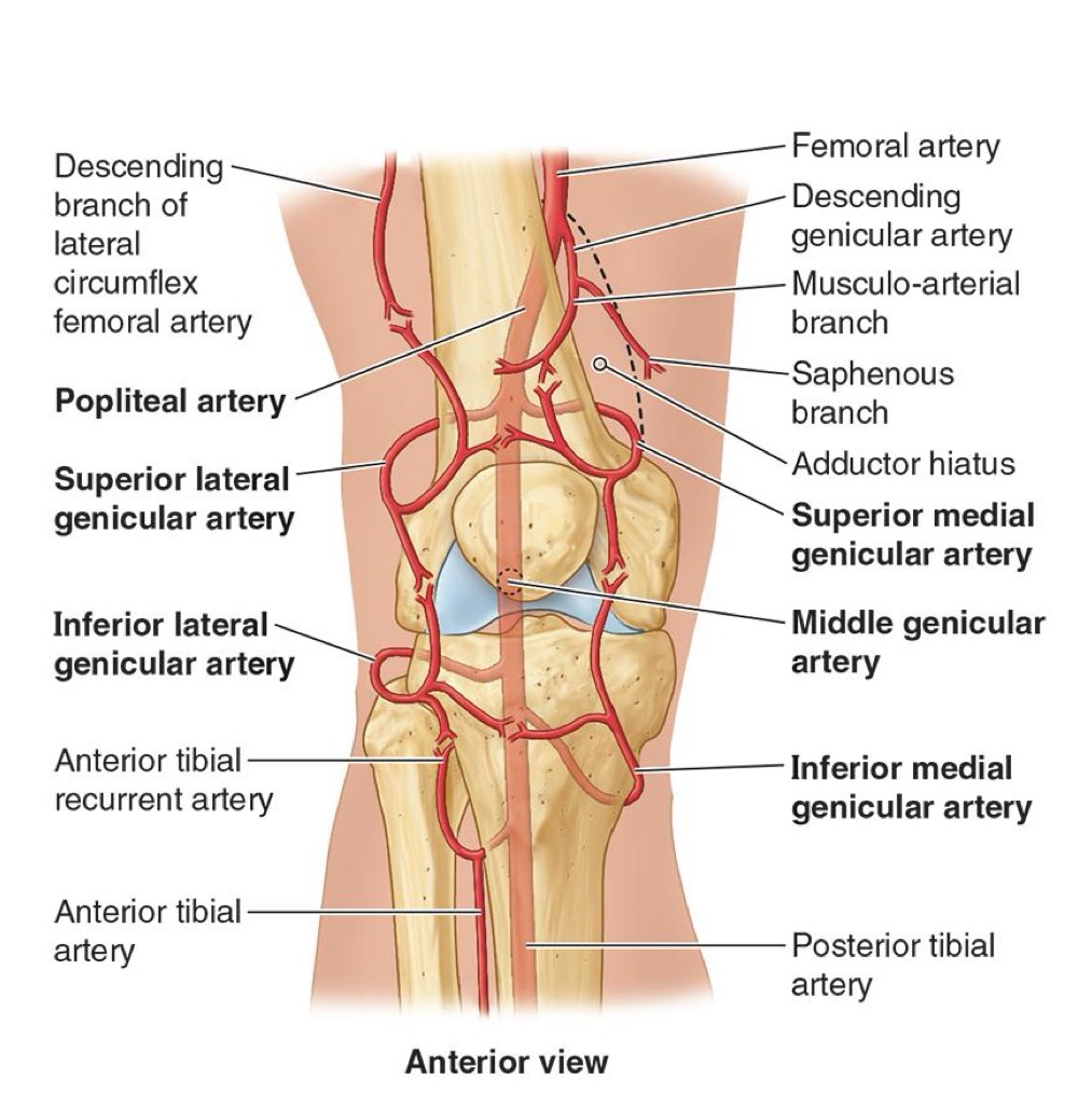
POPLITEAL FOSSA: BRANCHES

  • Include (& contribute to the Genicular Anastomosis)

    • 5 GENICULAR BRANCHES

      • SUP Lateral → anastomoses w/ Descending Lateral Femoral Circumflex A.


      • SUP Medial → anastomoses w/ Descending Genicular A.


      • Middle → anastomoses w/ 4th Perforating A.


      • Inferior Lateral → anastomoses w/ ANT Tibial Recurrent (from ANT Tibial A.)


      • Inferior Medial


    • ANT Tibial A.

    • POST Tibial A.


<ul><li><p>Include (&amp; contribute to the Genicular Anastomosis)</p><ul><li><p><strong>5 GENICULAR BRANCHES</strong></p><ul><li><p><strong>SUP Lateral → anastomoses w/ Descending Lateral Femoral Circumflex A.</strong></p></li></ul><p></p><ul><li><p><strong>SUP Medial → anastomoses w/ Descending Genicular A.</strong></p></li></ul><p></p><ul><li><p><strong>Middle → anastomoses w/ 4th Perforating A.</strong></p></li></ul><p></p><ul><li><p><strong>Inferior Lateral → anastomoses w/ ANT Tibial Recurrent (from ANT Tibial A.)</strong></p></li></ul><p></p><ul><li><p><strong>Inferior Medial</strong></p></li></ul></li></ul><p></p><ul><li><p><strong>ANT Tibial A.</strong></p></li><li><p><strong>POST Tibial A.</strong></p></li></ul></li></ul><p></p><p></p>
8
New cards

GENICULAR ANASTOMOSIS

PICTURE

<p>PICTURE</p>
9
New cards

POPLITEAL FOSSA: VENOUS & LYMPH DRAINAGE

  • Popliteal Vein

    • Begins at the distal border of the POPLITEUS as a continuation of the POST Tibial Vein

    • Ends as the FEMORAL vein as it passes through the ADDuctor Hiatus

    • Tributaries Include:

      • POST Tibial

      • ANT Tibial Vein Begins

      • Genicular Veins


  • Lymph:

    • Superficial POPLITEAL lymph nodes lies → around Small Saphenous Vein

    • Deep Popliteal lymph nodes receive lymph from JOINT CAPSULE of knee + deep veins of leg

    • ALL lymph vessels go to DEEP INGUINAL lymph nodes


10
New cards

LEG: BONES + 3 FASCIAL COMPARTMENTS

  • BONES:

    • Tibia + Fibula

    • Connected by an interosseous membrane


  • 3 FASCIAL COMPARTMENTS:

    • ANT

    • LATERAL

    • POST (Superficial + Deep)

    • Separated by ANT + POST intermuscular septa
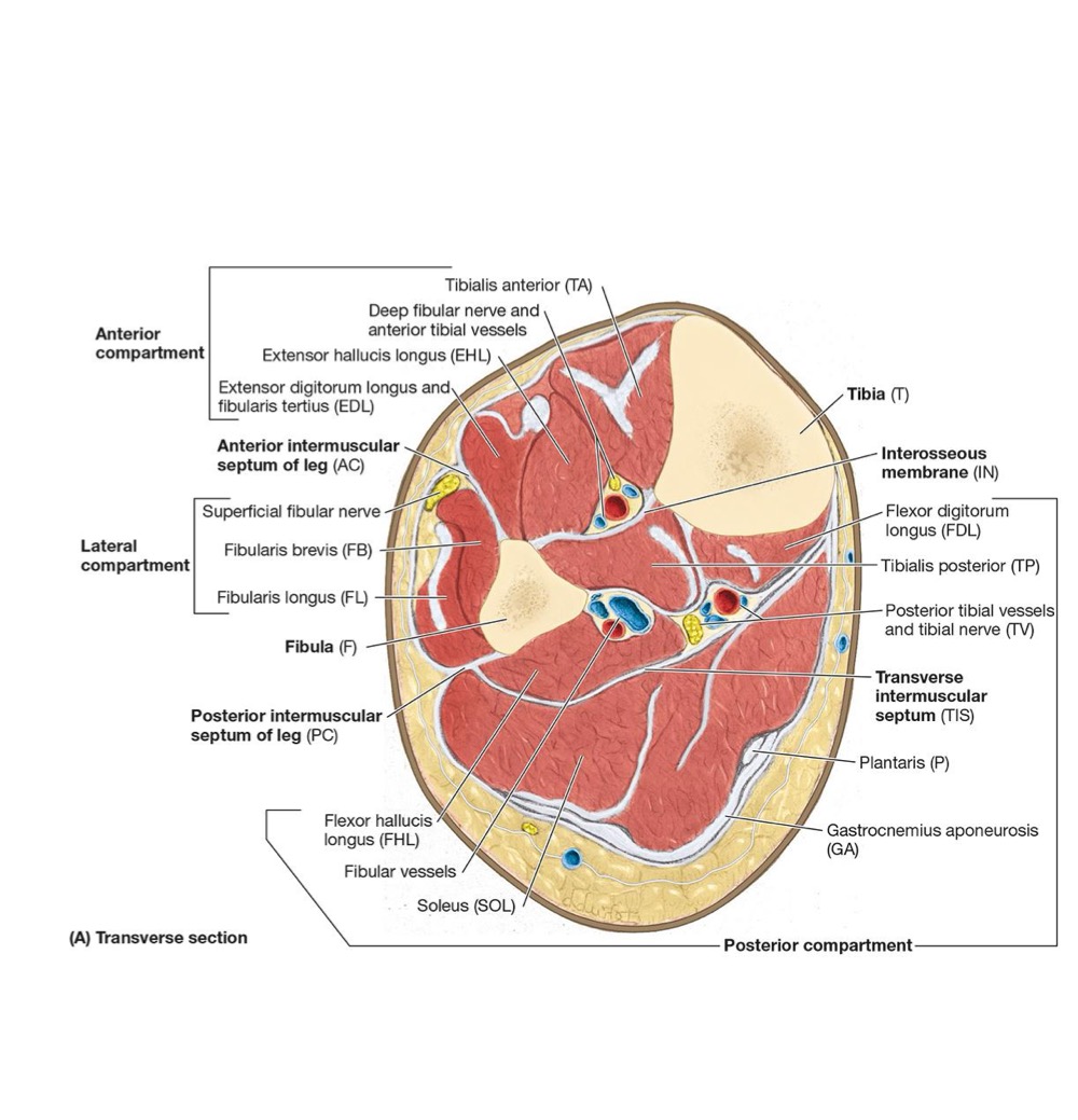


<ul><li><p>BONES:</p><ul><li><p>Tibia + Fibula</p></li><li><p>Connected by an interosseous membrane</p></li></ul></li></ul><p></p><ul><li><p>3 FASCIAL COMPARTMENTS:</p><ul><li><p>ANT</p></li><li><p>LATERAL</p></li><li><p>POST (Superficial + Deep)</p></li><li><p>Separated by ANT + POST intermuscular septa</p></li></ul></li></ul><p></p><p></p><p></p>
11
New cards

ANT COMPARTMENT OF LEG: DORSIFLEXOR (EXTENSOR) COMPARTMENT

  • DORSIFLEXOR (EXTENSOR) COMPARTMENT

    • ANT to the INTEROSSEOUS Membrane btw lateral surface of tibial shaft + medial surface of the shaft of the fibula

      • Inferiorly → 2 band-like thickenings

        • Superior Extensor Retinaculum

        • Inferior Extensor Retinaculum


  • Muscle Actions:

    • Dorsiflexion of ankle

    • Extension of toes

    • Inversion of ankle at subtalar joint


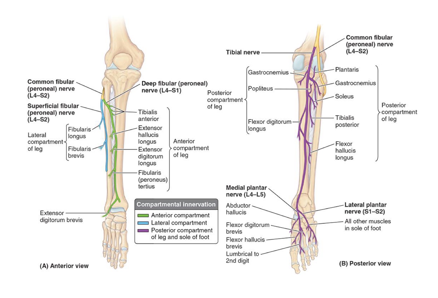
  • Muscle Nerve: Deep Fibular Nerve (from → Common Fibular Nerve)

  • Artery Supply: ANT Tibial Artery (from → Popliteal Artery)
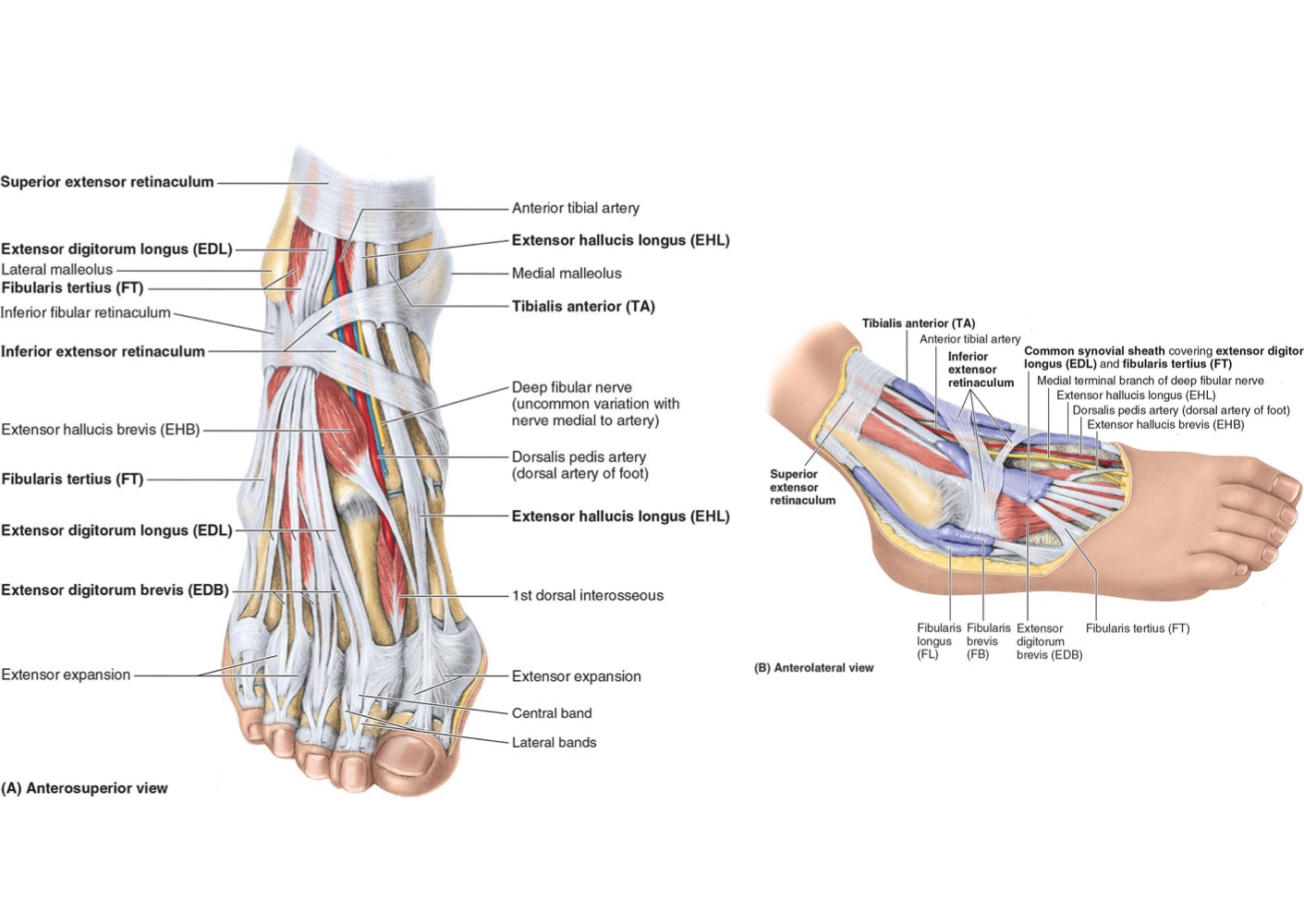

<ul><li><p><strong>DORSIFLEXOR (EXTENSOR) COMPARTMENT</strong></p><ul><li><p>ANT to the INTEROSSEOUS Membrane btw lateral surface of tibial shaft + medial surface of the shaft of the fibula</p><ul><li><p><strong>Inferiorly → 2 band-like thickenings</strong></p><ul><li><p><strong>Superior Extensor Retinaculum</strong></p></li><li><p><strong>Inferior Extensor Retinaculum</strong></p></li></ul></li></ul></li></ul></li></ul><p></p><ul><li><p><strong>Muscle Actions:</strong></p><ul><li><p><strong>Dorsiflexion of ankle</strong></p></li><li><p><strong>Extension of toes</strong></p></li><li><p><strong>Inversion of ankle at subtalar joint</strong></p></li></ul></li></ul><p></p><ul><li><p><strong>Muscle Nerve: Deep Fibular Nerve (from → Common Fibular Nerve)</strong></p></li><li><p><strong>Artery Supply: ANT Tibial Artery (from → Popliteal Artery)</strong></p></li></ul><p></p>
12
New cards

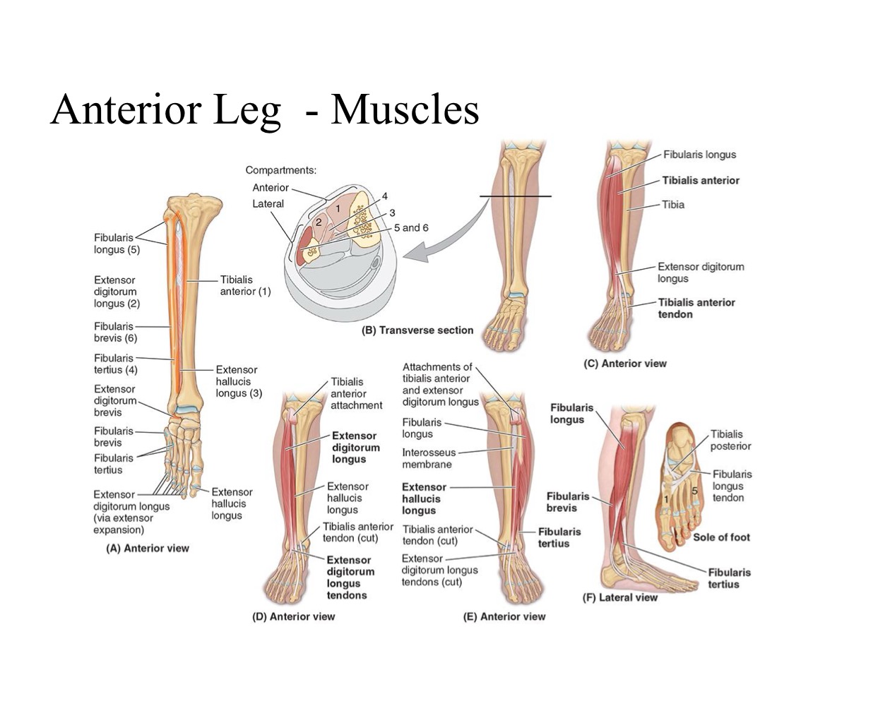
ANTERIOR LEG MUSCLES- DORSIFLEXION OF ANKLE: TIBIALIS ANTERIOR

  • PA: Lateral condyle + Lateral superiors surface of tibia & interosseous membrane


  • DA: Medial cuneiform + base of 1st Metatarsal


  • N: Deep Fibular Nerve


  • A: Dorsiflexes ankle → Inverts subtalar joint

<ul><li><p><strong>PA: </strong>Lateral condyle + Lateral superiors surface of tibia &amp; interosseous membrane</p></li></ul><p></p><ul><li><p><strong>DA: </strong>Medial cuneiform + base of 1st Metatarsal</p></li></ul><p></p><ul><li><p><strong>N: </strong>Deep Fibular Nerve</p></li></ul><p></p><ul><li><p><strong>A: </strong>Dorsiflexes ankle → Inverts subtalar joint</p></li></ul><p></p>
13
New cards

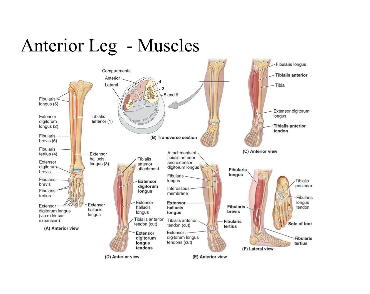
ANTERIOR LEG MUSCLES- DORSIFLEXION OF ANKLE: EXTENSOR DIGITORUM LONGUS

  • PA: Lateral condyle tibia + Superior medial surface of fibula + Interosseous membrane


  • DA: Middle & distal phalanges of lateral 4 digits (toes)


  • N: Deep Fibular Nerve


  • A: Extends lateral 4 toes + Dorsiflexes ankle → Inverts subtalar joint

<ul><li><p><strong>PA: </strong>Lateral condyle tibia + Superior medial surface of fibula + Interosseous membrane</p></li></ul><p></p><ul><li><p><strong>DA: </strong>Middle &amp; distal phalanges of lateral 4 digits (toes)</p></li></ul><p></p><ul><li><p><strong>N: </strong>Deep Fibular Nerve</p></li></ul><p></p><ul><li><p><strong>A: </strong>Extends lateral 4 toes + Dorsiflexes ankle → Inverts subtalar joint</p></li></ul><p></p>
14
New cards

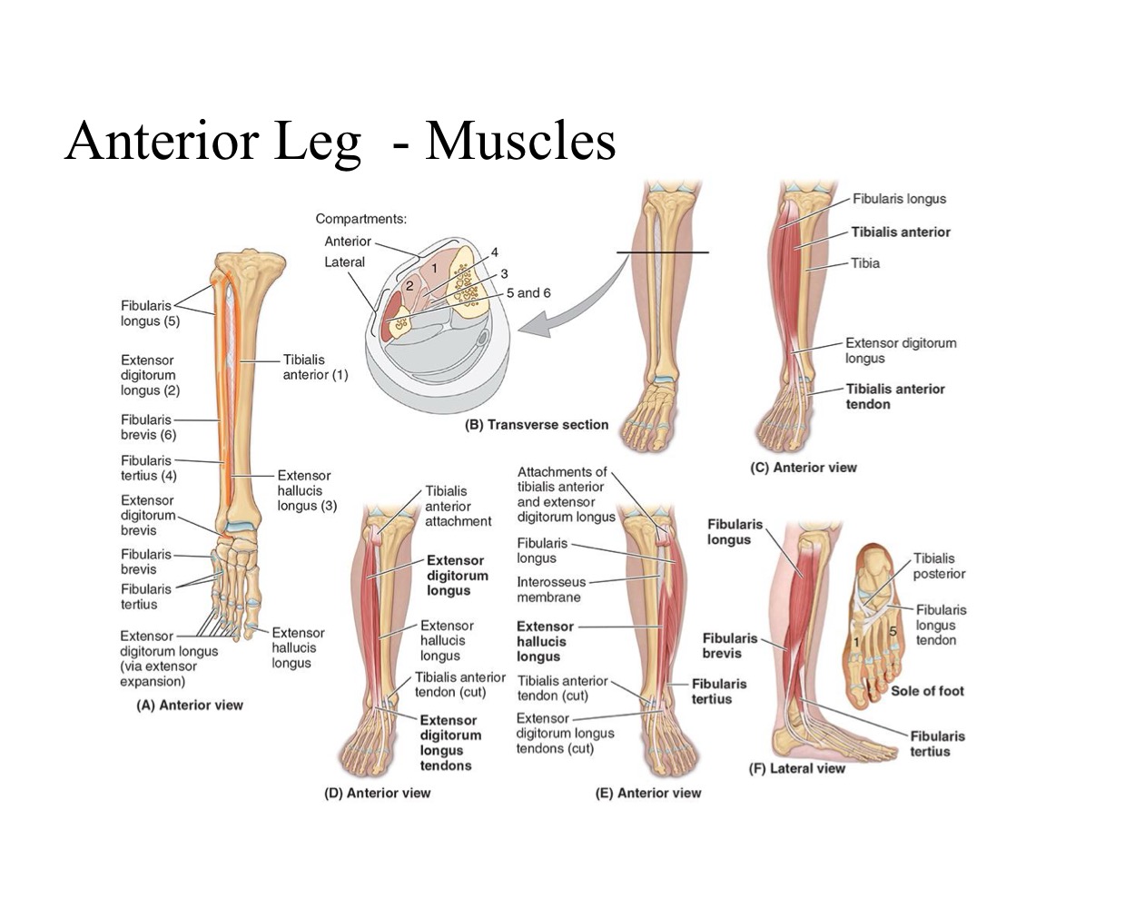
ANTERIOR LEG MUSCLES- DORSIFLEXION OF ANKLE: EXTENSOR HALLUCIS LONGUS

  • PA: Middle part of fibula & interosseous membrane


  • DA: Base of distal phalanx of great toe (Hallux)


  • N: Deep Fibular Nerve


  • A: Extends great toe + Dorsiflexes ankle joint

<ul><li><p><strong>PA: </strong>Middle part of fibula &amp; interosseous membrane</p></li></ul><p></p><ul><li><p><strong>DA: </strong>Base of distal phalanx of great toe (Hallux)</p></li></ul><p></p><ul><li><p><strong>N: </strong>Deep Fibular Nerve</p></li></ul><p></p><ul><li><p><strong>A: </strong>Extends great toe + Dorsiflexes ankle joint</p></li></ul><p></p>
15
New cards

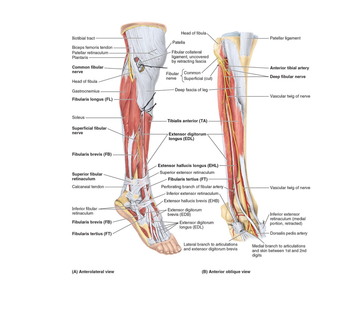
ANTERIOR LEG MUSCLES- DORSIFLEXION OF ANKLE: FIBULARIS TERTIUS

  • PA: Inferior third fibula & interosseous membrane


  • DA: Dorsum of base of 5th metatarsal


  • N: Deep Fibular Nerve


  • A: Dorsiflexes ankle + Everts subtalar joint

<ul><li><p><strong>PA: </strong>Inferior third fibula &amp; interosseous membrane</p></li></ul><p></p><ul><li><p><strong>DA: </strong>Dorsum of base of 5th metatarsal</p></li></ul><p></p><ul><li><p><strong>N: </strong>Deep Fibular Nerve</p></li></ul><p></p><ul><li><p><strong>A: </strong>Dorsiflexes ankle + Everts subtalar joint</p></li></ul><p></p>
16
New cards

LATERAL COMPARTMENT OF LEG: EVERTOR COMPARTMENT

  • Bound by lateral surface of fibula + ANT & POST intermuscular septa + deep fascia of leg

  • Ends Inferiorly at SUPERIOR Fibular Retinaculum


  • 2 muscles innervated → Superficial Fibular Nerve

  • Tendons pass POSTERIORLY to Lateral Malleolus

  • Blood Supply:

    • Perforating Branches of ANT Tibial Artery

    • Perforating Branches of Fibular Artery


17
New cards

LATERAL LEG MUSCLES → EVERTORS: FIBULARIS (PERONEUS) LONGUS (FL)

  • PA: Head & Superior 2/3 of fibula


  • DA: Base of 1st Metatarsal


  • N: Superficial Fibular Nerve


  • A:

    • Everts Subtalar joint

    • Weakly plantar flexes ankle joint


18
New cards

LATERAL LEG MUSCLES → EVERTORS: FIBULARIS (PERONEUS) BREVIS (FB)

  • PA: Head & Superior 2/3 of fibula


  • DA: Base of 1st Metatarsal


  • N: Superficial Fibular Nerve


  • A: Everts Subtalar joint + weakly plantar flexes ankle joint


19
New cards

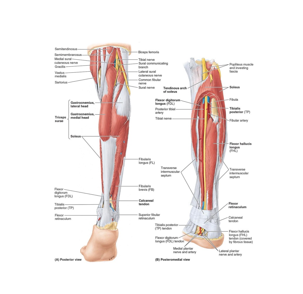
POSTERIOR COMPARTMENT OF LEG: PLANTARFLEXION COMPARTMENT

  • POST compartment = Superficial & Deep subcompartments → separated by Transverse intermuscular septum)

  • INFERIORLY, the Flexor Retinaculum (btw MEDIAL Malleolus & Calcaneus) hold deep muscle tendons + POsT Tibial Artery as they bend around Medial Malleolus


  • ALL innervated by → TIBIAL NERVE

  • Supplied by → POST TIBIAL & FIBULAR ARTERIES

  • POSTERIOR Muscle Actions include:

    • Plantarflexioin & Inversion


  • Calcaneal Tendon → MOST POWERFUL TENDON in body


“ You stroll with SOLEUS but win the long jump w/ GAESTROCNEMIUS” - per Dr. Mudnt
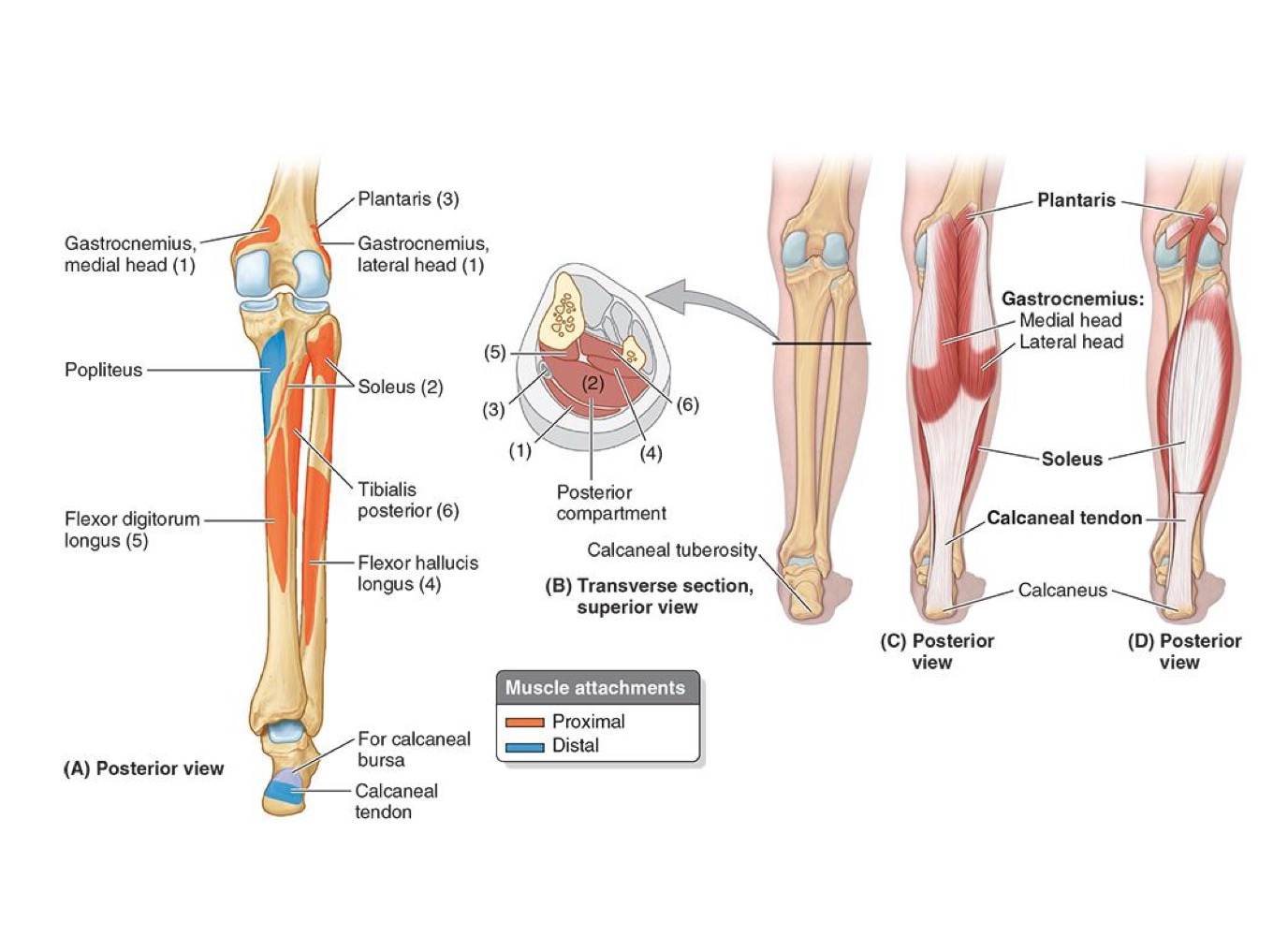
<ul><li><p>POST compartment = Superficial &amp; Deep subcompartments → separated by Transverse intermuscular septum)</p></li><li><p><strong>INFERIORLY, the Flexor Retinaculum (btw MEDIAL Malleolus &amp; Calcaneus) hold deep muscle tendons + POsT Tibial Artery as they bend around Medial Malleolus</strong></p></li></ul><p></p><ul><li><p><strong>ALL</strong> innervated by → <strong>TIBIAL NERVE</strong></p></li><li><p><strong>Supplied </strong>by → <strong>POST TIBIAL &amp; FIBULAR ARTERIES</strong></p></li><li><p><strong>POSTERIOR Muscle Actions include:</strong></p><ul><li><p>Plantarflexioin &amp; Inversion</p></li></ul></li></ul><p></p><ul><li><p><strong>Calcaneal Tendon → MOST POWERFUL TENDON in body</strong></p></li></ul><p></p><p>“ You stroll with SOLEUS but win the long jump w/ GAESTROCNEMIUS” - per Dr. Mudnt</p>
20
New cards

Ignore

Ignore


21
New cards

SUPERFICIAL POSTERIOR LEG MUSCLES- TRICEPS SURAE (PLANTARFLEXORS) : GASTROCNEMIUS

  • PA:

    • Lateral Head = Lateral aspect of lateral femoral condyle

    • Medial Head = Popliteal surface of femur; superior to medial condyle


  • DA: Calcaneus via Calcaneal Tendon


  • N: Tibial Nerve


  • A: Plantarflexes ankle joint when knee is extended + Raises heal during walking + Flexes knee joint
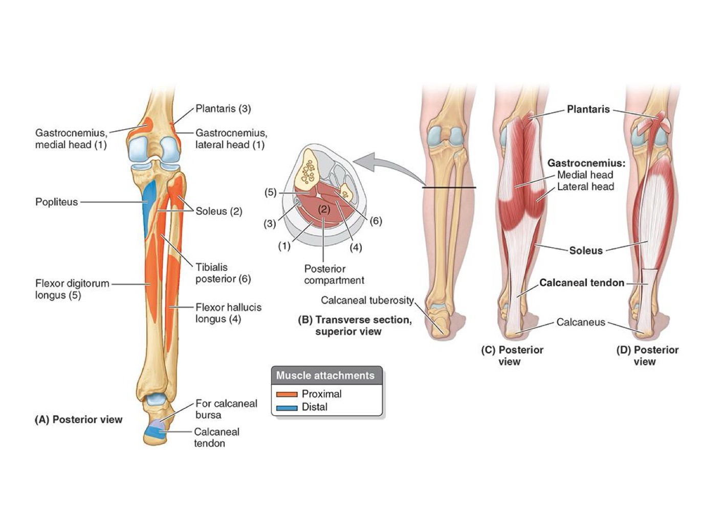

<ul><li><p><strong>PA:</strong></p><ul><li><p>Lateral Head = Lateral aspect of lateral femoral condyle</p></li><li><p>Medial Head = Popliteal surface of femur; superior to medial condyle</p></li></ul></li></ul><p></p><ul><li><p><strong>DA: Calcaneus via Calcaneal Tendon</strong></p></li></ul><p></p><ul><li><p><strong>N: Tibial Nerve</strong></p></li></ul><p></p><ul><li><p><strong>A: </strong>Plantarflexes ankle joint when knee is extended + Raises heal during walking + Flexes knee joint</p></li></ul><p></p>
22
New cards

SUPERFICIAL POSTERIOR LEG MUSCLES- TRICEPS SURAE (PLANTARFLEXORS) : SOLEUS

  • PA:

    • Superior Posterior Fibula + Soleal line & middle 1/3 tibia


  • DA: Calcaneus via Calcaneal Tendon


  • N: Tibial Nerve


  • A: Plantarflexes ankle independent of knee position; stabilizes ankle joint
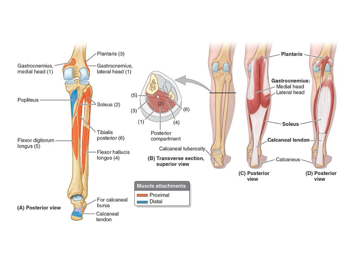

<ul><li><p><strong>PA:</strong></p><ul><li><p>Superior Posterior Fibula + Soleal line &amp; middle 1/3 tibia</p></li></ul></li></ul><p></p><ul><li><p><strong>DA: Calcaneus via Calcaneal Tendon</strong></p></li></ul><p></p><ul><li><p><strong>N: Tibial Nerve</strong></p></li></ul><p></p><ul><li><p><strong>A: </strong>Plantarflexes ankle independent of knee position; stabilizes ankle joint</p></li></ul><p></p>
23
New cards

SUPERFICIAL POSTERIOR LEG MUSCLES- TRICEPS SURAE (PLANTARFLEXORS) : PLANTARIS

  • ABSENT IN 5-10%


  • PA:

    • Lateral Supracondylar line of femur = Oblique popliteal ligament


  • DA: Calcaneus via Calcaneal Tendon


  • N: Tibial Nerve


  • A: Weakly assist in plantarflexing ankle
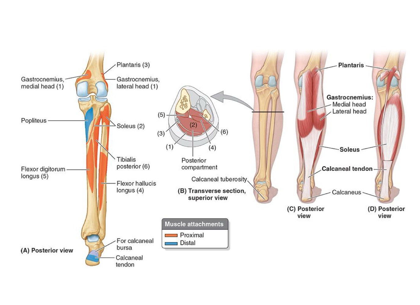


<ul><li><p>ABSENT IN 5-10%</p></li></ul><p></p><ul><li><p><strong>PA:</strong></p><ul><li><p>Lateral Supracondylar line of femur = Oblique popliteal ligament</p></li></ul></li></ul><p></p><ul><li><p><strong>DA: Calcaneus via Calcaneal Tendon</strong></p></li></ul><p></p><ul><li><p><strong>N: Tibial Nerve</strong></p></li></ul><p></p><ul><li><p><strong>A: </strong>Weakly assist in plantarflexing ankle</p></li></ul><p></p><p></p>
24
New cards

DEEP POSTERIOR LEG MUSCLES: POPLITEUS

  • PA: Lateral Femoral condyle


  • DA: Posterior surface of tibia (superior to soleal line)


  • N: Tibial Nerve


  • A: Weakly flexes KNEE joint to unlock knee by rotating femur 5° (on fixed TIBIA) + Medial rotates TIBIA unplanted limb
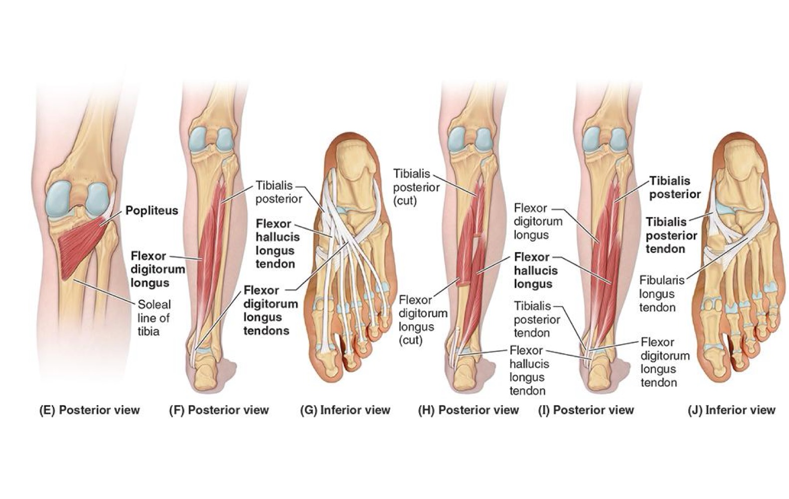

<ul><li><p><strong>PA: Lateral Femoral condyle</strong></p></li></ul><p></p><ul><li><p><strong>DA: Posterior surface of tibia (superior to soleal line)</strong></p></li></ul><p></p><ul><li><p><strong>N: Tibial Nerve</strong></p></li></ul><p></p><ul><li><p><strong>A: </strong>Weakly flexes KNEE joint to unlock knee by rotating femur 5° (on fixed TIBIA) + Medial rotates TIBIA unplanted limb</p></li></ul><p></p>
25
New cards

DEEP POSTERIOR LEG MUSCLES: FLEXOR HALLUCIS LONGUS

  • PA: INFERIOR 2/3 Fibula + Inferior Interosseous Membrane


  • DA: Base of Distal phalanx (great TOE = Hallux)


  • N: Tibial Nerve


A: Flexes great toe at all joints; weakly plantarflexes ankle joint; supports

medial longitudinal arch of foot
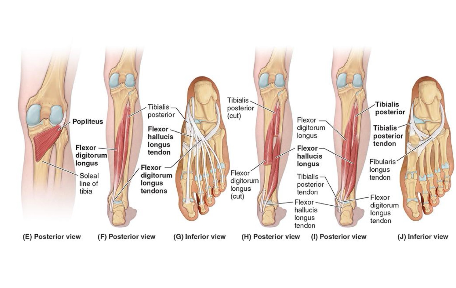

<ul><li><p><strong>PA: INFERIOR 2/3 Fibula + Inferior Interosseous Membrane</strong></p></li></ul><p></p><ul><li><p><strong>DA: Base of Distal phalanx (great TOE = Hallux)</strong></p></li></ul><p></p><ul><li><p><strong>N: Tibial Nerve</strong></p></li></ul><p></p><p><strong>A: Flexes great toe at all joints; weakly plantarflexes ankle joint; supports</strong></p><p><strong>medial longitudinal arch of foot</strong></p><p></p>
26
New cards

DEEP POSTERIOR LEG MUSCLES: FLEXOR DIGITORUM LONGUS

  • PA: POST SURFACE OF TIBIA + Fibula via broad Tendon


  • DA: Bases of distal phalanges of Lateral 4 digits


  • N: Tibial Nerve


  • A: Flexes 4 toes + Plantarflexes ankle joint + supports longitudal arches of foot
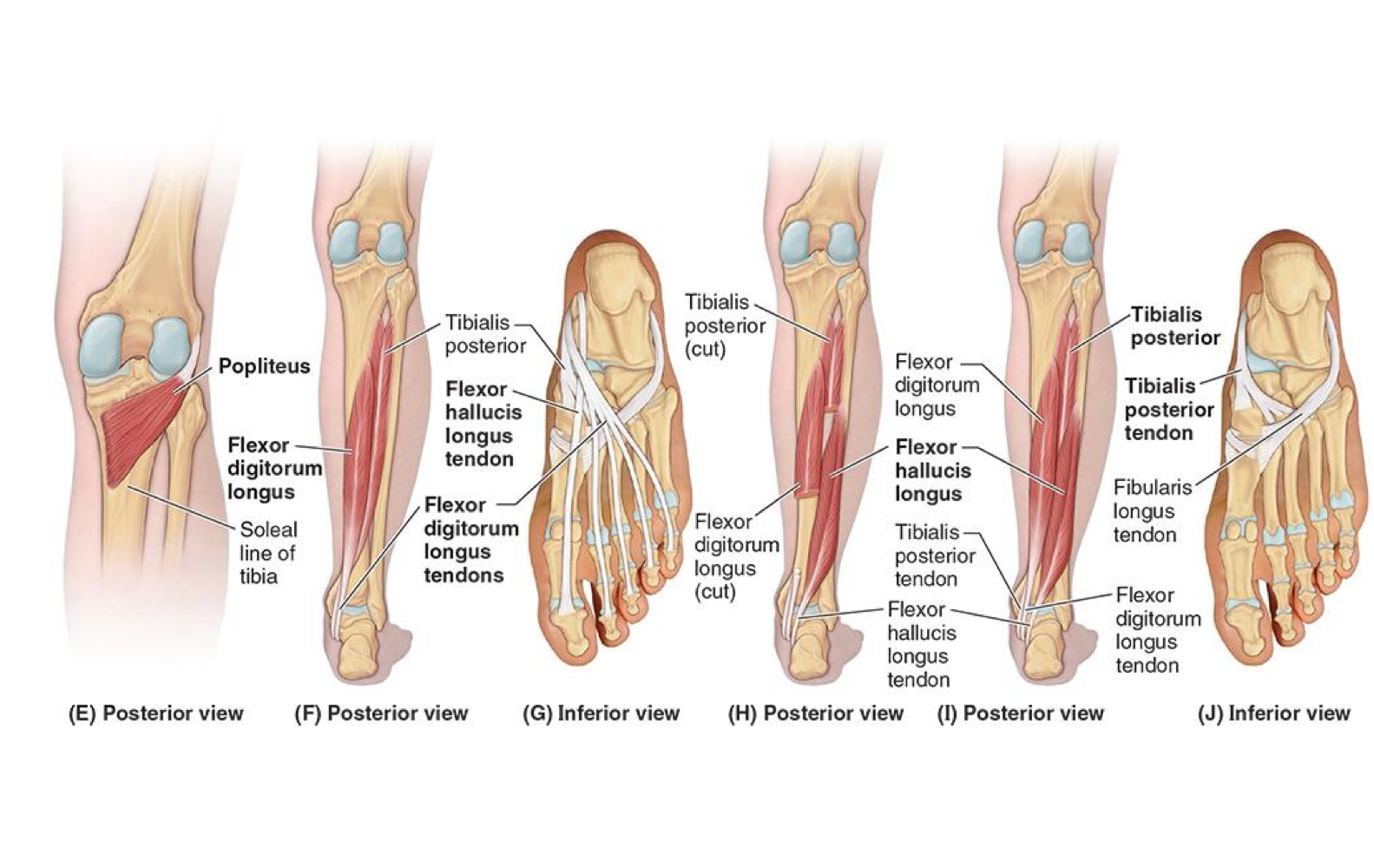


<ul><li><p><strong>PA: </strong>POST SURFACE OF TIBIA + Fibula via broad Tendon</p></li></ul><p></p><ul><li><p><strong>DA: </strong>Bases of distal phalanges of Lateral 4 digits</p></li></ul><p></p><ul><li><p><strong>N: Tibial Nerve</strong></p></li></ul><p></p><ul><li><p><strong>A: </strong>Flexes 4 toes + Plantarflexes ankle joint + supports longitudal arches of foot</p></li></ul><p></p><p></p>
27
New cards

DEEP POSTERIOR LEG MUSCLES: TIBIALIS POSTERIOR

  • PA: Interosseus membrane + POST TIBIA & FIBULA


  • DA: Tuberosity of Navicular + Cuneiform + cuboid + sustentaculum tali of calcaneus, bases of 2nd, 3rd & 4th metatarsals


  • N: Tibial Nerve


A: Plantarflexes ankle; inverts foot + maintains medial longitudinal arch
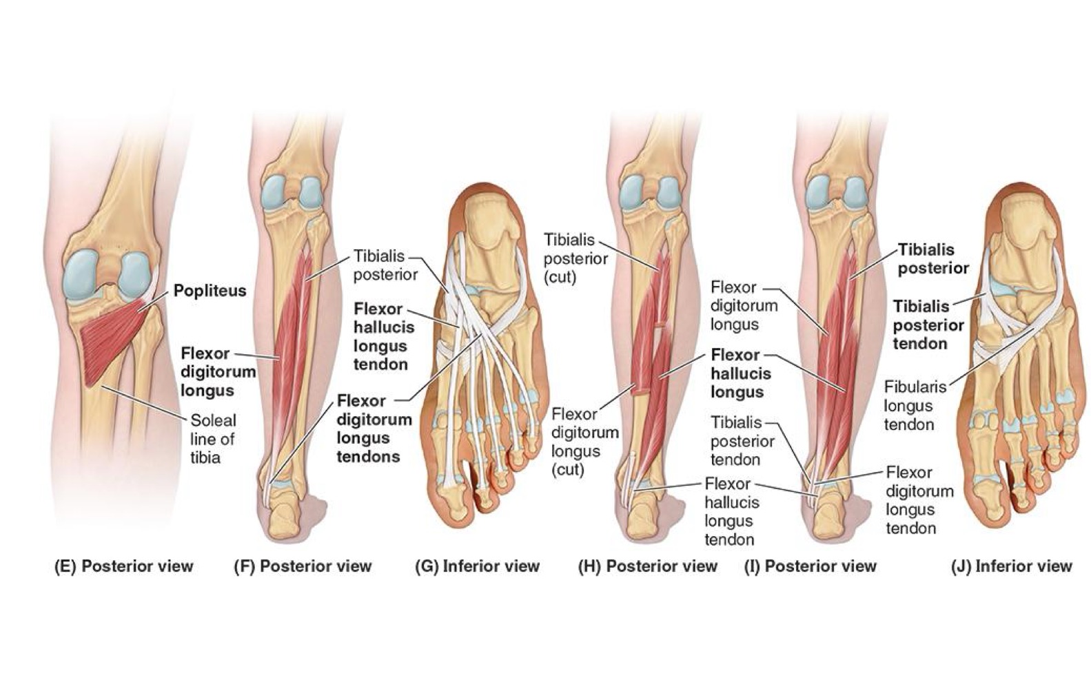

<ul><li><p><strong>PA: Interosseus membrane + POST TIBIA &amp; FIBULA</strong></p></li></ul><p></p><ul><li><p><strong>DA: Tuberosity of Navicular + Cuneiform + cuboid + sustentaculum tali of calcaneus, bases of 2nd, 3rd &amp; 4th metatarsals</strong></p></li></ul><p></p><ul><li><p><strong>N: Tibial Nerve</strong></p></li></ul><p></p><p><strong>A: Plantarflexes ankle; inverts foot + maintains medial longitudinal arch</strong></p><p></p>
28
New cards

NERVES OF LEG: SAPHENOUS NERVE

  • ORIGIN: FEMORAL NERVE


  • DISTRIBUTION: Supplies skin on Medial side of ANKLE + FOOT
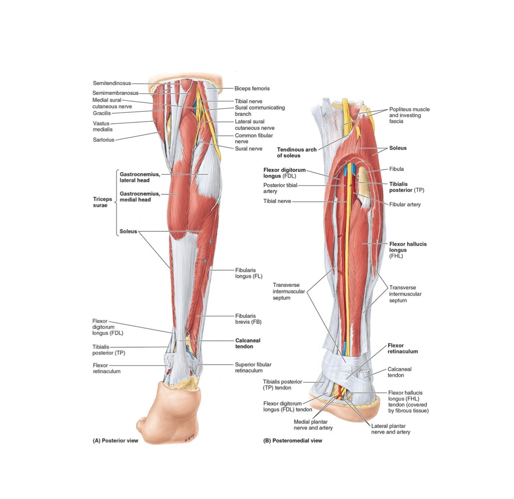


<ul><li><p><strong>ORIGIN: </strong>FEMORAL NERVE</p></li></ul><p></p><ul><li><p><strong>DISTRIBUTION: Supplies skin on Medial side of ANKLE + FOOT</strong></p></li></ul><p></p><p></p>
29
New cards

NERVES OF LEG: SURAL NERVE

  • ORIGIN: TIBIAL + COMMON FIBULAR NERVES


  • DISTRIBUTION: Supplies skin on POST & Lateral Leg & Foot
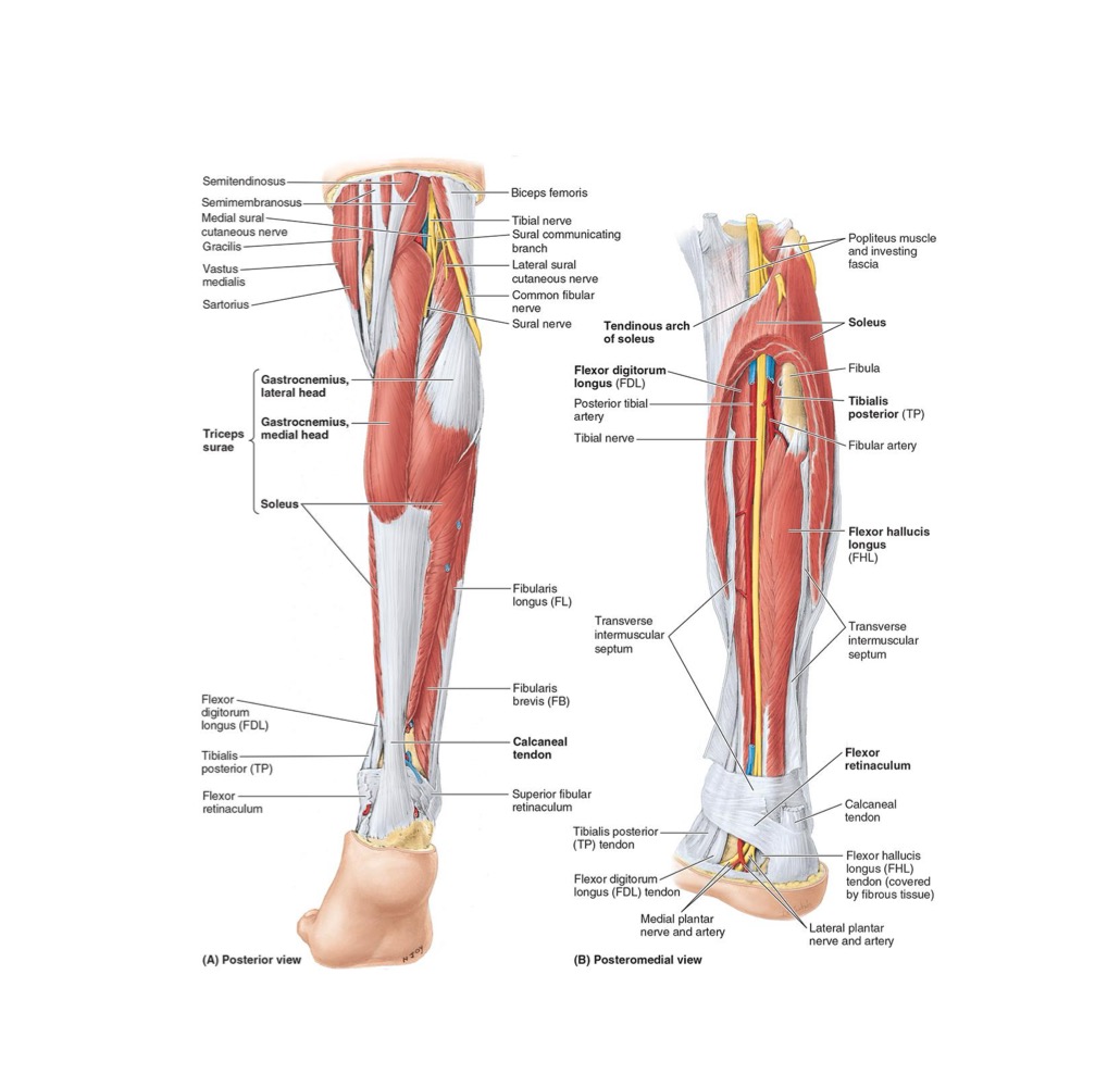


<ul><li><p><strong>ORIGIN: </strong>TIBIAL + COMMON FIBULAR NERVES</p></li></ul><p></p><ul><li><p><strong>DISTRIBUTION: </strong>Supplies skin on POST &amp; Lateral Leg &amp; Foot </p></li></ul><p></p><p></p>
30
New cards

NERVES OF LEG: TIBIAL NERVE

  • ORIGIN: Sciatic


  • DISTRIBUTION: Supplies POST Leg muscles & Knee joint
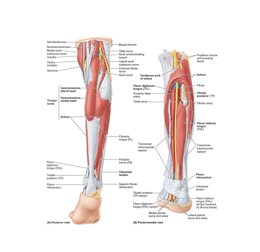


<ul><li><p><strong>ORIGIN: </strong>Sciatic</p></li></ul><p></p><ul><li><p><strong>DISTRIBUTION: </strong>Supplies POST Leg muscles &amp; Knee joint</p></li></ul><p></p><p></p>
31
New cards

NERVES OF LEG: COMMON FIBULAR NERVE

  • ORIGIN: Sciatic


  • DISTRIBUTION:

    • Supplies skin on lateral POST Leg via Sural

    • Knee joint via Articular Branch


<ul><li><p><strong>ORIGIN: </strong>Sciatic</p></li></ul><p></p><ul><li><p><strong>DISTRIBUTION:</strong></p><ul><li><p>Supplies skin on lateral POST Leg via Sural</p></li><li><p>Knee joint via Articular Branch</p></li></ul></li></ul><p></p><p></p>
32
New cards

NERVES OF LEG: SUPERFICIAL FIBULAR NERVE

  • ORIGIN: Common Fibular Nerve


  • DISTRIBUTION: Supplies Fibularis Longus & Fibularis Brevis → Supplies Skin on distal 1/3 on ANTERIOR Leg & dorsum of Foot
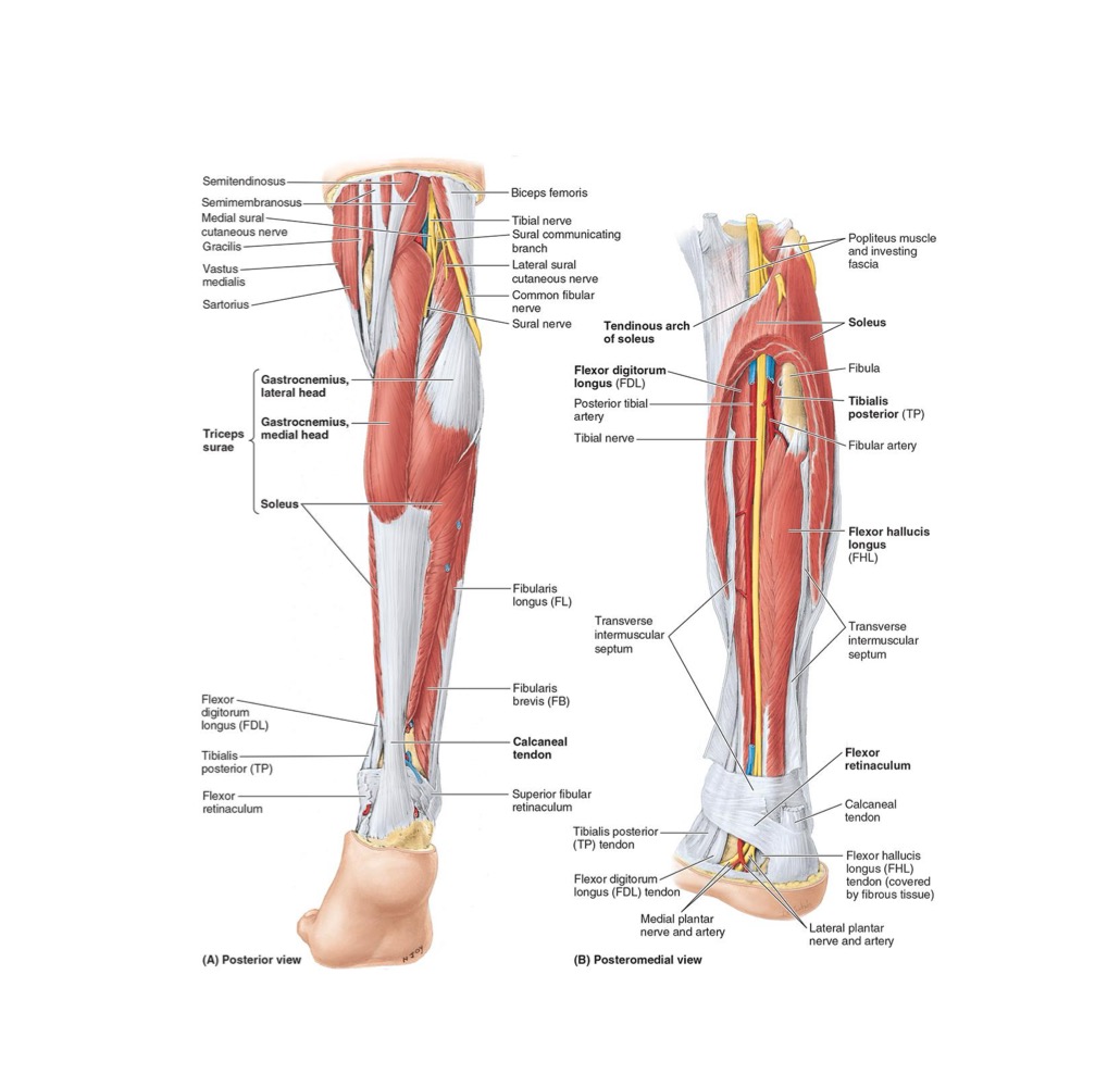

<ul><li><p><strong>ORIGIN: </strong>Common Fibular Nerve</p></li></ul><p></p><ul><li><p><strong>DISTRIBUTION: </strong>Supplies Fibularis Longus &amp; Fibularis Brevis → Supplies Skin on distal 1/3 on ANTERIOR Leg &amp; dorsum of Foot</p></li></ul><p></p>
33
New cards

NERVES OF LEG: DEEP FIBULAR NERVE

  • ORIGIN: Common Fibular Nerve


  • DISTRIBUTION: Supplies ANTERIOR Leg muscles → supplies SKIN between 1st & 2nd toes
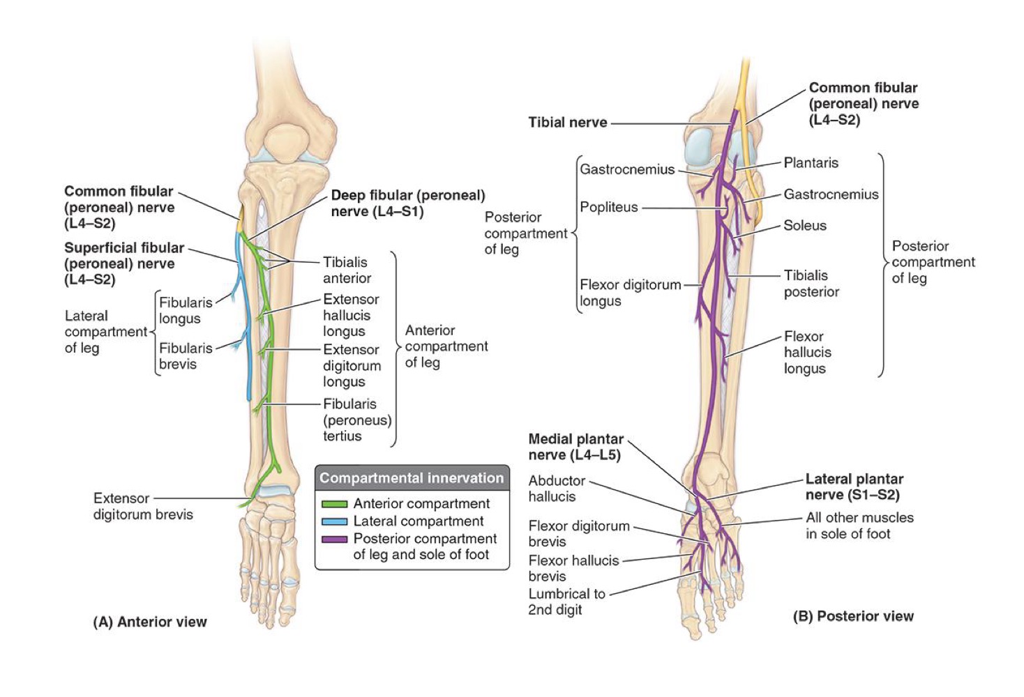

<ul><li><p><strong>ORIGIN: </strong>Common Fibular Nerve</p></li></ul><p></p><ul><li><p><strong>DISTRIBUTION: </strong>Supplies ANTERIOR Leg muscles → supplies SKIN between 1st &amp; 2nd toes</p></li></ul><p></p>
34
New cards
<p>ARTERIES OF LEG: <strong>POPLITEAL</strong></p>

ARTERIES OF LEG: POPLITEAL

  • ORIGIN: Femoral (once it passes through ADDUCTOR Hiatus)


  • DISTRIBUTION IN LEG: Superior, Middle & INF GENICULAR Arteries to Lateral + Medial aspects of Knee
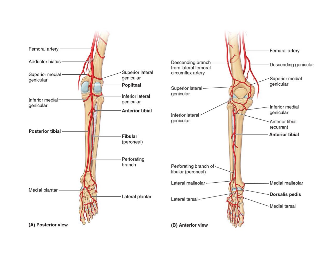
<ul><li><p><strong>ORIGIN: </strong>Femoral (once it passes through ADDUCTOR Hiatus)</p></li></ul><p></p><ul><li><p><strong>DISTRIBUTION IN LEG: </strong>Superior, Middle &amp; INF GENICULAR Arteries to Lateral + Medial aspects of Knee</p></li></ul><p></p>
35
New cards
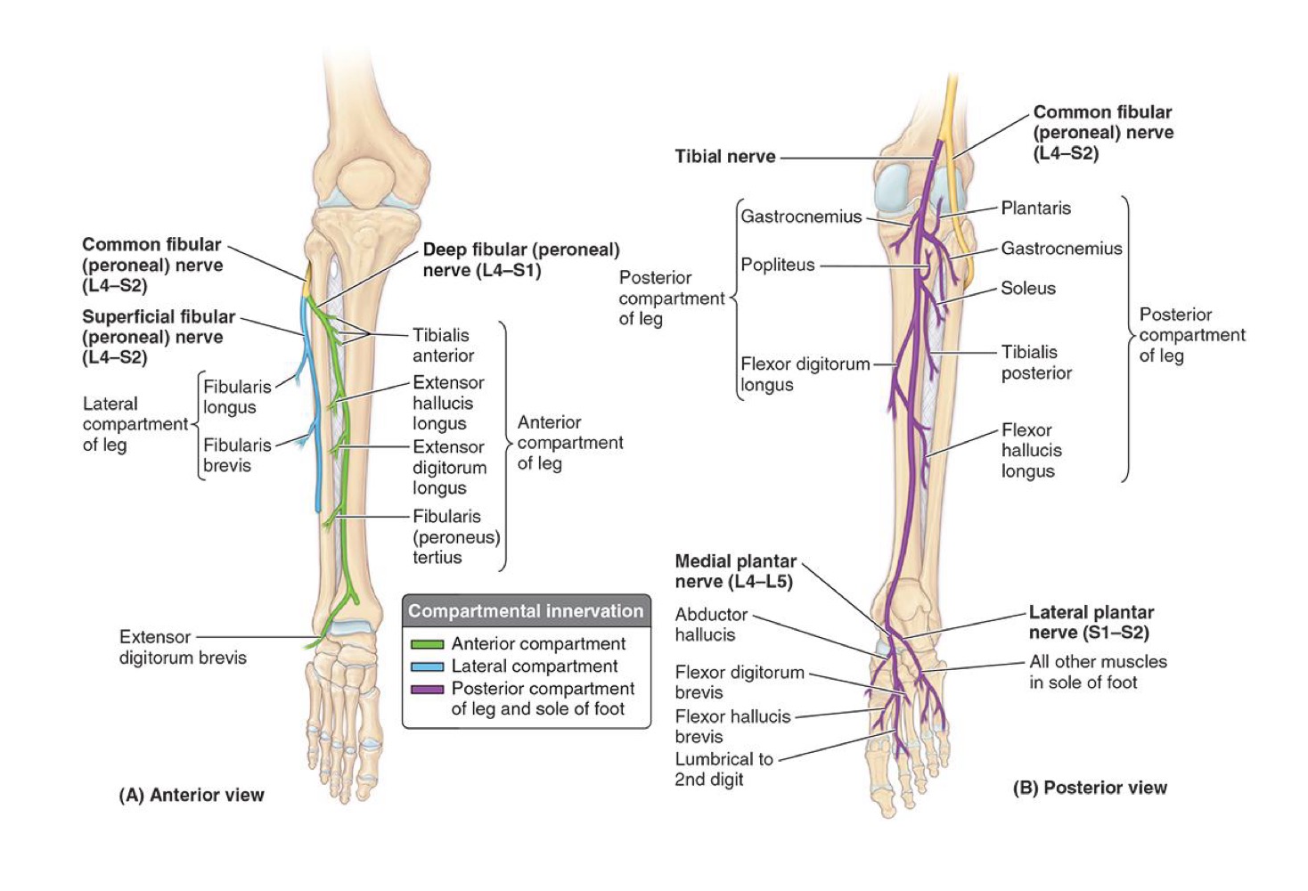
<p>ARTERIES OF LEG: <strong>ANTERIOR TIBIAL ARTERY</strong></p>

ARTERIES OF LEG: ANTERIOR TIBIAL ARTERY

  • ORIGIN: Popliteal


  • DISTRIBUTION IN LEG: Anterior (ANT) leg compartment
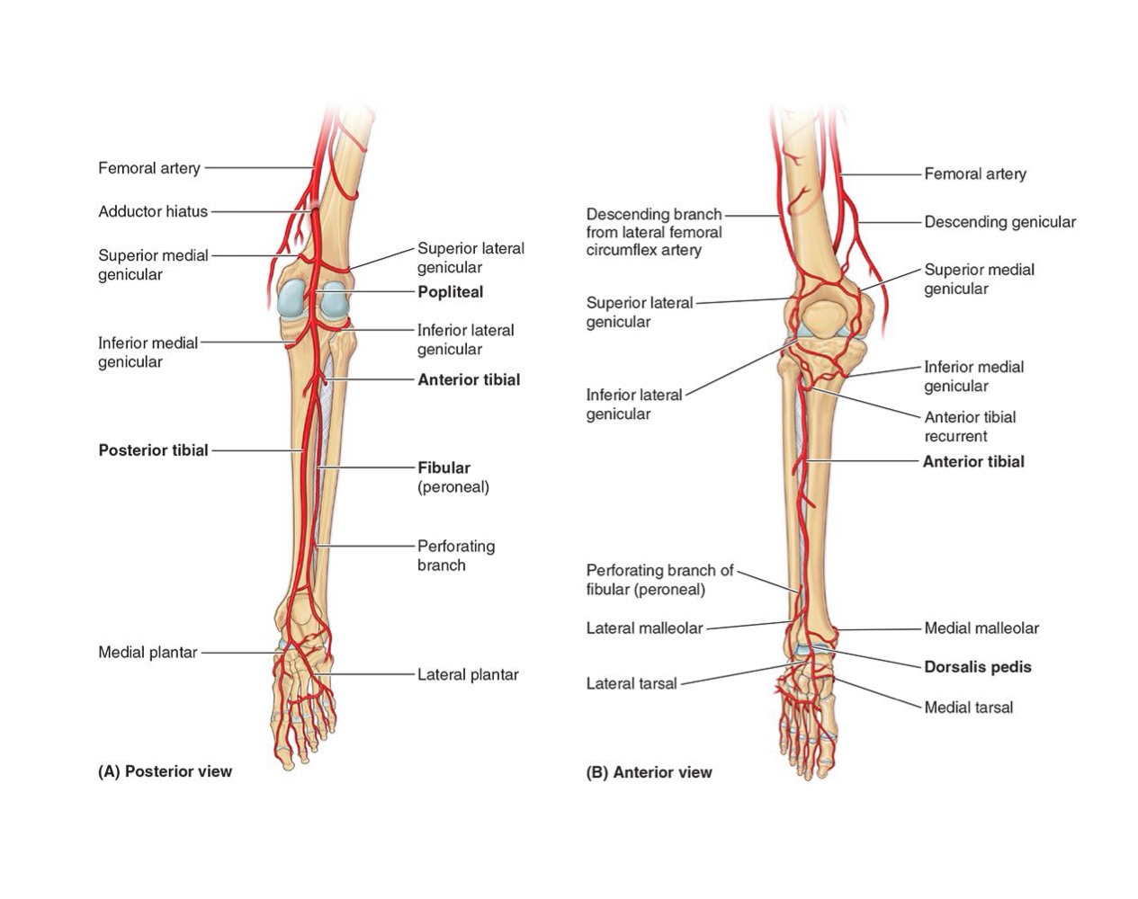

<ul><li><p><strong>ORIGIN: </strong>Popliteal</p></li></ul><p></p><ul><li><p><strong>DISTRIBUTION IN LEG: </strong>Anterior (ANT) leg compartment</p></li></ul><p></p>
36
New cards

ARTERIES OF LEG: DORSALIS PEDIS

  • ORIGIN: Continuation of ANT tibial Artery distal to INF Retinaculum


  • DISTRIBUTION IN LEG:

    • Muscles on dorsum of foot

    • pierces 1st dorsal Interosseous muscle as Deep Plantar Artery to help form Plantar Arch


37
New cards

ARTERIES OF LEG: POSTERIOR TIBIAL ARTERY

  • ORIGIN: Popliteal


  • DISTRIBUTION IN LEG:

    • POST & Lateral Leg compartments

    • Circumflex Fibular Branch joins anastomoses around Knee

    • Nutrient artery passes to → Tibia


<ul><li><p><strong>ORIGIN: </strong>Popliteal</p></li></ul><p></p><ul><li><p><strong>DISTRIBUTION IN LEG:</strong></p><ul><li><p>POST &amp; Lateral Leg compartments</p></li><li><p>Circumflex Fibular Branch joins anastomoses around Knee</p></li><li><p>Nutrient artery passes to → Tibia</p></li></ul></li></ul><p></p>
38
New cards

ARTERIES OF LEG: FIBULAR ARTERY

  • ORIGIN: Posterior Tibial


  • DISTRIBUTION IN LEG:

    • POST Leg compartment

    • Perforating branches supply lateral compartment of Leg


39
New cards

LEG CLINICAL CONSIDERATIONS

  • POPLITEAL pulse & POST Tibial pulse


  • Popliteal Aneurysm = abnormal dilation of ALL or PART of popliteal artery


  • Compartment Syndrome = infection of a compartment causes INCREASE in INTRA-COMPARTMENT pressure


  • Shin Splints = TIBIALIS anterior sprain (tears in the periosteum covering shaft of tibia)


  • Footdrop = Injury the COMMON FIBULAR NERVE (most injured nerve in lower limb)


  • Ruptured calcaneal tendon

  • Calcaneal tendon reflex


40
New cards

Ignore

  • POPLITEAL pulse & POST Tibial pulse

  • Popliteal Aneurysm = abnormal dilation of ALL or PART of popliteal artery

  • Compartment Syndrome = infection of a compartment causes INCREASE in INTRA-COMPARTMENT pressure

  • Shin Splints = TIBIALIS anterior sprain (tears in the periosteum covering shaft of tibia)

  • Footdrop = Injury the COMMON FIBULAR NERVE (most injured nerve in lower limb)

  • Ruptured calcaneal tendon

  • Calcaneal tendon reflex