Anterior Abdominal Wall and GI Tract
 Call Kai
 Learn
Learn Practice Test
 Spaced Repetition
 Match
Match Flashcards
 Knowt Play
1/97

Earn XP
Description and Tags
1. Recall the regions and quadrants of the abdomen. 2. Describe the bones of the abdomen (lumbar vertebrae). 3. Describe the arrangement of the muscles that make up the anterior and lateral abdominal wall and describe their actions. 4. Distinguish the major segments of the gastrointestinal (GI) tract and recall the key features of each segment. 5. Describe the pattern of blood supply and venous drainage of the GI tract.
Name | Mastery | Learn | Test | Matching | Spaced | Call with Kai | Chat |
|---|
No analytics yet
Send a link to your students to track their progress
98 Terms
Describe the abdominal wall muscles
Flat muscles surrounding the abdominal cavity
What are the two functional groups of abdominal wall muscles?
anterolateral muscles, posterior muscles
What are the three key functions of the abdominal muscles?
protect the abdominal viscera, maintain posture and move the trunk
Name the groups of structures found in the abdominal cavity
abdominal wall, accessory organs of digestion, urinary system, arteries, veins and lymphatics
What are the accessory organs of digestion?
liver, gallbladder, pancreas
What makes up the urinary system?
kidneys, ureters
Whats a key role of the anterolateral abdominal muscles?
increase intra-abdominal pressure (e.g. for defecation and child-birth)
Whats a notable structural feature of the anterolateral abdominal muscles?
different fibre orientation
What are the two groups of anterolateral abdominal muscles?
Anterior (two on either side of midline), Lateral (three on either side of the midline)
What joins the two groups of anterolateral abdominal muscles?
rectus sheath
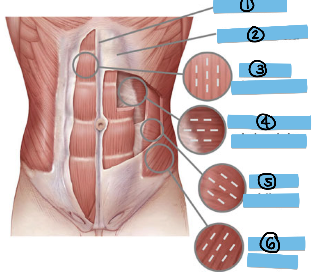
Label missing structures 1-3
linea alba, rectus fascia, rectus abominis
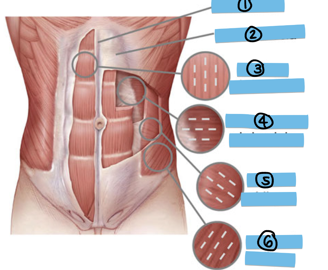
Label structures 4-6
transversus abdominis, internal oblique, external oblique
Is the external oblique muscle superficial or deep
most superficial muscle
define aponeurosis
sheet of pearly white fibrous tissue acting as a tendon in flat muscles and having a wide area of attachment
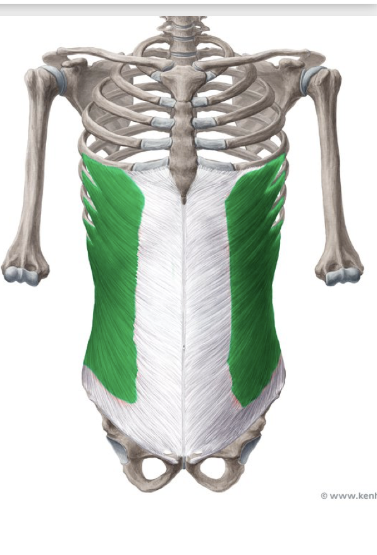
what structure is depicted in green?
external oblique muscle
Describe the main characteristics of the rectus abdominis muscle
long vertical muscles on either side of lines alba, inserted by tendinous intersections (ab lines), lies within the rectus sheath
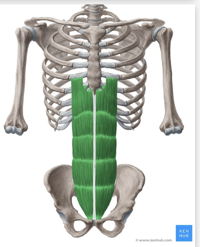
What structure is depicted in green?
rectus abdominis muscle
Describe the fibre orientation of internal oblique muscle
fibres oriented perpendicular to EOM (medially and superiorly),
What does linea alba mean
white line
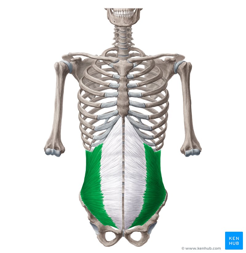
what structure is depicted in green
the internal oblique muscles
Is the transversus abdominis muscle superficial or deep?
its the deepest
What separates the transversus abdominis muscle from the abdominal cavity?
the transversalis fascia
describe the orientation of the transversus abdominis muscles fibres
horizontal

Describe how the aponeurosis of the internal oblique muscle divides
aponeurosis divides at rectus sheath anterior ½ forms anterior wall, posterior ½ forms posterior wall

describe the fibre orientation of the external oblique muscle
runs medially and inferiorly towards the midline
describe the external oblique muscles aponeurosis interactions with other structures in the abdominal wall
aponeurosis forms anterior wall of rectus sheath
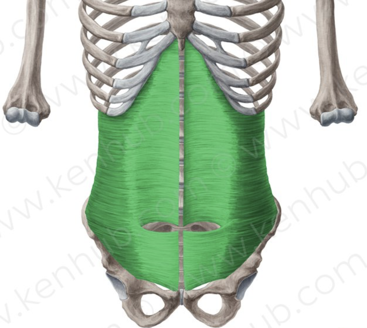
What structure is depicted in green
transversus abdominis muscle
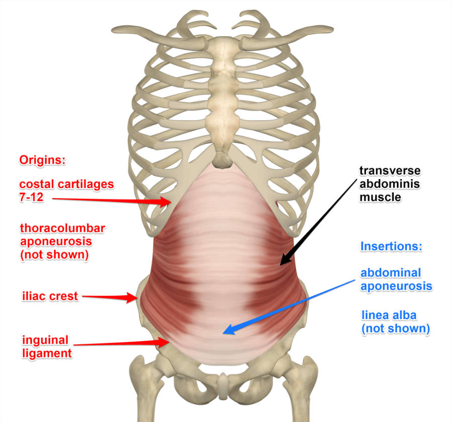
describe the interaction of the transversus abdominis muscle aponeurosis
aponeurosis forms the posterior wall of the rectus sheath
Define fascia
Thin casing of connective tissue that surrounds and holds structures
What is the rectus sheath formed by
The aponeurosis of the three lateral muscles enclosing the rectus abdominis
What forms the anterior wall of the rectus sheath
EOm + anterior ½ of IOm
What forms the posterior wall of the rectus sheath
the posterior ½ of IOm and TAm
What is the midline of the rectus sheath
linea alba
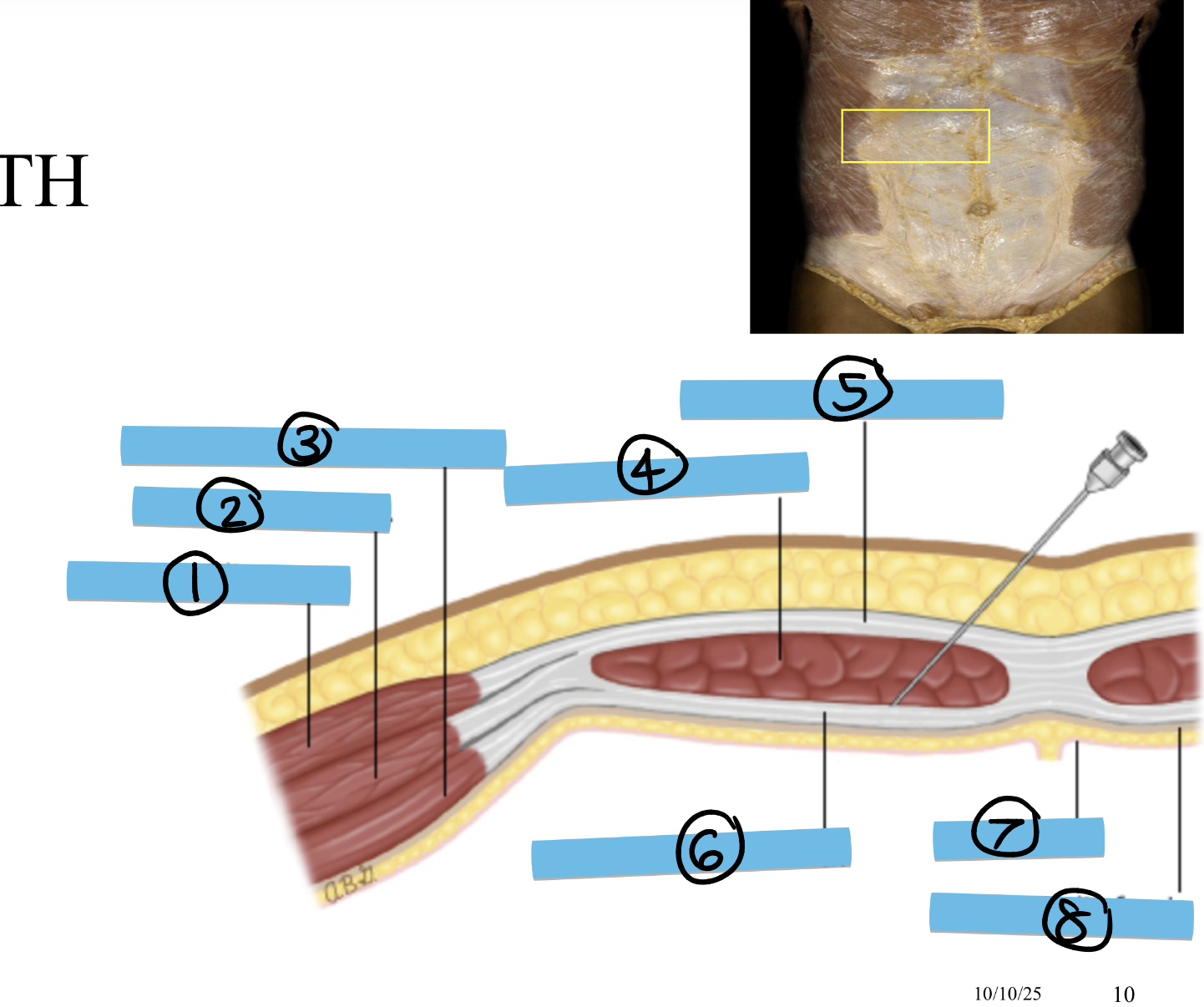
Label structures 1-4
external oblique m., internal oblique m., transverse abdominal m., rectus abdominal m.
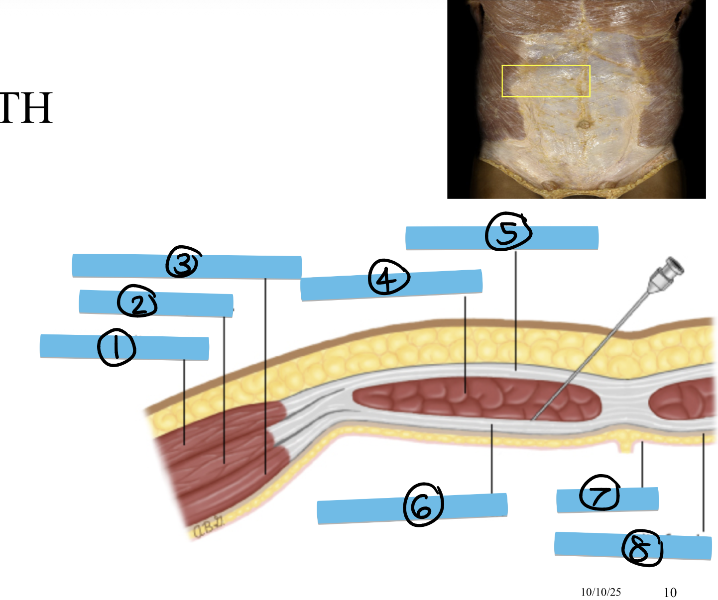
Label structures 5-8
Anterior rectus sheath, posterior rectus sheath, peritoneum, transversals fascia
What is the role of the GI tract
processing food and eliminating waste (supported by the accessory organs of digestion)
List the journey of the GI tract from mouth to anus
thoracic and abdominal oesophagus, stomach, small intestine, large intestine, upper 1/3 rectum
Describe the blood supply to the GI tract
from the abdominal aorta: coeliac trunk + superior mesenteric artery (SMA) + inferior mesenteric artery (IMA)
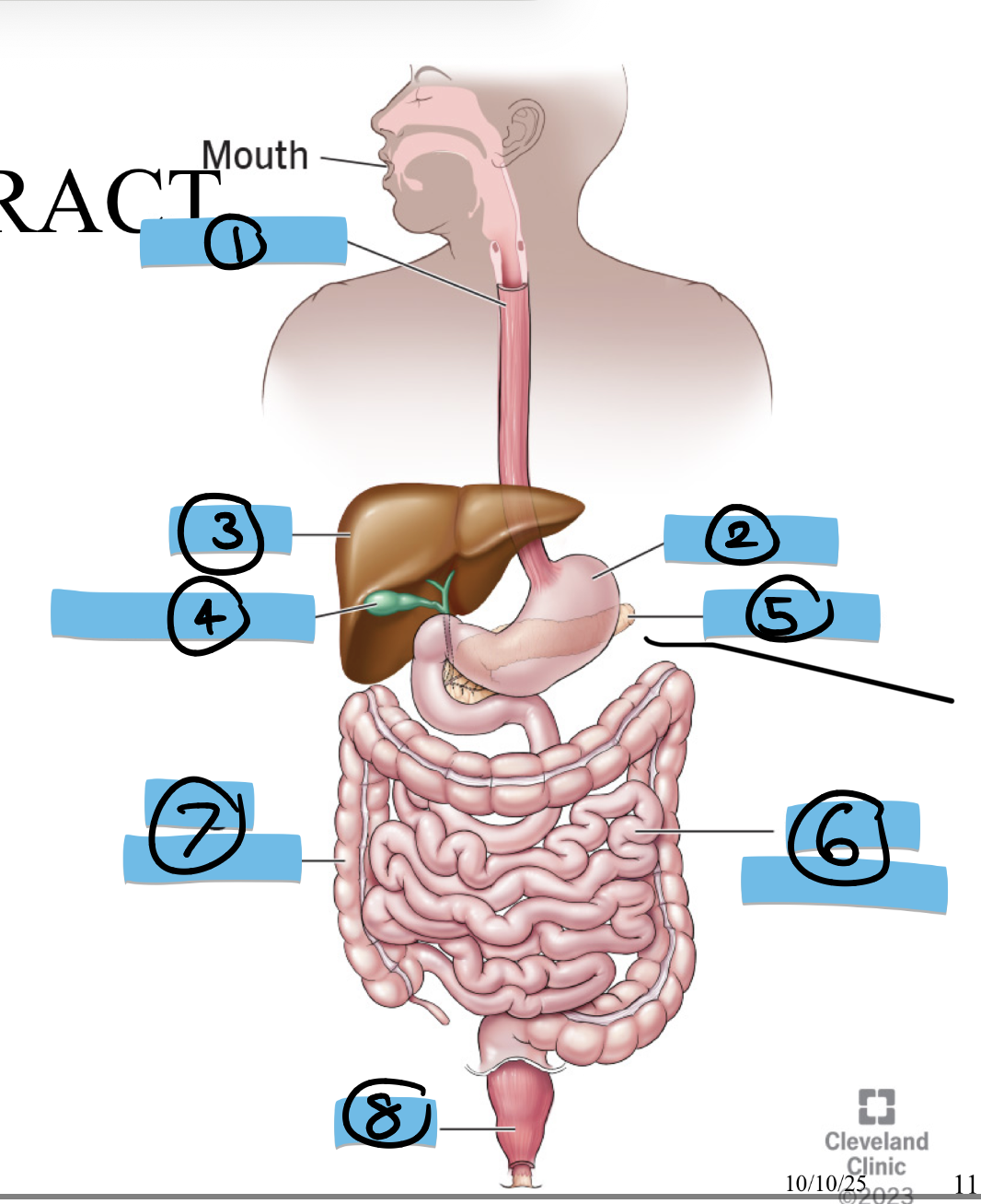
Label structures 1-8
eosophagus, stomach, liver, gallbladder, pancreas, small intestine, large intestine, anus
Where is the thoracic portion of the oesophagus found
the superior and posterior mediastina
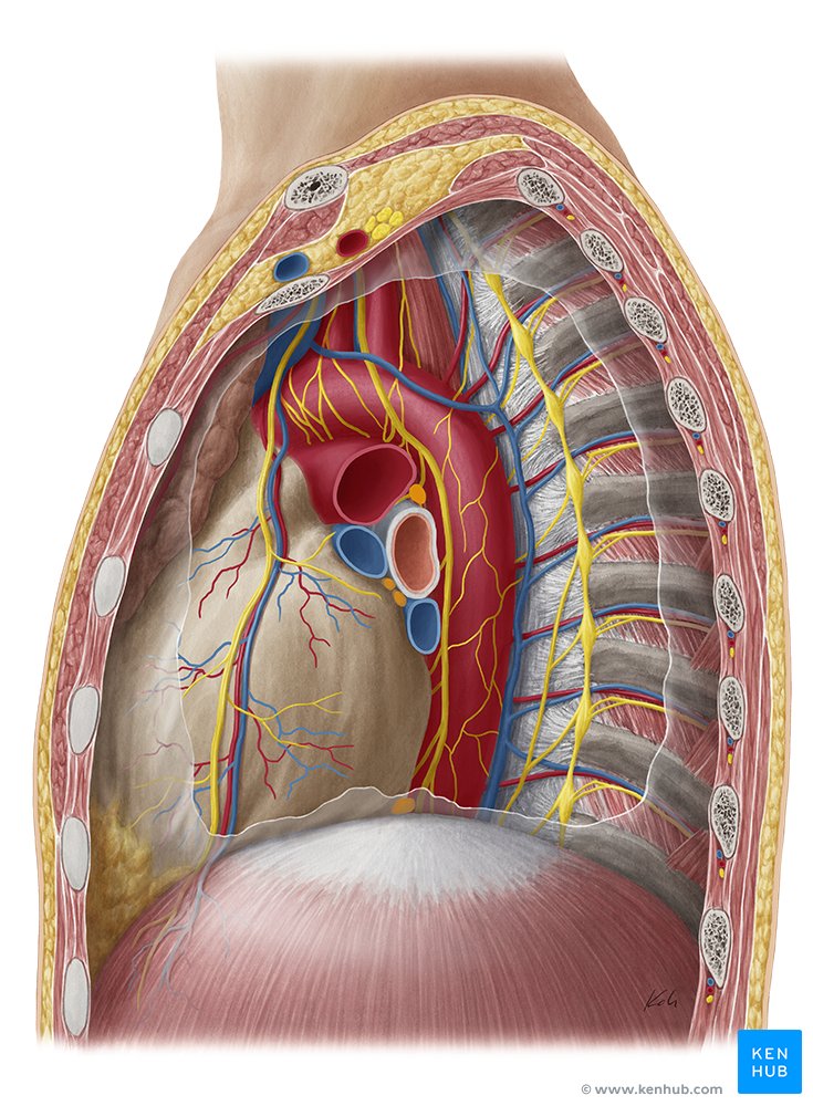
what is the mediastina?
a space in the middle of the thoracic cavity holding structures such as your heart
What major organ in the thoracic cavity is not part of the mediastinum
the lungs
Where does the thoracic portion of the oesophagus pierce the diaphragm?
At the oesophageal hiatus
What is notable about the abdominal portion of the oesophagus
very short
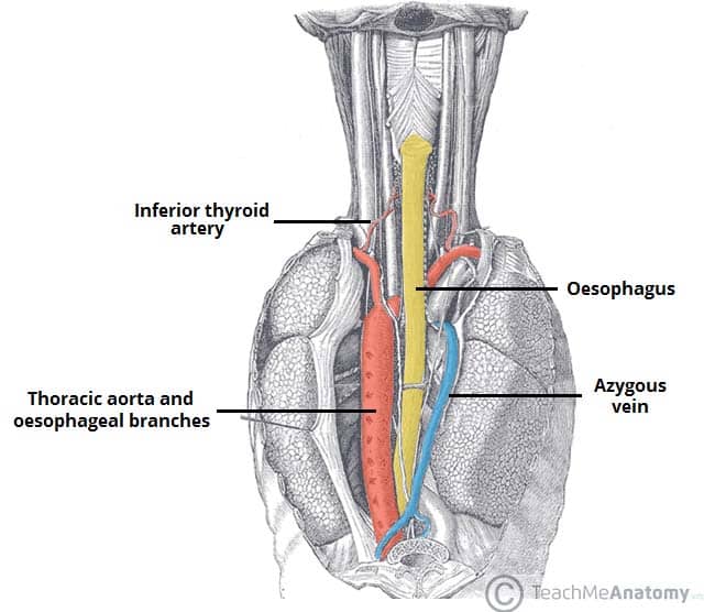
What does the thoracic portion of the oesophagus run with?
The vagal trunks
At what point does the abdominal portion of the oesophagus connect with the stomach?
The cardiac sphincter
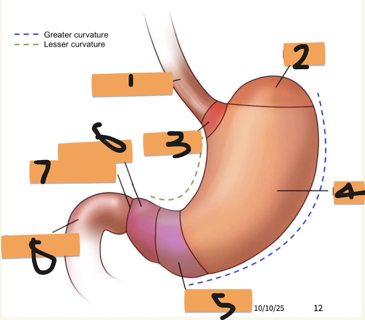
Label structures 1-4
oesophagus, fundus, cardia, body

label structures 5-8
antrum, pyloric canal, pyloric sphincter, duodenum
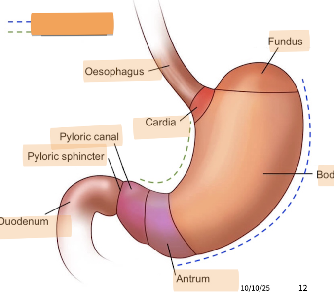
Label the green and blue dotted lines
lesser curvature (green), greater curvature (blue)

what makes up the pyloric region of the stomach?
antrum, canal and sphincter
What are the three parts of the small intestine (in order from stomach)?
duodenum, jejunum, ileum
why is the duodenum distinct?
because it is the first short, C-shaped part of the small intestine that connects the stomach to the rest of the gut
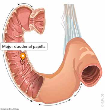
What is the major duodenal papilla?
a small bump where vital digestive juices from the liver, gallbladder, and pancreas enter the tract
What is the blood supply to the oesophagus and stomach?
coeliac trunk
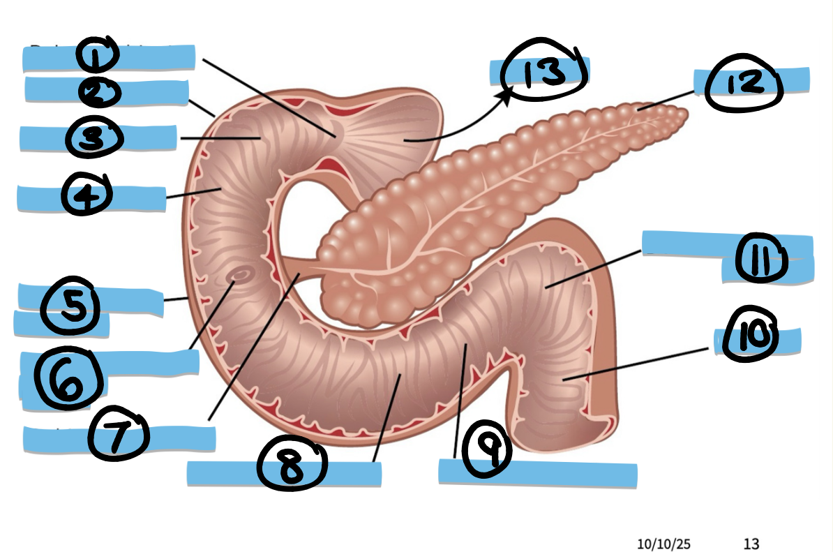
Label structures 1-4
pyloric sphincter, superior region, duodenal bulb, circular fold
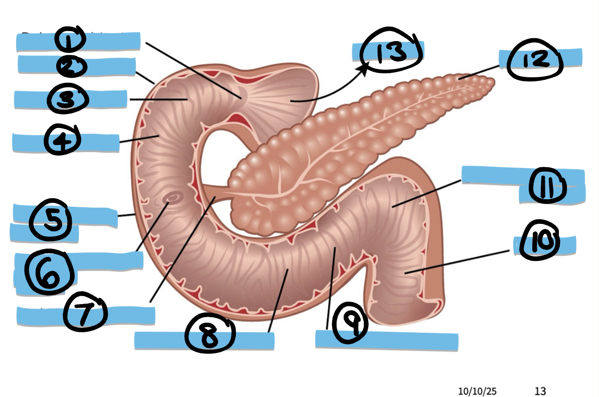
Label structures 5-8
descending region, major duodenal papilla, sphincter of odds, transverse region

Label structures 9-13
ascending region, jejunum, duodenojejunal flexure, pancreas, stomach
The jejunum and the ileum are very similar how are they distinguished?
By their blood supply
How do you identify the fundus of an organ and what are the latin roots that explain this?
add liquid, close openings, hold perpendicular, where ever liquid settles is fundus (from latin “bottom”) NB is not the most inferior part of the organ
What is the role of the mesentery?
encloses small intestines and parts of colon (transverse and sigmoid) + attaches them to the posterior abdominal wall
What is the blood supply to the small intestine?
coeliac trunk + SMA

Label structures 1-3
parietal peritoneum, visceral peritoneum, mesentery
What is the colon divided into?
ascending, transverse, descending, and sigmoid
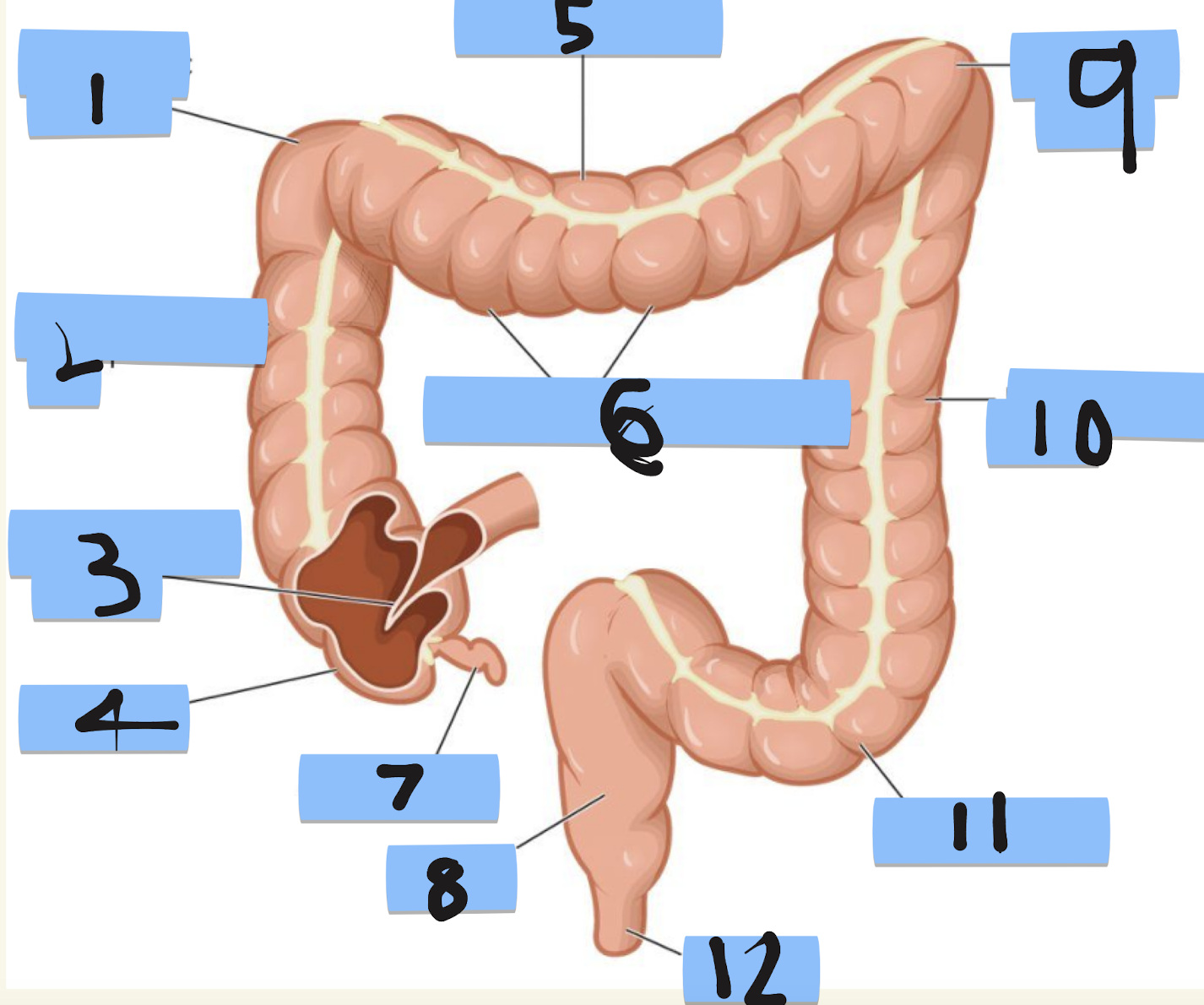
Label structures 1-4
right colic flexure, ascending colon, illiocaecal valve, caecum
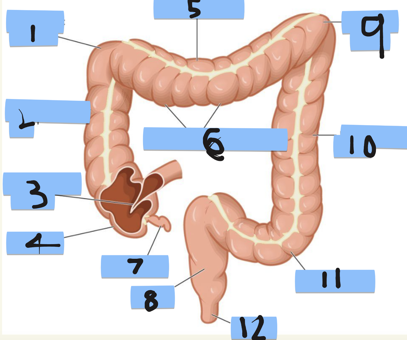
label structures 5-8
transverse colon, segment/hastrum, appendix, rectum
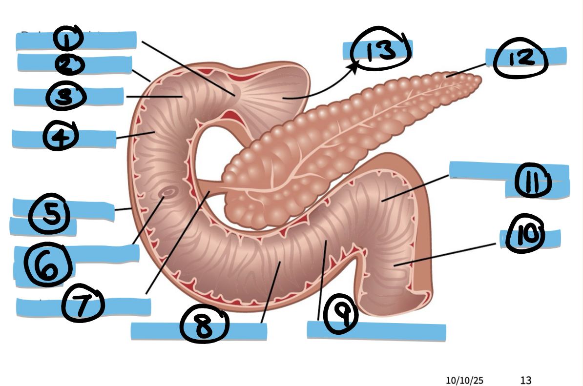
Label structures 9-13
ascending region, jejunum, duodenajejunum flexure, pancreas, stomach
What are the features of the large intestine?
epiploic appendices, taenae coli and haustrations
How do you identify the haustra?
they are the bulging of the large intestine walls
How do you identify the tenia coli?
The white line on the large intestine
How do you identify the epiploic appendices
they are the fatty tags on the haustra
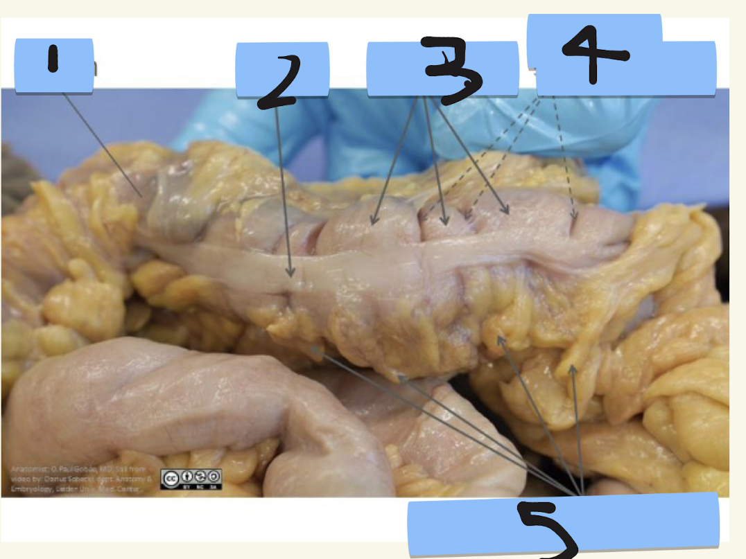
Label structures 1-5
colon, tenia, haustra, place of semilunar folds, epilopic appendices
What are the foregut, midgut and hindgut and why are they important to learn?
Different embryological origins of GI tract, important for understanding blood supply
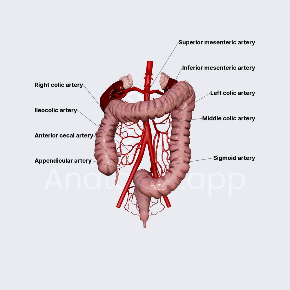
what is the blood supply to the large intestine?
SMA + IMA
What are the structural boundaries of the foregut?
oesophagus to duodenum
What is the blood supply of the foregut?
Coeliac trunk
What are the structural boundaries of the midgut?
From major duodenal papilla to 2/3 of transverse colon
What is the blood supply of the midgut?
Superior mesenteric artery (SMA)
What are the structural boundaries of the hindgut?
From last 1/3 of transverse colon to upper 1/3 rectum (+ anal canal)

What is the blood supply to the hindgut?
Inferior mesenteric artery (IMA)
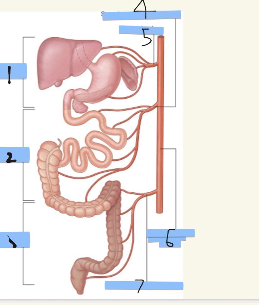
Label structures 1-7
foregut, midgut, hindgut, superior mesenteric artery, celiac trunk, abdominal aorta, inferior mesenteric artery
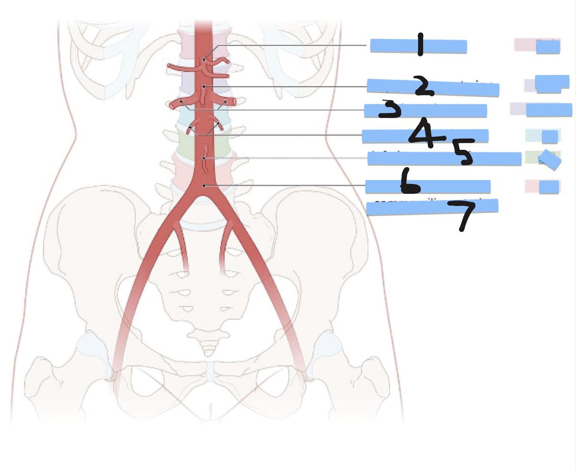
Label structures 1-7 and corresponding regions
coeliac trunk (T12), superior mesenteric artery (L1), renal arteries (L1/2), gonadal arteries (L2), Inferior mesenteric arteries (L3), bifurcation into L/R common iliac arteries (L4)
What does L1 stand for
first lumbar vertebrae
What does T12 stand for?
the twelfth (and final) thoracic vertebra
How can you use the duodenum to find the superior mesenteric artery and what is the medical consequence of this position
SMA is over the duodenum, if there’s lots of food in the duodenum SMA can get blocked, if there’s a blood clot in SMA the duodenum can get blocked
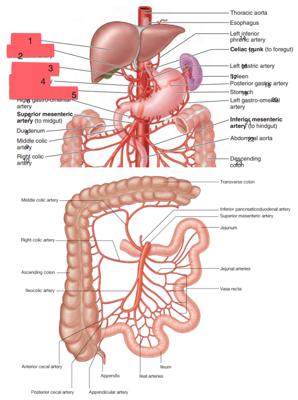
Label structures 1-5
liver, right inferior phrenic artery, right gastric artery, splenic artery, gastroduodenal artery
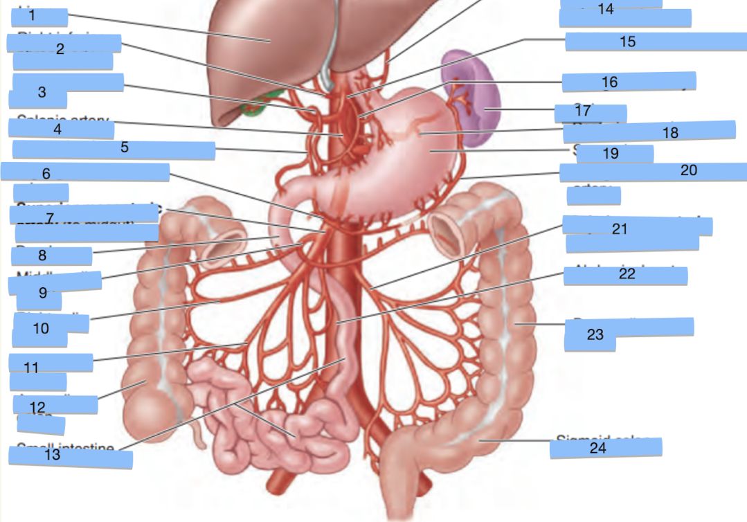
label structures 6-10
right gastro-omnetal artery, superior mesenteric artery, duodenum, middle colic artery, right colic artery
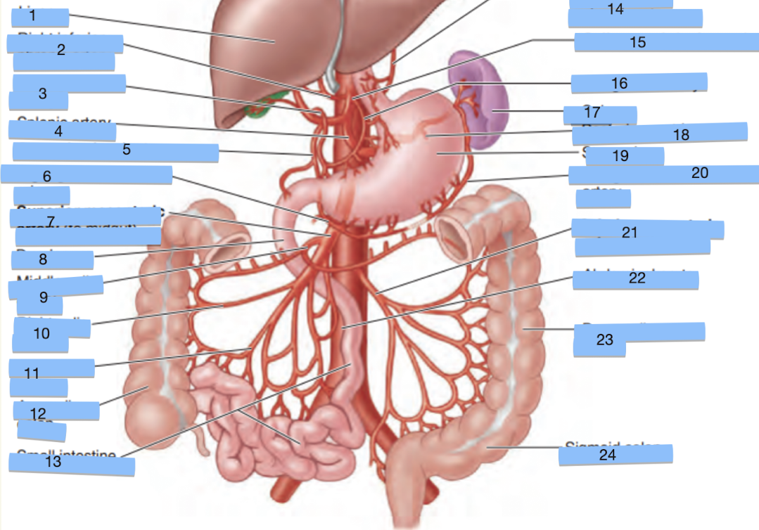
Label structures 11-15
ileocolic artery, ascending colon, small intestine, thoracic aorta, esophagus
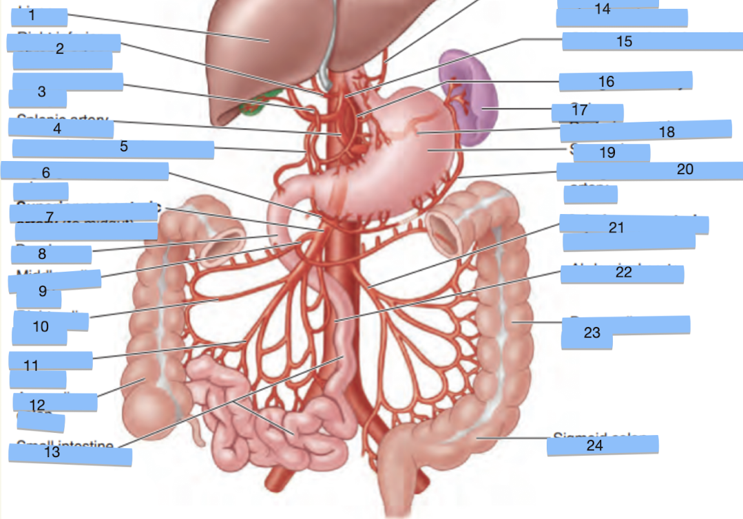
Label structures 14-18
left inferior phrenic artery, celiac trunk, left gastric artery, spleen, posterior gastric artery
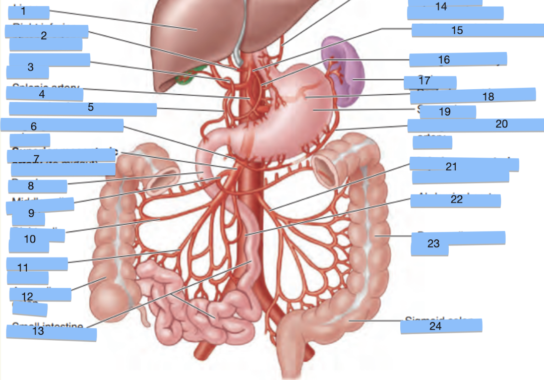
label structures 19-24 (I think I messed this one up do your best)
stomach, let gastro-mental artery, inferior mesenteric artery, abdominal aorta, descending colon, sigmoid colon
What are the branches of the superior mesenteric artery?
Inferior pancreaticoduodenal artery → pancreas and last part of duodenum
Jejunal and ileal arteries → jejunum and Ileum
Ileocolic artery → ileocaecal junction
Right colic artery → ascending colon
Middle colic artery → ascending colon and 2/3 of transverse colon
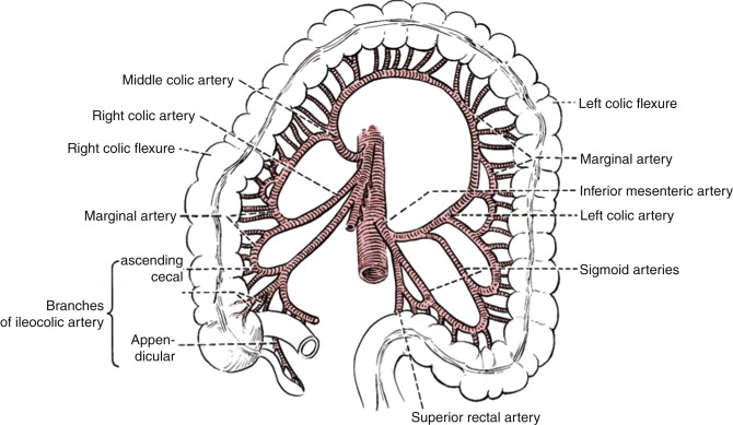
What are the branches of the inferior mesenteric artery?
Left colic artery, Sigmoid arteries, Superior rectal artery
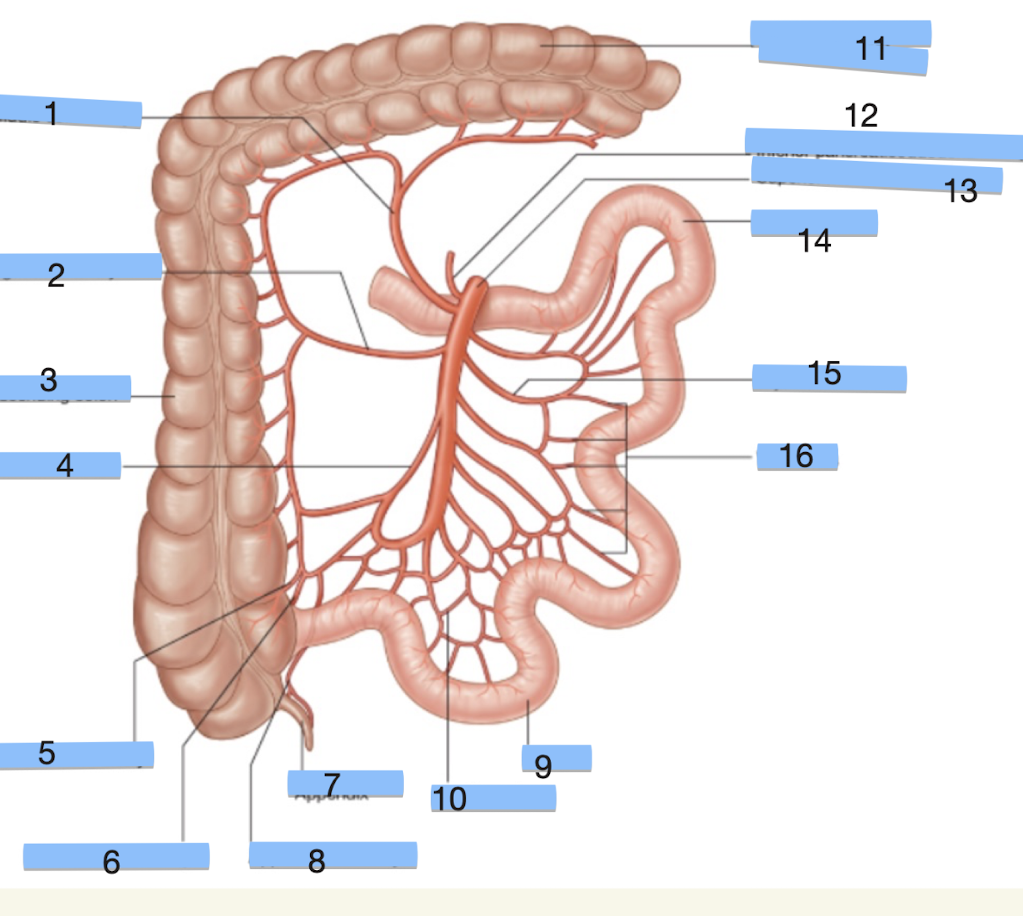
Label structures 1-5 (SMA)
middle colic artery, right colic artery, ascending colon, ileocolic artery, anterior cecal artery
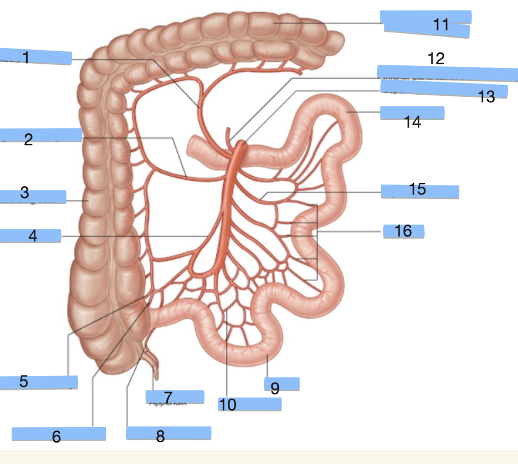
Label structures 6-10 (SMA)
posterior cecal artery, appendix, appendicular artery, ileum, ileal artery

Label strucutres 11-16 (SMA)
transverse colon, inferior pancreaticoduodenal artery, superior mesenteric artery, jejunum, jejunal arteries, vasa recta
is the inferior mesenteric artery over or under the duodenum
under
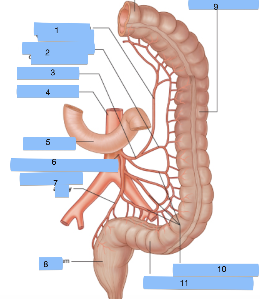
Label structures 1-5 (IMA)
ascending branch of left colic artery, descending branch of left colic artery, left colic artery, abdominal aorta, duodenum
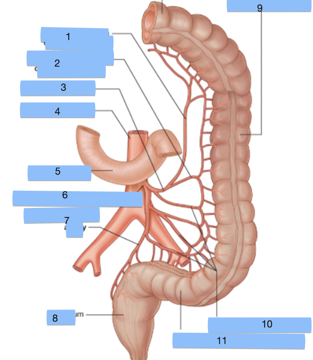
Label structures 6-11 (SMA)
inferior mesenteric artery superior rectal artery, rectum, descending colon, sigmoid arteries and sigmoid colon
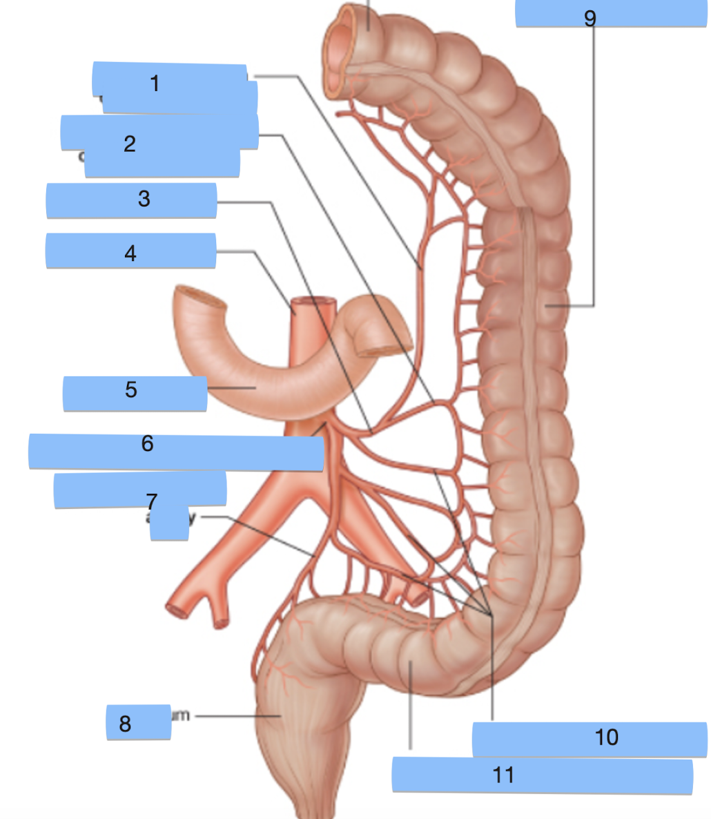
What structural label has been cut out of the top of this image?
transverse colon