Biopsychology: Nervous System
 Call Kai
 Learn
Learn Practice Test
 Spaced Repetition
 Match
Match Flashcards
 Knowt Play
1/81

Earn XP
Description and Tags
Name | Mastery | Learn | Test | Matching | Spaced | Call with Kai | Chat |
|---|
No analytics yet
Send a link to your students to track their progress
82 Terms
Neuraxis
Imaginary line oriented toward the spinal cord.
Dorsal
Toward the back or the top of the head
Ventral
Toward the chest or bottom of the head
Anterior
Toward the nose end/front of the body
Posterior
Toward the tail end or back of the head

Which area is the yellow arrows?
Dorsal


Which area is the red arrows?
Posterior
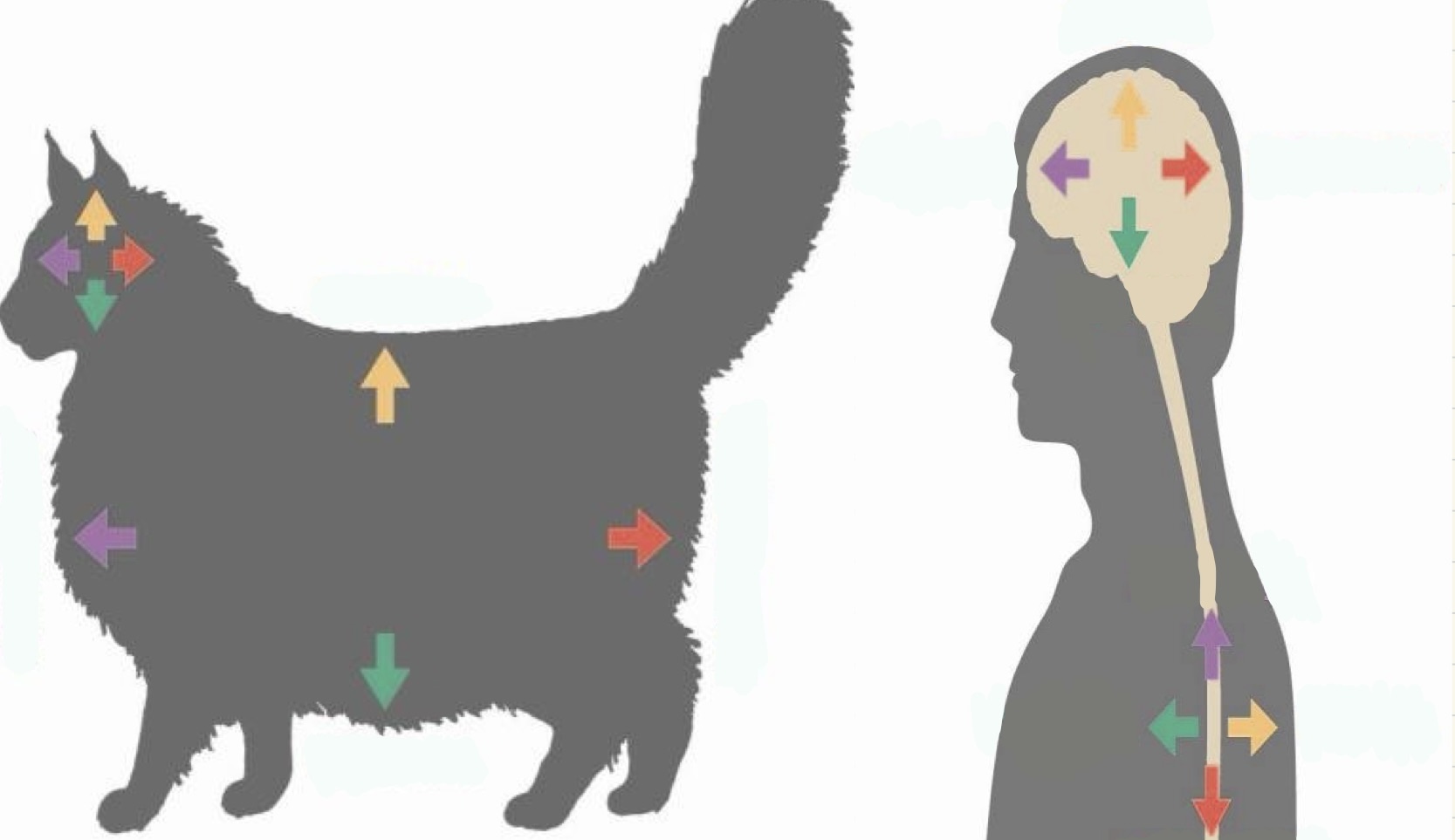

Which area is the green arrows?
Ventral


What are is the purple arrows?
Anterior

Rostral
Synonym for anterior, but used to describe relationships.
Caudal
Synonym for posterior, but used to describe relationships.
Transverse plane
Vertical cut done to the brain that creates a frontal section.
Sagittal Plane
Vertical cut done to the brain that separates it into left and right.
Horizontal Plane
Horizontal cut done to the Brian that separates the top and bottom.

What plane is W?
The sagittal plane


What plane is X?
Transverse plane (front section)


What plane is Y?
Horizontal plane


What plane is Z?
Transverse plane (cross section)

Ipsilateral
Relationship term meaning on the same side of the body
Contralateral
Relationship term meaning projecting from one side of the body to the other,
Afferent nerves
Nerves that carry sensory signals to the central nervous system.
Efferent nerves
Nerves that carry motor signals from the central nervous system to the skeletal muscles or internal organs.
Central Nervous System
The portion of the nervous system within the skull and spine.
Peripheral Nervous System
The portion of the nervous system outside the skull and spine.
Autonomic Nervous System
The part of the peripheral nervous system that participates in the regulation of the body’s internal environment.
Somatic Nervous System
The part of the peripheral nervous system that interacts with the external environment. Skeletal muscles.
Sympathetic Nervous System
Part of the autonomic nervous system that controls the body's rapid "fight-or-flight" response to stress and danger
Parasympathetic Nervous System
Part of the autonomic nervous system that controls resting, healing, and digesting
Temporal Lobe
The lobe processes hearing, language, memory, and emotions. It sits near your ears and temples on both sides of the brain

What lobe is this?
Temporal Lobe

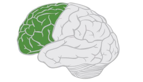
Frontal Lobe
The lobe that manages higher-level executive functions, voluntary movement, and behavioral control. Located directly behind the forehead, this brain region acts as the command center for key tasks

What lobe is this?
Frontal Lobe
Parietal Lobe
The lobe that processes and integrates sensory information like touch, pain, temperature, and body position. Located at the top and back of the brain.
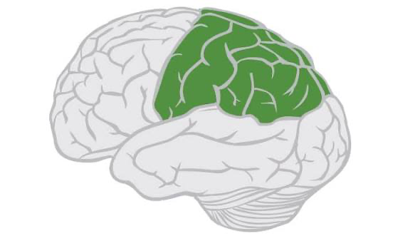
What lobe is this?
Parietal Lobe

Occipital Lobe
The lobe that processes visual information from the eyes. Located at the very back of the brain.
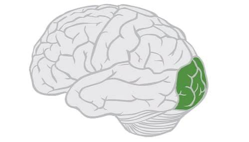
What lobe is this?
Occipital lobe

Primary Motor Cortex
Controls and executes voluntary muscle movements throughout the body. Located in the frontal lobe.
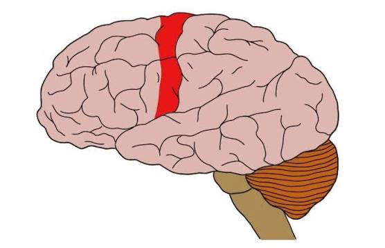
What cortex is this?
Primary motor cortex

Motor Association Cortex
The region of the frontal lobe that plans, prepares, and organizes complex voluntary muscle movements before they are executed
Primary Somatosensory Cortex
The main area of the brain that processes touch, pain, temperature, and body position. It sits in the upper, middle part of the cerebral cortex.

What cortex is this?
Primary Somatosensory Cortex

Somatosensory Association Cortex
The region of the brain responsible for interpreting sensory information and recognizing objects through touch. Located in the parietal lobe of the brain.
Primary Visual Cortex
Processes basic visual information from the eyes. Located in the occipital lobe at the very back of the brain.

What cortex is this?
Primary Visual Cortex

Visual Association Cortex
The region of the brain that interprets and gives meaning to raw visual signals received from the primary visual cortex. Located in the occipital lobe surrounding the primary visual cortex.
Primary Auditory Cortex
The main region of the brain that handles basic sound processing and conscious sound perception. Located in the temporal lobe of the brain.
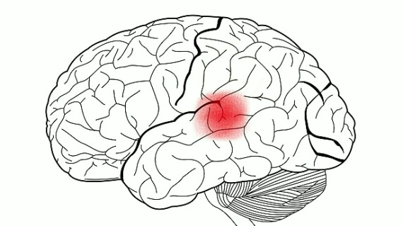
What cortex is this?
Primary Auditory Cortex

Auditory Association Cortex
The region of the brain that processes complex sounds, meaning, and memories of what you hear. Located in the temporal lobe of the brain, directly adjacent to and surrounding the primary auditory cortex.
Primary Olfactory Cortex
The group of brain regions on the bottom surface of the forebrain and temporal lobe that receives direct sensory input from the olfactory bulb. Located on the ventral (inferior) surface of the forebrain and temporal lobe.
Olfactory Association Area
Processes higher-order odor information to enable conscious perception, identification, and multi-sensory flavor experiences. Located in the inferior (ventral) surface of the orbitofrontal cortex, situated in the basal frontal lobe right above the orbits (eye sockets)
The Cerebrum
Contains frontal lobe, parietal lobe, temporal lobe, and occipital lobe. The largest part of the brain and controls higher-level functions like thinking, voluntary movement, and processing sensory information.
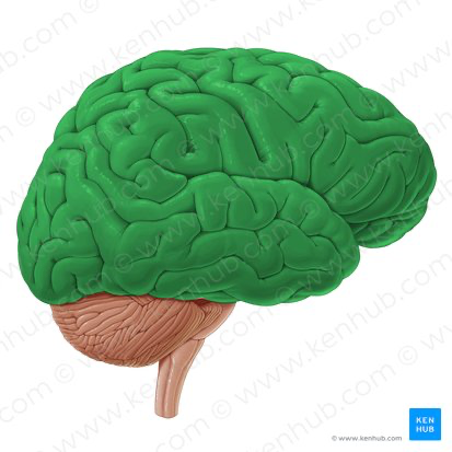
What brain structure is this?
The cerebrum

Cerebellum
The region of the brain responsible for coordinating voluntary muscle movements, maintaining balance, and supporting motor learning. Located at the lower back of the brain, directly below the cerebrum and behind the upper part of the brain stem.

What brain structure is this?
Cerebellum

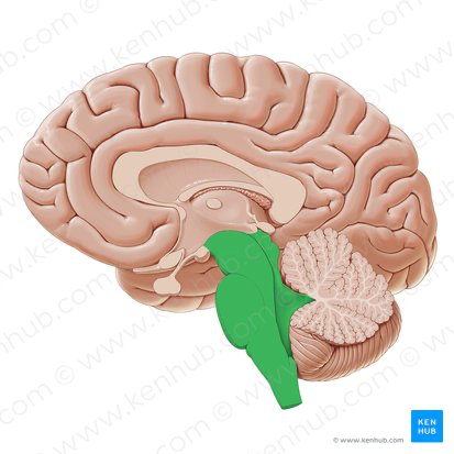
Brainstem
The body's vital control center and a communication highway between the brain and the spinal cord. Located at the base of the brain, inside the lower back part of the skull, where it connects the rest of the brain to the spinal cord.

What brain structure is this?
Brainstem
Ventricles
Produce, store, and circulate Cerebrospinal Fluid (CSF) through the central nervous system.
Right and left lateral ventricles
Located inside the left and right cerebral hemispheres of the cerebrum. Each large, C-shaped cavity extends protrusions (horns) into the frontal, temporal, and occipital lobes.
Third ventricle
Located along the midline of the brain in the diencephalon. It sits as a narrow, funnel-shaped cavity wedged directly between the left and right halves of the thalamus and the hypothalamus.
Fourth ventricle
Located in the hindbrain/brainstem region. It is positioned anteriorly (in front) against the pons and the upper portion of the medulla oblongata, and posteriorly (behind) against the cerebellum.
Meninges
Three protective membranes that cover and protect the brain and spinal cord
Dura mater
The thick, tough outermost layer attached to the skull and vertebral canal. Means “tough mother”
Arachnoid mater
The delicate, web-like middle layer.
Pia mater
The thin, tender innermost layer that tightly hugs the surface of the brain and spinal cord.
Arachnoid membrane
The middle, spiderweb-like layer of the meninges that covers and protects the brain and spinal cord
Subarachnoid space
The fluid-filled area located between the middle protective membrane (arachnoid mater) and the inner membrane (pia mater) surrounding the brain and spinal cord
Gray matter
Outlines the brain. Shaped like a butterfly or H-letter in the center of the cord. It contains nerve cell bodies, interneurons, and unmyelinated fibers that process reflex and sensory signals.
White Matter
Surrounds the gray matter. It consists of bundles of myelinated axons (nerve fibers) forming ascending and descending pathways that shuttle signals between the body and the brain
Cranial Nerves
A set of 12 pairs of nerves that connect your brain directly to your head, neck, and body, bypassing the spinal cord.
Spinal Nerves
31 pairs of mixed peripheral nerves that connect the spinal cord to the rest of the body to transmit sensory, motor, and autonomic signals. 8 in the neck region, 12 in the chest region, 5 in the hips/legs, and 1 for the tail.
Cerebral cortex
The wrinkled, outermost layer of the brain that handles high-level thinking, memory, and voluntary movement.
Corpus Callosum
A large, thick band of nerve fibers that connects the left and right hemispheres of the brain
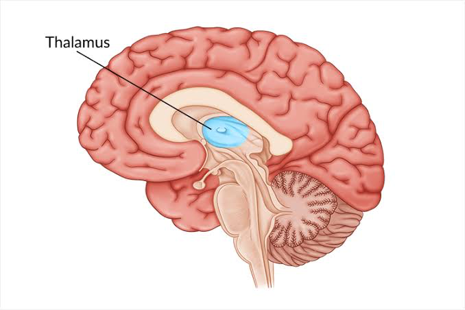
Thalamus
The brain's main information relay station, routing and processing sensory, motor, and cognitive signals. Sits just above your brainstem and processes nearly all incoming sensory (like touch, sight, and sound) and motor (movement) signals before sending them to the cerebral cortex for final interpretation.
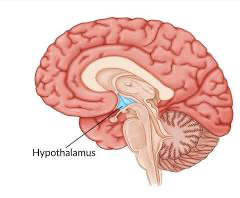
Hypothalamus
A tiny structure at the base of the brain that controls body balance, hormones, and basic survival needs. It sits deep in the center of the brain, below the thalamus and right above the pituitary gland.

Midbrain/Mesencephalon
The topmost part of the brainstem that links the higher forebrain to the lower hindbrain and spinal cord. Acts as a major relay center for movement, eye control, and sensory signals.

Tectum
The "roof" or dorsal part of the midbrain, which sits in the upper portion of the brainstem. It coordinates quick reflex responses to sights and sounds.
Tegmentum
A thick network of nerve fibers and cell clusters that forms the central floor of the brainstem. Coordinates vital functions including movement control, pain processing, wakefulness, and basic sensory reflexes.

Pons
Act as a major relay station and bridge connecting the cerebrum, cerebellum, and medulla oblongata to coordinate vital bodily messages.
Medulla Oblongata
The lower part of the brainstem that connects your brain to your spinal cord, controlling vital, involuntary life-support functions. (Breathing, heart rate, swallowing, etc.)
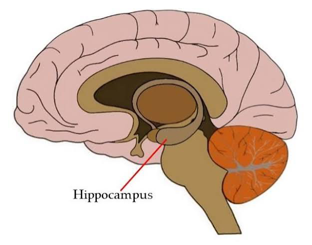
Hippocampus
A seahorse-shaped region deep inside the brain's temporal lobe that plays a central role in learning, memory consolidation, and spatial navigation.

Limbic System
A group of connected structures deep inside the brain that control emotions, memory, and basic survival behaviors

Amygdala
An almond-shaped cluster of neurons located deep within the brain's temporal lobe that processes emotions and survival responses. One in each hemisphere. As a key part of the limbic system, it acts as the brain's emotional and threat-detection center.